. 2021 Jul;11(7):1835-1852.
doi: 10.1016/j.apsb.2021.01.015. Epub 2021 Jan 23.

Targeted inhibition of GRK2 kinase domain by CP-25 to reverse fibroblast-like synoviocytes dysfunction and improve collagen-induced arthritis in rats

Affiliations

Targeted inhibition of GRK2 kinase domain by CP-25 to reverse fibroblast-like synoviocytes dysfunction and improve collagen-induced arthritis in rats

Chenchen Han et al. Acta Pharm Sin B. 2021 Jul.

Abstract

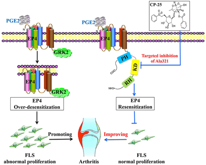
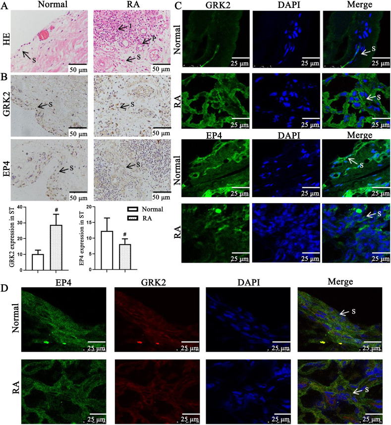
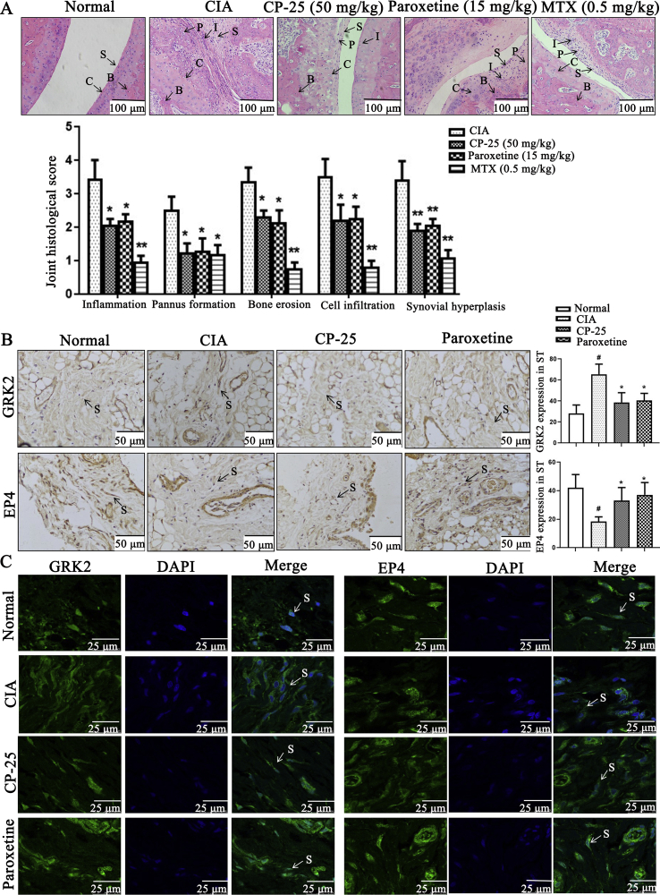
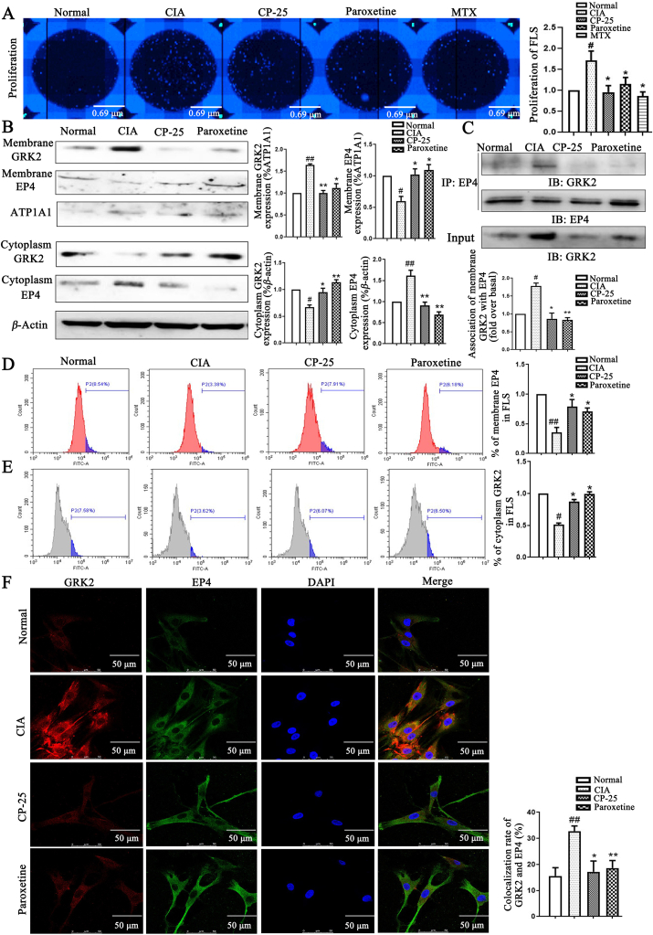
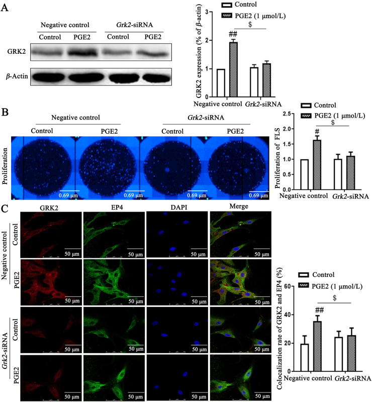
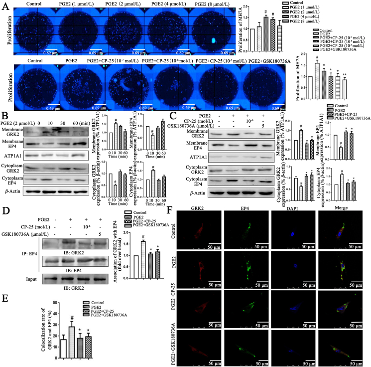
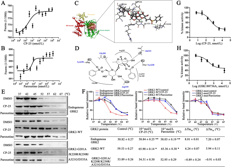
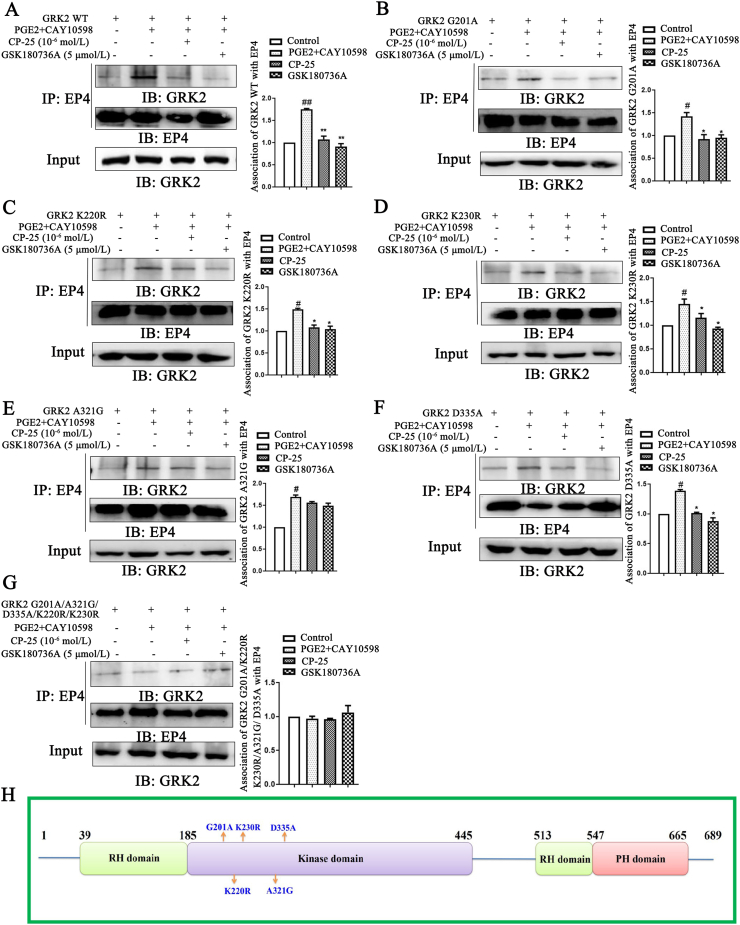
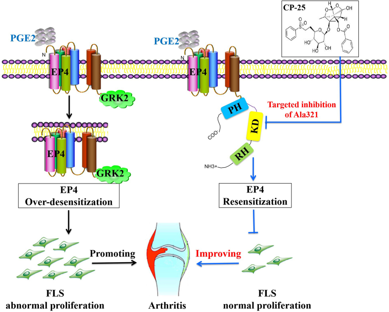
Rheumatoid arthritis (RA) is an autoimmune disease and is mainly characterized by abnormal proliferation of fibroblast-like synoviocytes (FLS). The up-regulated cellular membrane expression of G protein coupled receptor kinase 2 (GRK2) of FLS plays a critical role in RA progression, the increase of GRK2 translocation activity promotes dysfunctional prostaglandin E4 receptor (EP4) signaling and FLS abnormal proliferation. Recently, although our group found that paeoniflorin-6'-O-benzene sulfonate (CP-25), a novel compound, could reverse FLS dysfunction via GRK2, little is known as to how GRK2 translocation activity is suppressed. Our findings revealed that GRK2 expression up-regulated and EP4 expression down-regulated in synovial tissues of RA patients and collagen-induced arthritis (CIA) rats, and prostaglandin E2 (PGE2) level increased in arthritis. CP-25 could down-regulate GRK2 expression, up-regulate EP4 expression, and improve synovitis of CIA rats. CP-25 and GRK2 inhibitors (paroxetine or GSK180736A) inhibited the abnormal proliferation of FLS in RA patients and CIA rats by down-regulating GRK2 translocation to EP4 receptor. The results of microscale thermophoresis (MST), cellular thermal shift assay, and inhibition of kinase activity assay indicated that CP-25 could directly target GRK2, increase the protein stability of GRK2 in cells, and inhibit GRK2 kinase activity. The docking of CP-25 and GRK2 suggested that the kinase domain of GRK2 might be an important active pocket for CP-25. G201, K220, K230, A321, and D335 in kinase domain of GRK2 might form hydrogen bonds with CP-25. Site-directed mutagenesis and co-immunoprecipitation assay further revealed that CP-25 down-regulated the interaction of GRK2 and EP4 via controlling the key amino acid residue of Ala321 of GRK2. Our data demonstrate that FLS proliferation is regulated by GRK2 translocation to EP4. Targeted inhibition of GRK2 kinase domain by CP-25 improves FLS function and represents an innovative drug for the treatment of RA by targeting GRK2.

Keywords: CP-25; Fibroblast-like synoviocyte; G protein coupled receptor kinase 2; MH7A; Prostaglandin E4 receptor; Rheumatoid arthritis.


Conflict of interest statement

The authors have no financial conflicts of interest.

Figures

Image 1
Graphical abstract
Fig. 1
Figure 1
G protein coupled receptor kinase 2 (GRK2) and prostaglandin E4 receptor (EP4) expression in synovial tissues (ST) of rheumatoid arthritis (RA) patients. (A) Representative micrographs of haematoxylin and eosin (HE)-stained histological sections of synovial tissues. The histology section shows the synoviocytes (S), the pannus (P), the inflammatory cells (I) (n = 8). Scale bars = 50 μm. (B) GRK2 and EP4 expression in synovial tissues paraffin section of RA patients were detected by immunohistochemistry (n = 8). Data are expressed as mean ± standard deviation (SD). #P < 0.05 vs. normal group. Arrows indicated the fibroblast-like synoviocytes (FLS) of RA. Scale bars = 50 μm. (C) GRK2 and EP4 expression in synovial tissues paraffin section of RA patients were detected by laser scanning confocal microscope (n = 8). Arrows indicated the FLS of RA. Scale bars = 25 μm. (D) GRK2/EP4 expression in synovial tissues frozen section of RA patients were detected by laser scanning confocal microscope (n = 8). Arrows indicated the FLS of RA. Scale bars = 25 μm.
Fig. 2
Figure 2
GRK2 and EP4 expression in synovial tissues of CIA rats. (A) Representative micrographs of HE-stained histological sections of the joints are shown. The histology section of the joints shows the synoviocytes (S), the pannus formation (P), the inflammatory cells (I), the bone (B), and the cartilage (C) (n = 6). Data are expressed as mean ± SD. P < 0.05, P < 0.01 vs. CIA group. Scale bars = 100 μm. (B) The effect of CP-25 on the expression of GRK2 and EP4 in the synovial cells of CIA rats was detected by immunohistochemistry assay (n = 6). Arrows indicated the FLS of CIA rats. Scale bars = 50 μm. (C) The effect of CP-25 on the distribution of GRK2 and EP4 in the synovial cells of CIA rats was detected by laser scanning confocal microscope (n = 6). Arrows indicated the FLS of CIA rats. Scale bars = 25 μm. Data are expressed as mean ± SD. #P < 0.05 vs. normal group; ∗P < 0.05, ∗∗P < 0.01 vs. CIA group.
Fig. 3
Figure 3
CP-25 treatment improves FLS proliferation of CIA rats by down-regulating GRK2 translocation. (A) CP-25 inhibited FLS abnormal proliferation was detected by high content cell imaging system (n = 6). (B) The effect of CP-25 on the expression of GRK2 and EP4 on membrane and cytoplasm of FLS (n = 4). (C) FLS lysates were used anti-EP4 antibody by co-immunoprecipitate and subjected to Western blot to visualize GRK2 to evaluate the association with EP4 (n = 4). (D) EP4 membrane expression of FLS by flow cytometry (n = 6). (E) GRK2 cytoplasm expression of FLS by flow cytometry (n = 6). (F) The co-expression of GRK2 and EP4 was detected by laser confocal microscopy (n = 6). Scale bars = 50 μm. Data are expressed as mean ± SD. #P < 0.05, ##P < 0.01 vs. normal group; P < 0.05, P < 0.01 vs. CIA group.
Fig. 4
Figure 4
GRK2 knockdown strongly inhibits the proliferation of FLS, possibly via PGE2–EP4 signaling pathway. (A) Grk2 siRNA was transfected into FLS, the inhibition rate of GRK2 was detected by Western blot (n = 4). (B) FLS proliferation was detected by high content cell imaging system images (n = 6). (C) The co-expression of GRK2 and EP4 was detected by laser confocal microscopy (n = 6). Scale bars = 50 μm. Data are expressed as mean ± SD. #P < 0.05, ##P < 0.01 vs. control group; $P < 0.05 vs. negative control group with PGE2.
Fig. 5
Figure 5
CP-25 inhibits PGE2-induced MH7A proliferation by down-regulating GRK2 translocation. (A) CP-25 inhibited PGE2-induced MH7A proliferation (n = 3). (B) PGE2 induced EP4 receptor short-term desensitization and GRK2 membrane translocation. The membrane expression of EP4 and GRK2 in MH7A treated by PGE2 were detected by Western blot (n = 3). (C) The effect of CP-25 on the expression of GRK2 and EP4 on membrane of MH7A stimulated by PGE2 (n = 3). (D) PGE2 stimulated MH7A lysates were used anti-EP4 antibody by co-immunoprecipitate and subjected to Western blot to visualize GRK2 to evaluate the association with EP4 (n = 3). (E) and (F) The co-expression of GRK2 and EP4 was detected by laser confocal microscopy (n = 6). Scale bars = 50 μm. Data are expressed as mean ± SD. #P < 0.05 vs. control group; P < 0.05, P < 0.01 vs. PGE2.
Fig. 6
Figure 6
CP-25 directly binds to the kinase domain of GRK2 and inhibits GRK2 activity. (A) and (B) Microscale thermophoresis (MST) of CP-25 and paroxetine. GRK2 (0.49 mg/mL) were incubated with increasing concentrations of CP-25 (A) and paroxetine (B). The interactions of CP-25 or paroxetine and GRK2 were quantified by MST and binding data were plotted applying the Kd equation. (C) Molecular docking modeling of compound CP-25 and GRK2, the small molecule and the critical interaction of 3KRW are represented by sticks. Panel is a view into the active site cavity. (D) Schematic representation of the binding mode of CP-25 in the GRK2 binding site of 3KRW. (E) Cellular thermal shift assay (CETSA) presented the thermal stability of endogenous GRK2, wild-type GRK2 (GRK2-WT), and GRK2 G201A/A321G/D335A/K220R/K230R mutant proteins under treatment with CP-25 (10−6 mol/L) and paroxetine (10−6 mol/L). (F) CETSA curve and the thermal stability to reach 50% of temperature (Tm50) value was performed using GraphPad Prism software. Data are expressed as mean ± SD. P < 0.05, P < 0.01 vs. control group. (G) and (H) Determining IC50 for CP-25 and GRK2 inhibitor. IC50 were determined using the ADP-Glo™ Kinase Assay. Curve fitting was performed using GraphPad Prism software. All data are expressed as mean ± SD.
Fig. 7
Figure 7
Study on the binding sites of CP-25 and GRK2. HEK 293T cells transfection with pIRES-EGFP-GRK2 WT (A), pIRES-EGFP-GRK2 G201A (B), pIRES-EGFP-GRK2 K220R (C), pIRES-EGFP-GRK2 K230R (D), pIRES-EGFP-GRK2 A321G (E), pIRES-EGFP-GRK2 D335A (F), and pIRES-EGFP-GRK2 G201A/K220R/K230R/A321G/D335A (G) plasmids were challenged with CAY10598 (1 μmol/L) following a 1 h pre-treatment with PGE2 (10 μmol/L) for 30 min in the presence and absence of CP-25 (10−6 mol/L) or GSK180736A (5 μmol/L). The association of GRK2 mutants and EP4 was determined by co-IP using the EP4-specific antibody and subsequent blotting with GRK2-specific (n = 3). Data are expressed as mean ± SD. #P < 0.05, ##P < 0.01 vs. control group; P < 0.05, P < 0.01 vs. PGE2+CAY10598 group. (H) Location of mutations made in the GRK2 domain. Light green bars represent the regulator of G-protein signaling (RGS) homology (RH) domain, light purple bars represent kinase domain and pink bars represent Pleckstrin homology (PH) domain. G201, K220, K230, A321, and D335 residues are those that were mutated in this study.
Fig. 8
Figure 8
Schematic illustration of CP-25 improves EP4 desensitization mediated FLS dysfunction via stabilizing KD and controlling Ala321 of GRK2. PGE2 level in FLS of CIA rats was up-regulated, promoting PGE2 binding to the EP4 receptor, inducing the increasing of GRK2 translocation, leading to EP4 over-desensitization, thereby promoting FLS dysfunction. CP-25 can stabilize the kinase domain of GRK2 to directly inhibit GRK2 kinase, leading to decreasing of GRK2 translocation to EP4 by controlling Ala321 of GRK2, thereby promoting EP4 resensitization, which was an important mechanism of CP-25 in improving FLS dysfunction.

References

    1. Cross M., Smith E., Hoy D., Carmona L., Wolfe F., Vos T. The global burden of rheumatoid arthritis: Estimates from the Global Burden of Disease 2010 study. Ann Rheum Dis. 2014;73:1316–1322. - PubMed
    1. Benson R.A., McInnes I.B., Brewer J.M., Garside P. Cellular imaging in rheumatic diseases. Nat Rev Rheumatol. 2015;11:357–367. - PubMed
    1. Schumacher H.R. Are we being open enough to all approaches to therapy of rheumatoid arthritis?. J Clin Rheumatol. 2013;19:167–171. - PubMed
    1. Lee D.M., Kiener H.P., Agarwal S.K., Noss E.H., Watts G.F.M., Chisaka O. Cadherin-11 in synovial lining formation and pathology in arthritis. Science. 2007;315:1006–1010. - PubMed
    1. Chen Q., Wei W. Effects and mechanisms of glucosides of chaenomeles speciosa on collagen-induced arthritis in rats. Int Immunopharm. 2003;3:593–608. - PubMed

LinkOut - more resources