

AncPhore: A versatile tool for anchor pharmacophore steered drug discovery with applications in discovery of new inhibitors targeting metallo- β-lactamases and indoleamine/tryptophan 2,3-dioxygenases
- PMID: 34386329
- PMCID: PMC8343198
- DOI: 10.1016/j.apsb.2021.01.018
AncPhore: A versatile tool for anchor pharmacophore steered drug discovery with applications in discovery of new inhibitors targeting metallo- β-lactamases and indoleamine/tryptophan 2,3-dioxygenases
Abstract
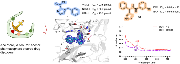
We herein describe AncPhore, a versatile tool for drug discovery, which is characterized by pharmacophore feature analysis and anchor pharmacophore (i.e., most important pharmacophore features) steered molecular fitting and virtual screening. Comparative analyses of numerous protein-ligand complexes using AncPhore revealed that anchor pharmacophore features are biologically important, commonly associated with protein conservative characteristics, and have significant contributions to the binding affinity. Performance evaluation of AncPhore showed that it had substantially improved prediction ability on different types of target proteins including metalloenzymes by considering the specific contributions and diversity of anchor pharmacophore features. To demonstrate the practicability of AncPhore, we screened commercially available chemical compounds and discovered a set of structurally diverse inhibitors for clinically relevant metallo-β-lactamases (MBLs); of them, 4 and 6 manifested potent inhibitory activity to VIM-2, NDM-1 and IMP-1 MBLs. Crystallographic analyses of VIM-2:4 complex revealed the precise inhibition mode of 4 with VIM-2, highly consistent with the defined anchor pharmacophore features. Besides, we also identified new hit compounds by using AncPhore for indoleamine/tryptophan 2,3-dioxygenases (IDO/TDO), another class of clinically relevant metalloenzymes. This work reveals anchor pharmacophore as a valuable concept for target-centered drug discovery and illustrates the potential of AncPhore to efficiently identify new inhibitors for different types of protein targets.
Keywords: AMPC, asian mouse phenotyping consortium; AP, anchor pharmacophore; AR, aromatic ring; AUC, area under the curve; Anchor pharmacophore; BACE1, beta-secretase 1; BRD4, bromodomain-containing protein 4; CA, carbonic anhydrase; CA2, carbonic anhydrase 2; CDK2, cyclin-dependent kinase 2; CTS, cathepsins; CV, covalent bonding; CatK, cathepsin K; EF, enrichment factor; EX, exclusion volume; GA, genetic algorithm; HA, hydrogen-bond acceptor; HD, hydrogen-bond donor; HIV-P, human immunodeficiency virus protease; HIV1-P, human immunodeficiency virus type 1 protease; HY, hydrophobic; IDO1, indoleamine 2,3-dioxygenase 1; IMP, imipenemase; Indoleamine 2,3-dioxygenase; LE, ligand efficiency; MAPK14, mitogen-activated protein kinase 14; MB, metal coordination; MBL, metallo-β-lactamase; MIC, minimum inhibitory concentration; MMP, matrix metalloproteinase; MMP13, matrix metallopeptidase 13; Metallo-β-lactamase; Metalloenzyme; NDM, new delhi MBL; NE, negatively charged center; NP, without anchor pharmacophore features; PO, positively charged center; RMSD, root mean square deviation; ROC curve, receiver operating characteristic curve; ROCK1, rho-associated protein kinase 1; RT, reverse transcriptase; RTK, receptor tyrosine kinase; SBL, serine beta lactamase; SSEL, secondary structure element length; STK, serine threonine kinase; TDO, tryptophan 2,3-dioxygenase; TDSS, torsion-driving systematic search; TNKS2, tankyrase 2; Tryptophan 2,3-dioxygenase; VEGFR2, vascular endothelial growth factor receptor 2; VIM, verona integron-encoded MBL; Virtual screening.
© 2021 Chinese Pharmaceutical Association and Institute of Materia Medica, Chinese Academy of Medical Sciences. Production and hosting by Elsevier B.V.
Conflict of interest statement
The authors declare no competing financial interest.
Figures
Similar articles
-
Metal binding pharmacophore click-derived discovery of new broad-spectrum metallo-β-lactamase inhibitors.Eur J Med Chem. 2023 Sep 5;257:115473. doi: 10.1016/j.ejmech.2023.115473. Epub 2023 May 13. Eur J Med Chem. 2023. PMID: 37209449
-
Sulfamoyl Heteroarylcarboxylic Acids as Promising Metallo-β-Lactamase Inhibitors for Controlling Bacterial Carbapenem Resistance.mBio. 2020 Mar 17;11(2):e03144-19. doi: 10.1128/mBio.03144-19. mBio. 2020. PMID: 32184250 Free PMC article.
-
Small Molecule Carboxylates Inhibit Metallo-β-lactamases and Resensitize Carbapenem-Resistant Bacteria to Meropenem.ACS Infect Dis. 2020 Jun 12;6(6):1366-1371. doi: 10.1021/acsinfecdis.9b00459. Epub 2020 Apr 3. ACS Infect Dis. 2020. PMID: 32227874 Free PMC article.
-
Epidemiology and Characteristics of Metallo-β-Lactamase-Producing Pseudomonas aeruginosa.Infect Chemother. 2015 Jun;47(2):81-97. doi: 10.3947/ic.2015.47.2.81. Epub 2015 Jun 30. Infect Chemother. 2015. PMID: 26157586 Free PMC article. Review.
-
Principles and current strategies targeting metallo-β-lactamase mediated antibacterial resistance.Med Res Rev. 2020 Sep;40(5):1558-1592. doi: 10.1002/med.21665. Epub 2020 Feb 25. Med Res Rev. 2020. PMID: 32100311 Review.
Cited by
-
MeDBA: the Metalloenzyme Data Bank and Analysis platform.Nucleic Acids Res. 2023 Jan 6;51(D1):D593-D602. doi: 10.1093/nar/gkac860. Nucleic Acids Res. 2023. PMID: 36243971 Free PMC article.
-
Exploring potential SARS-CoV-2 Mpro non-covalent inhibitors through docking, pharmacophore profile matching, molecular dynamic simulation, and MM-GBSA.J Mol Model. 2023 Apr 13;29(5):138. doi: 10.1007/s00894-023-05534-3. J Mol Model. 2023. PMID: 37055578 Free PMC article.
-
Computational study on the mechanism of small molecules inhibiting NLRP3 with ensemble docking and molecular dynamic simulations.BMC Pharmacol Toxicol. 2025 Mar 3;26(1):49. doi: 10.1186/s40360-025-00851-0. BMC Pharmacol Toxicol. 2025. PMID: 40033437 Free PMC article.
-
CovalentInDB 2.0: an updated comprehensive database for structure-based and ligand-based covalent inhibitor design and screening.Nucleic Acids Res. 2025 Jan 6;53(D1):D1322-D1327. doi: 10.1093/nar/gkae946. Nucleic Acids Res. 2025. PMID: 39441070 Free PMC article.
-
CovPDB: a high-resolution coverage of the covalent protein-ligand interactome.Nucleic Acids Res. 2022 Jan 7;50(D1):D445-D450. doi: 10.1093/nar/gkab868. Nucleic Acids Res. 2022. PMID: 34581813 Free PMC article.
References
-
- Du J., Guo J., Kang D., Li Z., Wang G., Wu J. New techniques and strategies in drug discovery. Chin Chem Lett. 2020;31:1695–1708.
-
- Yang S.Y. Pharmacophore modeling and applications in drug discovery: challenges and recent advances. Drug Discov Today. 2010;15:444–450. - PubMed
-
- Schaller D., Šribar D., Noonan T., Deng L., Nguyen T.N., Pach S. Next generation 3D pharmacophore modeling. WIREs Comput Mol Sci. 2020;10:e1468.
-
- Taminau J., Thijs G., De Winter H. Pharao: Pharmacophore alignment and optimization. J Mol Graph Model. 2008;27:161–169. - PubMed
LinkOut - more resources
Full Text Sources
Research Materials
Miscellaneous
