. 2021 Jul 19:21:101510.
doi: 10.1016/j.jcot.2021.101510. eCollection 2021 Oct.

Imaging of traumatic peripheral nerve injuries

Affiliations

Imaging of traumatic peripheral nerve injuries

Ankur Goyal et al. J Clin Orthop Trauma. .

Abstract

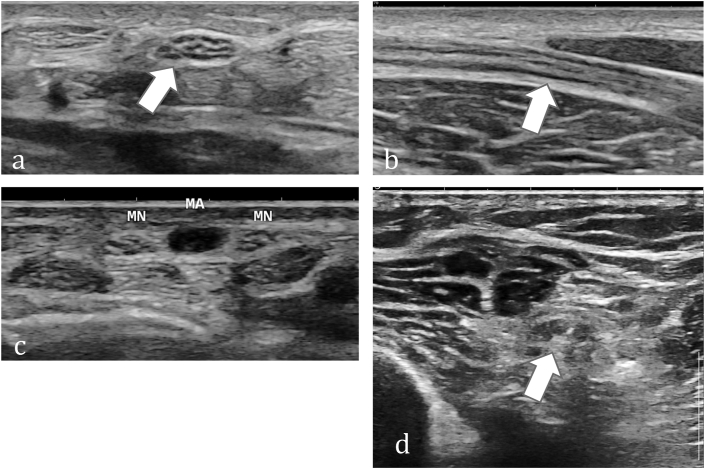
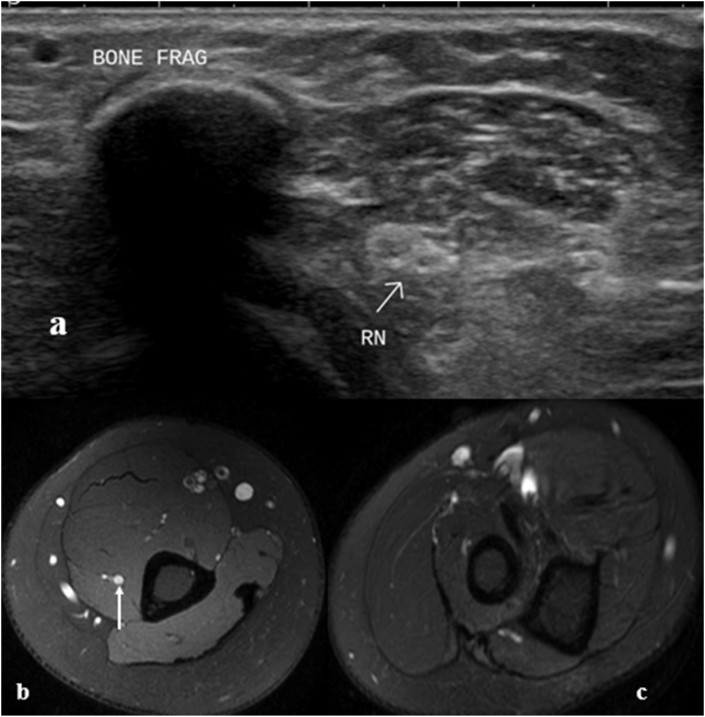
Nerves are commonly injured in case of blunt or penetrating trauma to the extremities. Patients with nerve injuries have profound consequences and thus a timely decision for operative management is a very important. Conventionally, management decisions have been based on clinical findings, patient course and electrophysiological studies. However, imaging modalities have an enormous role not only in localizing and grading of the nerve injuries but also in the follow-up of the nerve recovery. High-resolution ultrasound (HUS) is the modality of choice for evaluation of peripheral nerves. Magnetic resonance neurography (MRN) plays a complementary role, enabling better assessment of muscle changes and deeper nerves. Corresponding to the injured layer of the cross-section of the nerve, imaging manifestations differ in different grades of injury. Since imaging cannot detect ultrastructural changes at the microscopic level, thus there may be overlap in the imaging findings. Herewith, we discuss the imaging findings in different grades of nerve injury and propose a simple 3-tier grading for imaging (HUS and MRN) assessment of peripheral nerve injuries.

Keywords: High-resolution ultrasound (HUS); Magnetic resonance; Neurography (MRN); Peripheral nerve injuries.


Conflict of interest statement

The Authors declare that there is no conflict of interest.

Figures

Fig. 1
Fig. 1
Examples of patient positioning while evaluating peripheral nerves. (a) and (b) show the arm and elbow positioning while evaluating the median nerve. (c) depicts abducted arm for examination of medially located ulnar nerve while (d) shows the positioning for examination of the ulnar nerve at the elbow (patient back towards the examiner). (e) demonstrates the examination of radial nerve in the posterior arm while (f) shows examination of radial nerve in the lateral aspect of distal arm. (g) depicts prone positioning of the patient for assessing sciatic nerve and its divisions. (h) demonstrates evaluation of the tibial nerve posterior to the medial malleolus.
Fig. 2
Fig. 2
Imaging appearance of normal peripheral Nerves on ultrasound. (a) Axial ultrasound image shows speckled appearance in cross-section of the median nerve with hypoechoic fascicles and echogenic connective tissue. (b) Shows fascicular pattern in the longitudinal section of the median nerve.(c) Axial ultrasound image in a different patient depicts duplicated median nerve (MN) with persistent median artery (MA) in between.(d) Axial ultrasound image in a different patient depicts speckled appearance in cross-section of the sciatic nerve.
Fig. 3
Fig. 3
Imaging appearance of Normal peripheral Nerves on MRI (a) Axial T1W, (b) T2W (c) T1W fat-suppressed (FS) and (d) T2W FS turbo-spin-echo (TSE) images show the fascicular pattern of normal tibial (block arrow) and peroneal (thin arrow) nerves. (e) Diffusion-weighted color-coded fiber tractography image depicts the fibers of these nerves after delineating the corresponding regions of interest (ROI). (f) Diffusion tensor imaging (DTI) image shows tibial nerve overlapped with the T2W FS image shows corresponding color-coded FA map (inset in f) (FA value – 0.52 and ADC value 1.39 × 10−3 mm2/s).(g) Coronal thick maximal intensity projection (MIP) image from volume diffusion-weighted imaging in a different patient with old right sciatic nerve injury depicts neuroma-in-continuity in the gluteal region (block arrow) with atrophy and reduced signal intensity of RT sciatic nerve compared to the left (thin arrows).
Fig. 4
Fig. 4
Schematic representation of different grades of nerve injuries (Sunderland). (a) Normal appearing Nerve in grade 1 injury, (b) mild bulkiness (nerve edema) in grade II injury, (c) Focal bulkiness and fascicular thickening in grade III injury, (d) Focal fascicular disruption in grade 4 injury and (e) nerve transection with discontinuity of epineurium in grade 5 injury. The individual nerve fascicles are covered by perineurium (arrows in a) while the white area between the fascicles represents the connective tissue of the inner epineurium. Outer epineurium covers the entire nerve (arrowheads in a).
Fig. 5
Fig. 5
Imaging findings in Neuropraxia (Sunderland Grade 1 injury).(a) Axial ultrasound image in a patient with sensory loss in radial nerve distribution after humeral shaft fracture shows normal appearing radial nerve adjacent to the bony fragment. The sensory loss significantly improved spontaneously within two months. Findings are consistent with neuropraxia with normal ultrasound appearance. (b) Axial T2W FS image in a different patient shows abnormal T2 hyperintensity in the right radial nerve (c) Caudal section shows absence of any muscle edema. Diffuse T2 hyperintensity with absent denervation changes suggested Grade 1 injury.
Fig. 6
Fig. 6
Imaging findings in Axonotmesis (Sunderland Grade 2 injury). (a) Longitudinal ultrasound image shows diffuse hypoechogenicity of ulnar nerve in cubital fossa (rectangle). Distal part shows preserved fascicular pattern. (b) Axial US image shows normal honeycombed appearance in the distal part. (c) Focused assessment of the abnormal part longitudinal image shows diffuse hypoechogenicity and bulkiness. (d) Axial US image better demonstrates the bulkiness of the hypoechoic ulnar nerve along with thickening of perineural soft tissues, suggesting grade 2 injury. (e) Axial T2W FS image in a different patient with left wrist drop after humeral shaft fracture shows mildly bulky and hyperintense radial nerve. (f) Caudal section shows edema in supinator muscle. Findings are consistent with at least grade 2 injury.
Fig. 7
Fig. 7
Imaging findings in Sunderland Grade 3 injury. (a) Axial ultrasound image in a patient with ulnar nerve injury shows normal appearing ulnar nerve proximal to the cubital tunnel. (b) Axial ultrasound image at the injury site shows focal bulkiness and hypoechogenicity with perineural thickening at the site of injury. (c) Longitudinal US image in a different patient shows focal effacement of fascicles (block arrow) in median nerve, corresponding to grade 3 or 4 injury. (d) Oblique coronal T1W MR Image shows focal bulkiness of peroneal nerve (arrow) at the site of injury adjacent to the fibular head (asterisk), suggesting grade 3 injury. (e) Axial US image in a different patient shows focal changes in the radial nerve (arrow) with loss of speckled pattern corresponding to the hypoechoic scar tract of penetrating injury. (f) Axial T2W FS image in a different patient shows abnormal focal T2 hyperintensity in the ulnar nerve with thickened fascicles and surrounding perineural edema, suggesting grade 3 ulnar nerve injury behind the medial epicondyle.
Fig. 8
Fig. 8
Imaging findings in Sunderland Grade 4 injury. (a) Longitudinal ultrasound image in a patient with ulnar claw hand after trauma shows focal bulkiness and disruption of the nerve fascicles (block arrow, area between the 2 cursors), suggesting grade 4 injury. (b) Longitudinal ultrasound image in a patient with peroneal nerve injury after knee replacement surgery, shows focal bulkiness and disruption of the nerve fascicles (block arrows) suggesting grade 4 injury. (c) Corresponding axial section shows hypoechogenicity, bulkiness and loss of speckled pattern, concordant with fascicular disruption.
Fig. 9
Fig. 9
Imaging findings in Sunderland Grade 4 injury at the origin of peroneal nerve. (a) Axial US image at sciatic nerve division and (b) distally show bulky hypoechoic peroneal component (TN: Tibial nerve and CPN: Peroneal nerve). ((c) Longitudinal extended field-of-view panoramic US image shows fascicular sciatic nerve proximally (asterisk) and hypoechoic bulky peroneal nerve (oval), abnormal right from its origin. (d) Focused longitudinal image of peroneal nerve segment shows loss of fascicular pattern (oval), but continuity is preserved and fascicular pattern is seen in distal part (block arrow). (e) Radiograph shows fracture of lateral tibial condyle and fibular head and neck with internal fixation implant in situ.
Fig. 10
Fig. 10
Ultrasound findings in Sunderland Grade 5 injury. (a) Longitudinal US image shows complete transection of ulnar nerve after penetrating injury (tract seen, denoted by asterisk) with retracted neuromas (block arrows) (b) Axial US image shows multiple punctate echogenic foci, suggestive of retained foreign bodies (glass pieces in this case) in the gap between the neuromas(c) Longitudinal ultrasound image shows typical imaging appearance of end neuroma with round bulbous hypoechoic retracted ending. (d) Longitudinal ultrasound image in a different patient shows complete discontinuity of the median nerve with break in epineurium (block arrow) and intervening hematoma.
Fig. 11
Fig. 11
MR Imaging findings in Sunderland Grade 5 injury. (a) Axial T2W FS image shows thickened median (thick arrow) and ulnar nerves (thin arrow). (b) Caudal section shows that the nerves are not visualized, rather there is hyperintense scar tissue (block arrow). (c) Distally, bulky nerves are seen with muscle denervation changes. (d) Oblique Sagittal T2w image depicts the nerve transections with end neuromas (arrows).

Similar articles

Cited by

References

    1. Noble J., Munro C.A., Prasad V.S., Midha R. Analysis of upper and lower extremity peripheral nerve injuries in a population of patients with multiple injuries. J Trauma. 1998 Jul;45(1):116–122. - PubMed
    1. Smith P.J. Nerve injuries and their repair—a critical appraisal Sir Sydney Sunderland.560 pages, 158 illustrations. Churchill Livingstone, Edinburgh, 1991. ISBN 0–443–04161–X. Price £79.50. J Hand Surg. 1992;17(1):120–121.
    1. Chung T., Prasad K., Lloyd T.E. Peripheral neuropathy: clinical and electrophysiological considerations. Neuroimaging Clin N Am. 2014;24(1):49–65. - PMC - PubMed
    1. Friedman W.A. The electrophysiology of peripheral nerve injuries. Neurosurg Clin N Am. 1991;2(1):43–56. - PubMed
    1. Oh Sj M.D. first ed. Williams & Wilkins; Baltimore: 1998. Principles of Clinical Electromyography: Case Studies; p. 604.

LinkOut - more resources