. 2021 Dec 17;31(1):133-145.
doi: 10.1093/hmg/ddab228.

Charcot-Marie-tooth disease causing mutation (p.R158H) in pyruvate dehydrogenase kinase 3 (PDK3) affects synaptic transmission, ATP production and causes neurodegeneration in a CMTX6 C. elegans model

Affiliations

Charcot-Marie-tooth disease causing mutation (p.R158H) in pyruvate dehydrogenase kinase 3 (PDK3) affects synaptic transmission, ATP production and causes neurodegeneration in a CMTX6 C. elegans model

Ramesh K Narayanan et al. Hum Mol Genet. .

Abstract

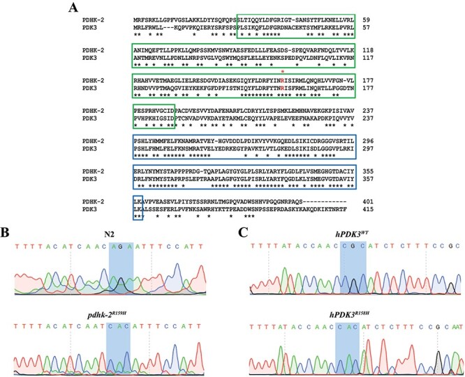
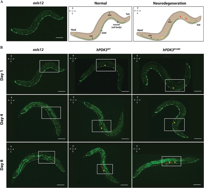
Charcot-Marie-Tooth (CMT) is a commonly inherited, non-fatal neurodegenerative disorder that affects sensory and motor neurons in patients. More than 90 genes are known to cause axonal and demyelinating forms of CMT. The p.R158H mutation in the pyruvate dehydrogenase kinase 3 (PDK3) gene is the genetic cause for an X linked form of axonal CMT (CMTX6). In vitro studies using patient fibroblasts and iPSC-derived motor neurons have shown that this mutation causes deficits in energy metabolism and mitochondrial function. Animal models that recapitulate pathogenic in vivo events in patients are crucial for investigating mechanisms of axonal degeneration and developing therapies for CMT. We have developed a C. elegans model of CMTX6 by knocking-in the p.R158H mutation in pdhk-2, the ortholog of PDK3. In addition, we have developed animal models overexpressing the wild type and mutant form of human PDK3 specifically in the GABAergic motor neurons of C. elegans. CMTX6 mutants generated in this study exhibit synaptic transmission deficits, locomotion defects and show signs of progressive neurodegeneration. Furthermore, the CMTX6 in vivo models display energy deficits that recapitulate the phenotype observed in patient fibroblasts and iPSC-derived motor neurons. Our CMTX6 animals represent the first in vivo model for this form of CMT and have provided novel insights into the cellular function and metabolic pathways perturbed by the p.R158H mutation, all the while closely replicating the clinical presentation observed in CMTX6 patients.


Figures

Figure 1
Figure 1
Knocking-in and overexpression of CMTX6 causing mutation in C. elegans. (A) Alignment of full-length amino acid sequences of human PDK3 (415 amino acids) and the C. elegans ortholog, PDHK-2 (401 amino acids) using ClustalW. Highlighted boxed regions indicate conserved protein domains [Green: BCDHK_Adom3 (Mitochondrial branched-chain alpha-ketoacid dehydrogenase kinase; Blue: HTPase_PDK-like (Histidine kinase-like ATPase domain of pyruvate dehydrogenase kinase)]. The conserved amino acid position in human where the reported mutation is present is highlighted by an FX1. Conserved amino acid residues between PDK3 and PDHK-2 are indicated by *. (B) Sequence trace of pdhk-2 from N2 and knock-in mutant generated using CRISPR-Cas9. The highlighted region indicates the nucleotide changes used to introduce histidine at amino acid residue 159 in the knock-in model. More information on CRISPR modification is available in the supplementary information. (C) Sequence trace confirming the human wild type and mutant PDK3. Highlighted region shows the codon change from CGC (encoding an arginine in wild type hPDK3) to CAC (encoding a Histidine in the mutant p.R158H PDK3).
Figure 2
Figure 2
The knock in and overexpression of the PDK3 mutation causing CMTX6 affects the body width in C. elegans. Sample image of an N2 animal and the methodology used for measuring body width (A) using Image J software. The squares indicate the location along the animal body selected to construct the yellow line, which represents the width (from the end of the vulva on the ventral side to the dorsal side) of the animal. The length of the yellow line has been calibrated within the software according to the reference scale bar. Scale bar is 0.25 mm. Violin plots of body width (B) observed when comparing CMTX6 animals and their respective controls. Black dotted line represents median value of the data sets and grey dots represents measured data points for individual animals. The number of animals used for the body width measurements: N2 (n = 30), pdhk-2R159H (n = 30), oxIs12 (n = 30), hPDK3WT (n = 30) and hPDK3R158H (n = 30). **** adjusted p-value < 0.0001.
Figure 3
Figure 3
Synaptic transmission defect in CMTX6 models of C. elegans. (A) Illustration of C. elegans neuromuscular junction and the actions of aldicarb and levamisole at the synapse. Aldicarb is a nematicide that cleaves acetylcholinesterase, an enzyme that regulates acetylcholine levels, resulting in paralysis and eventually death in worms. Aldicarb identifies potential deficits associated with synaptic transmission in C. elegans. Levamisole is an acetylcholine receptor blocker and allows identifying the role of pre-synaptic (neuron) or/and post-synaptic (muscle) compartment in the aldicarb induced phenotype. (B and C) C. elegans strains were treated with 1 mM aldicarb. CMTX6 animals showed significant sensitivity to aldicarb when compared to their respective controls (N2 and oxIs12), which indicates deficits in synaptic transmission. C. elegans overexpressing human mutant PDK3 (hPDK3R158H) showed the highest sensitivity to aldicarb followed by those expressing human wild type PDK3 (hPDK3WT) and knock-in mutant (pdhk-2R159H). 20–35 animals were used in one biological replicate per genotype with the numbers of biological replicates used are as follows: N2 = 12; oxIs12 = 3; pdhk-2R159H = 9, hPDK3WT = 3, and hPDK3R158H = 10. The mean ± SEM is presented. * p-value < 0.05, two-tailed unpaired t-test. (D and E) C. elegans strains treated with 0.2 mM levamisole. No difference was observed in the time at which the CMTX6 animals paralysed in response to levamisole treatment when compared to controls, suggesting the involvement of the pre-synaptic compartment (neuron) in the aldicarb induced phenotype observed in the CMTX6 mutants. 20–35 animals per biological replicate per genotype with the numbers of biological replicates used are as follows: N2 = 6; oxIs12 = 3; pdhk-2R159H = 3, hPDK3WT = 3 and hPDK3R158H = 3. Error bars indicate mean ± SEM.
Figure 4
Figure 4
CMTX6 models of C. elegans recapitulate ATP deficits observed in CMTX6 patients and are susceptible to paraquat induced oxidative stress. PCR based mitochondrial DNA copy number quantification shows that there is no change in mitochondrial DNA copy number at day 1 (A) and day 4 (B) old animals. (C) ATP production in day 1 old C. elegans strains. ATP production is significantly reduced in knock-in pdhk-2R159H animals. There was no significant difference in ATP production in overexpression CMTX6 animal lysates when compared to oxIs12 animals. N = 1500 to 3000 age synchronised day 1 old animals per replicate per genotype. A total of three biological replicates were used for the ATP production assay. Average luminescence of ATP normalised to 1 μg of total protein is presented. * adjusted p-value < 0.05. (D and E) 0.2 M paraquat assay. 1 h post treatment, CMTX6 animals showed increased sensitivity to oxidative stress with knock-in mutants showing a significant increase in the percentage of mortality while the overexpression animal models of CMTX6 displayed mortality greater than 50%. N = 6 animals per genotype per replicate was used for mitochondrial DNA copy number quantification. A total of 3 biological replicates were used. For paraquat assay N = 25 to 38 animals per genotype per replicate was used with a total 3 biological replicates used in the experiment. * adjusted p-value = 0.0353 and **** adjusted p-value < 0.0001. Error bars indicate ± SEM.
Figure 5
Figure 5
Overexpression of human wild type and mutant PDK3 in motor neurons leads to neurodegeneration in C. elegans. (A) Neurodegeneration scoring paradigm. Representative image of day 1 old oxIs12 animals and schematic illustrating normal and neurodegenerative phenotypes. The morphology of the ventral nerve cord (VNC) of C. elegans was analysed for assessing the impact of over expression of wild type and mutant PDK3 on neuron morphology. The dorsal nerve cord (DNC) was not included in scoring due to the reduced GFP expression. In the normal animal, the axon of the ventral nerve cord is intact, which is indicated by continuous GFP expression, with all the neurons (cell bodies) still visible. The presence of blebbing or beading of the axons as indicated by yellow asterisk FX2 and/or the complete loss of axon (indicated by red asterisk FX3) and/or neuron (yellow arrowhead) in the VNC of C. elegans is scored as neurodegeneration phenotype. A-Anterior, P-posterior, D-Dorsal and V-ventral; Scale bar = 0.3 mm. (B) Representative images of day 1, day 4 and day 8 old wild type (oxIs12) and overexpression CMTX6 animals (hPDK3WT and hPDK3R158H). Boxed region highlights the difference between oxIs12 and CMTX6 overexpression animals. CMTX6 over expression mutants show signs of axon degeneration at day 1. Yellow asterisk FX4 indicate region of the axon showing blebbing or beading (degeneration). There was no sign of complete loss of axon or neuron (cell body) in day 1 old animals. Regions of axon loss (loss or interruption of GFP signal along the VNC is indicated by the red asterisk FX5. CMTX6 overexpression animals show increased signs of axon degeneration characterised by beading or blebbing and loss of GFP signal at day 4. Axon degeneration is more severe in the overexpression CMTX6 animals at day 8 with hPDK3R158H animals showing complete loss of axon (indicated by red asterisk FX6) and neuron cell body (yellow arrowhead). Number of animals used for live imaging are as follows: Day 1–31 oxIs12, 26 hPDK3WT and 27 hPDK3R158H animals; Day 8–46 oxIs12, 38 hPDK3WT and 45 hPDK3R158H animals; A-Anterior, P-posterior, D-Dorsal and V-ventral; Scale bar—0.3 mm.
Figure 6
Figure 6
Quantification of neuron and axon loss in overexpression CMTX6 animals reveals progressive neurodegeneration. (A) No neuron loss was observed in day 1 old CMTX6 and control animals. hPDK3R158H showed signs of neuron loss at day 4 and with the number of overexpression CMTX6 animals exhibiting neuron loss significantly higher than the oxIs12 animals at day 8. (B) Axon degeneration is more prominent in the overexpression animal models at day 4, with day 8 old overexpression CMTX6 animals showing complete loss of axon and several neurons. Animals used for live imaging are as follows: day 4 oxIs12 (n = 87), hPDK3WT (n = 98) and hPDK3R158H (n = 97). There is no error bar because of the statistical test used for analysis. A chi-square test was used to compare categorical data (presence of wild type neuron/axon or axon/neuron loss). Neuron loss: * p-value = 0.0406; Axon loss: * p-value < 0.05, **p-value = 0.0024, ns—not significant.
Figure 7
Figure 7
Overexpression animal models of CMTX6 show locomotion defects. (A) Body thrash quantification of CMTX6 animals. There was no significant difference in the number of body thrashes for the knock-in animals when compared to N2. CMTX6 animals expressing the human PDK3 gene (hPDK3WT and hPDK3R158H) in the GABAergic motor neurons, showed significant reduction in the number of body thrashes when compared to oxIs12 animals. Number of animals used for thrashing assay: n = 30 for N2, pdhk-2R159H and hPDK3R158H; hPDK3WT (31); oxIs12 (37). **** adjusted p-value < 0.0001, ns—not significant. (B) Quantification of body bends in the C. elegans mutants of CMTX6. pdhk-2R159H and hPDK3WT animal models of CMTX6 showed no difference in the number of body bends when compared to their respective controls. hPDK3R158H worms showed a reduction in the number of body bends when compared to hPDK3WT and oxIs12 animals. Grey spheres indicate data from individual animals. n = 32 N2, 40 pdhk-2R159H, 29 oxIs12, 24 hPDK3WT and 33 hPDK3R158H for the body bend experiment. * adjusted p-value = 0.031, **** adjusted p-value < 0.0001, ns—not significant. Error bars indicate ± SEM.

References

    1. Fowler, W.M., Abresch, R.T., Koch, T.R., Brewer, M.L., Bowden, R.K. and Wanlass, R.L. (1997) Employment profiles in neuromuscular diseases. Am. J. Phys. Med. Rehabil., 76, 26–37. - PubMed
    1. Schorling, E., Thiele, S., Gumbert, L., Krause, S., Klug, C., Schreiber-Katz, O., Reilich, P., Nagels, K. and Walter, M.C. (2019) Cost of illness in Charcot-Marie-tooth neuropathy results from Germany. Neurology, 92, E2027–E2037. - PubMed
    1. Drew, A.P., Zhu, D., Kidambi, A., Ly, C., Tey, S., Brewer, M.H., Ahmad-Annuar, A., Nicholson, G.A. and Kennerson, M.L. (2015) Improved inherited peripheral neuropathy genetic diagnosis by whole-exome sequencing. Mol Genet Genom Med, 3, 143–154. - PMC - PubMed
    1. Timmerman, V., Strickland, A.V. and Zuchner, S. (2014) Genetics of Charcot-Marie-tooth (CMT) disease within the frame of the human genome project success. Genes-Basel, 5, 13–32. - PMC - PubMed
    1. Kennerson, M.L., Yiu, E.M., Chuang, D.T., Kidambi, A., Tso, S.C., Ly, C., Chaudhry, R., Drew, A.P., Rance, G., Delatycki, M.B. et al. (2013) A new locus for X-linked dominant Charcot-Marie-tooth disease (CMTX6) is caused by mutations in the pyruvate dehydrogenase kinase isoenzyme 3 (PDK3) gene. Hum. Mol. Genet., 22, 1404–1416. - PMC - PubMed

Publication types

MeSH terms

Substances