. 2021 Dec;93(12):6765-6777.
doi: 10.1002/jmv.27270. Epub 2021 Aug 20.

Vaccination versus infection with SARS-CoV-2: Establishment of a high avidity IgG response versus incomplete avidity maturation

Affiliations

Vaccination versus infection with SARS-CoV-2: Establishment of a high avidity IgG response versus incomplete avidity maturation

Friedhelm Struck et al. J Med Virol. 2021 Dec.

Abstract

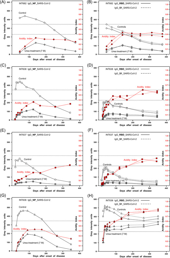
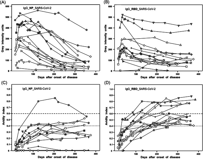
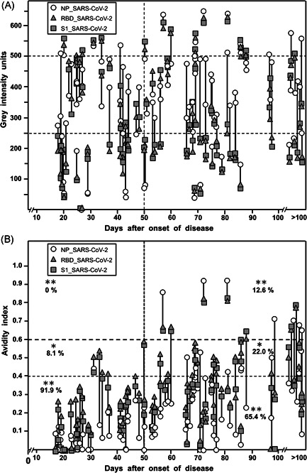
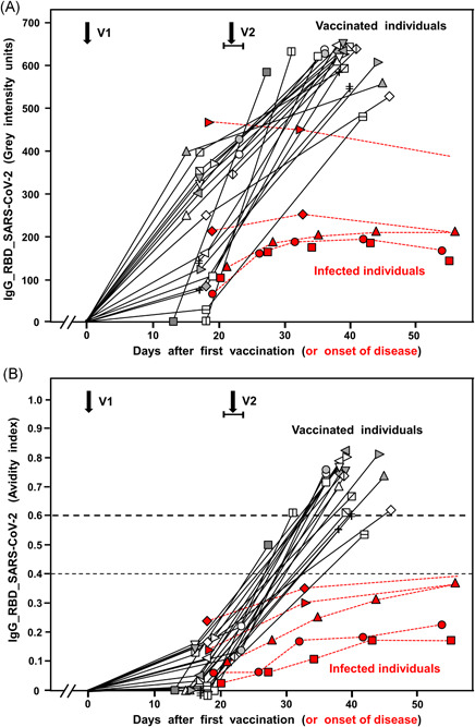
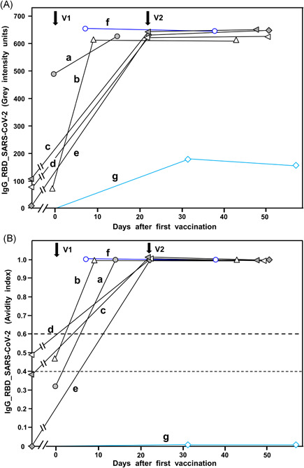
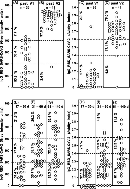
Avidity is defined as the binding strength of immunoglobulin G (IgG) toward its target epitope. Avidity is directly related to affinity, as both processes are determined by the best fit of IgG to epitopes. We confirm and extend data on incomplete avidity maturation of IgG toward severe acute respiratory syndrome coronavirus 2 (SARS-CoV-2) nucleoprotein (NP), spike protein-1 (S1), and its receptor-binding domain (RBD) in coronavirus disease 2019 (COVID-19) patients. In SARS-CoV-2-infected individuals, an initial rise in avidity maturation was ending abruptly, leading to IgG of persistently low or intermediate avidity. Incomplete avidity maturation might facilitate secondary SARS-CoV-2 infections and thus prevent the establishment of herd immunity. Incomplete avidity maturation after infection with SARS-CoV-2 (with only 11.8% of cases showing finally IgG of high avidity, that is, an avidity index > 0.6) was contrasted by regular and rapid establishment of high avidity in SARS-CoV-2 naïve individuals after two vaccination steps with the BioNTech messenger RNA (mRNA) Vaccine (78% of cases with high avidity). One vaccination step was not sufficient for induction of complete avidity maturation in vaccinated SARS-CoV-2 naïve individuals, as it induced high avidity only in 2.9% of cases within 3 weeks. However, one vaccination step was sufficient to induce high avidity in individuals with previous SARS-CoV-2 infection.

Keywords: SARS-CoV-2; avidity; protective immunity; receptor-binding domain; recomLine SARS-CoV-2 IgG; vaccination.


Conflict of interest statement

E. Soutschek and M. Motz are owners of Mikrogen GmbH. E. Soutschek is the present CEO of Mikrogen GmbH. F. Struck, E. Stachik, K. Wochinz‐Richter, S. Schulz, and P. Schreiner are employees of Mikrogen GmbH. The determination of avidity of antibodies, using immunoblots or other techniques that allow the parallel measurement of humoral immune reactions toward several antigens in one assay, has been patented by Mikrogen GmbH (WO 00/54055; PCT/EP00/01883). In addition, a new patent application for a method to determine the antibodies toward SARS‐CoV‐2 has been filed by Mikrogen GmbH and is pending (EP 2019/2550). Mikrogen GmbH develops and produces test systems for serological analysis of infectious diseases. G. Bauer is a member of the Medical Faculty of the University of Freiburg. He is the inventor of WO 00/54055; PCT/EP00/01883 and one of the coinventors of EP 2019/2550.

Figures

Figure 1
Figure 1
Kinetics of the serological responses to SARS‐CoV‐2 nucleoprotein (NP), receptor‐binding domain (RBD), and spike protein S1. Sera from four patients with COVID‐19 confirmed by positive RT‐PCR and clinical symptoms were tested for IgG toward SARS‐CoV‐2 NP (A, C, E, G) or RBD and S1 (B, D, F, H) using a line immunoassay with purified recombinant antigens, as described under Methods. The immunoassays were performed without (“Control”) and with urea treatment (7 M) for the determination of avidity. The determined gray intensity values (black) and the calculated avidity indices (red) are plotted toward the days after the onset of the disease. These findings demonstrate that, after a rapid initial increase, the concentrations of IgG directed toward NP, RBD, and S1 are declining. Incomplete avidity maturation seems to be characteristic for the IgG response toward SARS‐CoV‐2 NP, RBD, and S1, as the avidity indices remain at plateaus at low (avidity index < 0.4) or intermediate avidity (avidity index between 0.4 and 0.6). The avidities of IgG toward RBD and S1 under H are exceptional, showing an initial plateau of avidity after 50 days, followed by a secondary increase in avidity that is finalized at the second plateau of higher avidity. This pattern is indicative of secondary infection with SARS‐CoV‐2.COVID‐19, coronavirus disease 2019; IgG, immunoglobulin G; RT‐PCR, real‐time polymerase chain reaction; SARS‐CoV‐2, severe acute respiratory syndrome coronavirus 2
Figure 2
Figure 2
Summary of 16 long‐term kinetics of IgG responses toward NP and RBD after SARS‐CoV‐2 infection leading to COVID‐19. The responses of IgG directed toward NP (A) or RBD (B) in 16 cases of COVID‐19 cases were followed for about 12 months, and the corresponding avidity indices were calculated (IgG NP: C; IgG RBD: D). The results for the IgG levels described under A and B show a rapid increase in IgG toward NP or RBD, followed by a decline over time, characterized by a high degree of variability. Avidity indices (C, D) indicate that avidity maturation is not completed. In the case of IgG directed toward SARS‐CoV‐2 NP, only one serum reached high avidity (avidity index > 0.6). In the case of RBD, one 5/16 sera reached high avidity, whereas 11/16 sera remained at a plateau of intermediate avidity. COVID‐19, coronavirus disease 2019; IgG, immunoglobulin G; NP, nucleoprotein; RBD, receptor‐binding domain; SARS‐CoV‐2, severe acute respiratory syndrome coronavirus 2
Figure 3
Figure 3
IgG responses in 93 serum samples from patients with confirmed SARS‐CoV‐2 infection and COVID‐19. Ninety‐three sera from 70 patients with clinical signs of COVID‐19, quite predominantly showing ambulatory mild disease, and with positive SARS‐CoV‐2 RT‐PCR test were tested with the recomLine SARS‐CoV‐2 IgG test. The gray intensities (A), which reflect the concentrations of the respective antibodies and the avidity indices (B) are shown in correlation to the time after the onset of disease for each serum. (A) Reflectometric gray intensity values. The result shows a broad distribution of gray scales, which was similar at all time points. The percentages of sera showing values in the ranges 0–250, 250–500, and more than 500 gray intensity were not significantly different when sera were taken before or after 50 days after the onset of the disease were compared, with the exception of IgG toward NP at less than 250 units. Importantly, all sera that gave a positive result showed positivity toward all three antigens tested. In 93.5% of the sera with positive IgG toward S1, IgG toward RBD showed a value that was less than 20 gray intensity units different from that obtained for IgG S1, whereas, in 6.5% of the sera, IgG values toward S1 and RBD were different more than 20 gray intensity units (p < 0.001). In contrast, only 15% of sera showed IgG toward NP at a value that differed less than 20 gray intensity units from that of the respective IgG S1 value, and 84.9% showed a higher difference (p < 0.001). (B) Avidity indices. Whereas the gray intensity values (indicative of the respective concentrations of IgG) were broadly similar between sera taken within 50 days or after 50 days after the onset of the disease; the avidity of the IgG was increasing between these groups. However, no complete avidity maturation was reached. A total of 91.9% of the sera taken before Day 50 showed an avidity index below 0.4, whereas only 65.4% of the sera taken after 50 days showed similar low avidity (p < 0.001). As an indication of partial avidity maturation, 8.1% of sera taken before 50 days showed an avidity index between 0.4 and 0.6, in contrast to 22.0% of sera in the group where sera were taken after 50 days (p < 0.01). Avidity indices above 0.6 were only found at 12.6% in the group where sera had been taken later than 50 days after the onset of the disease (p < 0.001). In summary, an overall low degree of avidity maturation was seen, with a minority of samples of high avidity at late time points. These findings confirm incomplete avidity maturation after SARS‐CoV‐2 infection. COVID‐19, coronavirus disease 2019; IgG, immunoglobulin G; RT‐PCR, real‐time polymerase chain reaction; NP, nucleoprotein; SARS‐CoV‐2, severe acute respiratory syndrome coronavirus 2
Figure 4
Figure 4
IgG responses after vaccination toward SARS‐CoV‐2. The increase in gray intensity units (representing IgG concentration) (A) and in avidity (B) of IgG directed toward SARS‐CoV‐2 RBD after vaccination with the BioNTech/Pfizer vaccine are summarized for 21 cases (black curves). For comparison, five representative cases of natural SARS‐CoV‐2 infections (labeled in red) are included. V1 and V2 indicate the time of the first and second vaccination. V1 is defined as Day zero. V2 was varying between Days 21–23 after the first vaccination. The results show high variability of the IgG response after the first vaccination, followed by an additional stronger response after the second vaccination. Avidity was low after the first vaccination but reached intermediate avidity (2/20 cases) and high avidity (18/20 cases) after the second vaccination (p < 0.001). In comparison with natural infection, vaccination induces a strong and continuous IgG response (p < 0.001) with uninhibited avidity maturation, in contrast to incomplete avidity maturation after natural infection, characterized by plateau values in the low avidity range (p < 0.001). IgG, immunoglobulin G; NP, nucleoprotein; RBD, receptor‐binding domain; SARS‐CoV‐2, severe acute respiratory syndrome coronavirus 2
Figure 5
Figure 5
Analysis of special cases of vaccination. The figure shows the increase in gray intensity units (A) or avidity index (B) of special cases of vaccinated individuals. Cases a−e represent individuals with natural coronavirus infections several months before vaccination. Before vaccination, in four out of the five cases, the antibody titers derived from primary infection had declined to very low levels. One vaccination step was sufficient to induce a strong IgG response with extremely high avidity in all five cases. Case f represents a case without known preceding SARS‐CoV‐2 infection. However, the high response with high avidity already after the first vaccination step and the detection of IgG directed toward NP (not shown in the figure) indicates that this case is analogous to cases a‐e, that is, it seems to represent a vaccinated individual with (clinical inapparent) SARS‐CoV‐2 infection. Finally, Case g represents the only case seen so far in our study, which was characterized by a complete failure in avidity maturation after vaccination, paralleled by a low IgG response. V1 and V2 indicate the time point of the first and second vaccination. IgG, immunoglobulin G; SARS‐CoV‐2, severe acute respiratory syndrome coronavirus 2
Figure 6
Figure 6
IgG responses after vaccination and natural infection with SARS‐CoV‐2. Thirty‐nine sera taken between 9 and 22 days after first vaccination with the BioNTech/Pfizer vaccine (A) and 41 sera taken between 6 and 25 days after second vaccination with the BioNTech/Pfizer vaccine (B) were tested for IgG directed toward SARS‐CoV‐2 RBD. The determined gray intensity units are shown under A and B, whereas the avidity indices, determined by parallel treatment with 7 M urea after incubation of serum with test antigens, are shown under C (sera obtained after the first vaccination) and D (sera obtained after the second vaccination). For comparison, 37 sera were taken from COVID‐19 outpatients between 17 and 30 days after the onset of the disease (E), 42 sera from COVID‐19 outpatients taken between 31 and 60 days after the onset of the disease (F) and 34 sera from COVID‐19 outpatients taken between 61 and 140 days after the onset of the disease were tested for IgG directed toward RBD (gray intensity units). The avidity indices of these sera were determined and are shown (H−J). The figure shows a variable induction of IgG after the first vaccination (A), which extends to uniformly high values after the second vaccination (B) (p < 0.001). This is paralleled by low avidity indices after the first vaccination (with one exception) and high avidity in 78.0% of the samples taken after the second vaccination (p < 0.001). Though natural infection with SARS‐CoV‐2 can also induce IgG concentrations of higher levels, a broad range of intensities is maintained over long times without statistically significant changes (E−G). Though a certain degree of avidity maturation in the low and intermediate range was seen with time for sera from patients with natural SARS‐CoV‐2 infection, only a minority of sera (11.8%) reached high avidity values (>0.6) (H−J) at 61−140 days after natural infection, whereas 78% of sera taken from vaccinated individuals had reached avidity indices above 0.6 between 6 and 25 days after the second vaccination (p < 0.001). The median values calculated from the data presented in Figure 6 are summarized in Figure S20. The confirmation of the data presented in Figure 6A−D through the increase in the number of vaccinated subjects are presented in Figure S18. COVID‐19, coronavirus disease 2019; IgG, immunoglobulin G; RBD, receptor‐binding domain; SARS‐CoV‐2, severe acute respiratory syndrome coronavirus 2

References

    1. Zhao J, Yuan Q, Wang H, et al. Antibody responses to SARS‐CoV‐2 in patients of novel coronavirus disease 2019. Clin Infect Disease. 2020;71:2027‐2034. 10.1093/cid/ciaa344 - DOI - PMC - PubMed
    1. Jin Y, Wang M, Zuo Z, et al. Diagnostic value and dynamic variance of serum antibody in coronavirus disease. Int J Infect Dis. 2020;94:49‐52. - PMC - PubMed
    1. Xiao S‐Y, Wu Y, Liu H. Evolving status of the 2019 novel coronavirus infection: Proposal of conventional serologic assays for disease diagnosis and infection monitoring. J Med Virol. 2020;92:464‐467. - PMC - PubMed
    1. Wölfel R, Corman V, Guggemos W, et al. Virological assessment of hospitalized patients with COVID‐2019. Nature. 2020;581:465‐469. 10.1038/s41586-020-2196-x - DOI - PubMed
    1. Bauer G. The potential significance of high avidity IgG for protective immunity towards SARS CoV‐2. Int J Infect Dis. 2021;106:61‐64. - PMC - PubMed