. 2021 Sep 13;39(9):1245-1261.e6.
doi: 10.1016/j.ccell.2021.07.006. Epub 2021 Aug 12.

Targeting Aurora B kinase prevents and overcomes resistance to EGFR inhibitors in lung cancer by enhancing BIM- and PUMA-mediated apoptosis

Affiliations

Targeting Aurora B kinase prevents and overcomes resistance to EGFR inhibitors in lung cancer by enhancing BIM- and PUMA-mediated apoptosis

Kosuke Tanaka et al. Cancer Cell. .

Abstract

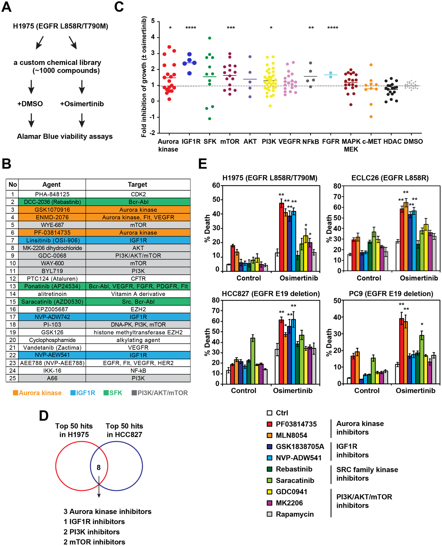
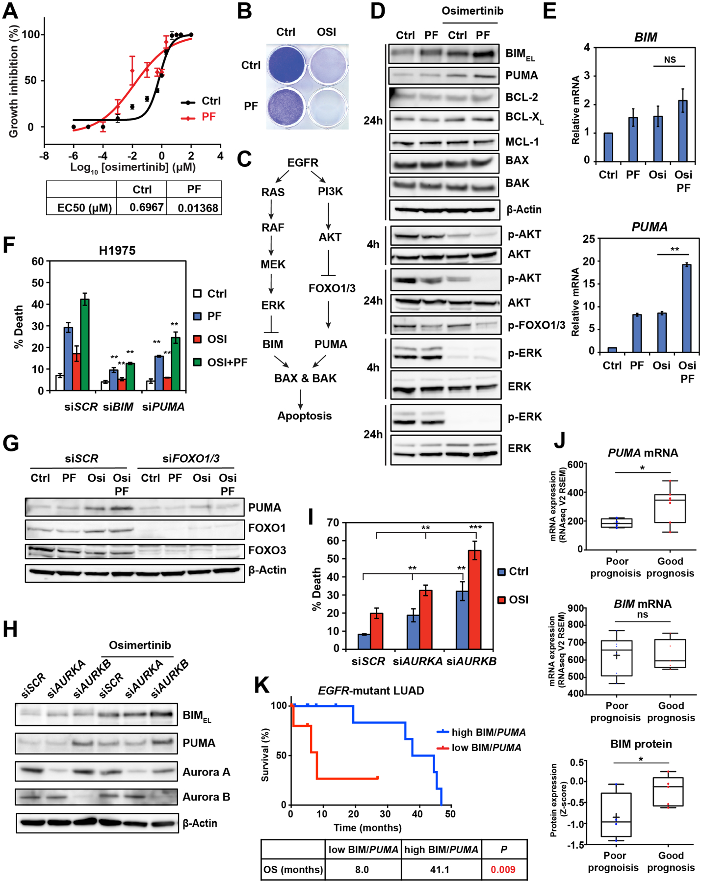
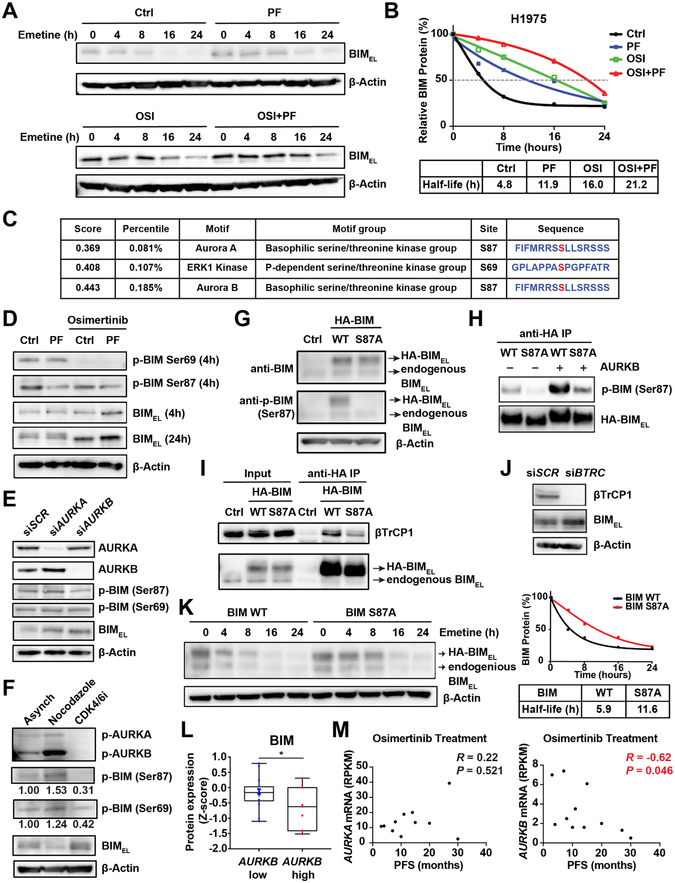
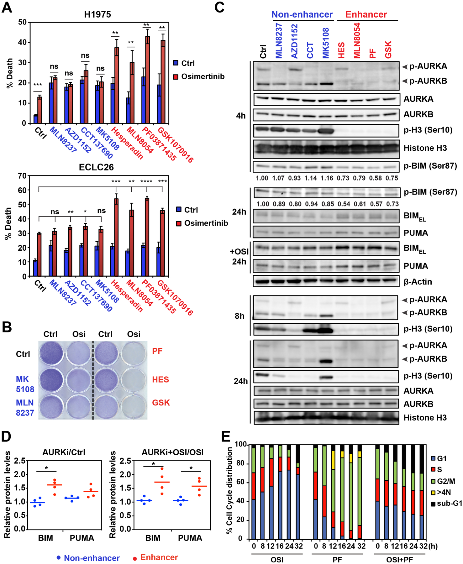
The clinical success of EGFR inhibitors in EGFR-mutant lung cancer is limited by the eventual development of acquired resistance. We hypothesize that enhancing apoptosis through combination therapies can eradicate cancer cells and reduce the emergence of drug-tolerant persisters. Through high-throughput screening of a custom library of ∼1,000 compounds, we discover Aurora B kinase inhibitors as potent enhancers of osimertinib-induced apoptosis. Mechanistically, Aurora B inhibition stabilizes BIM through reduced Ser87 phosphorylation, and transactivates PUMA through FOXO1/3. Importantly, osimertinib resistance caused by epithelial-mesenchymal transition (EMT) activates the ATR-CHK1-Aurora B signaling cascade and thereby engenders hypersensitivity to respective kinase inhibitors by activating BIM-mediated mitotic catastrophe. Combined inhibition of EGFR and Aurora B not only efficiently eliminates cancer cells but also overcomes resistance beyond EMT.

Keywords: Aurora B kinase; BCL-2 family; EMT; apoptosis; drug resistance; drug tolerance; epidermal growth factor receptor; lineage plasticity; lung cancer; mitotic catastrophe.


Conflict of interest statement

Declaration of interests H.A.Y. has consulted for AstraZeneca, Daiichi, Janssen, and Blueprint Medicine; she has received research funding from AstraZeneca, Daiichi, Janssen, Pfizer, Novartis, Cullinan, and Lilly. C.M.R. has consulted for AbbVie, Amgen, AstraZeneca, Epizyme, Genentech/Roche, Ipsen, Jazz, Lilly, and Syros; he serves on the scientific advisory boards of Bridge Medicines, Earli, and Harpoon Therapeutics. M.G.K. has consulted for AstraZeneca, Daiichi-Sankyo, Janssen, Novartis, Pfizer, Regeneron, and Sanofi/Genzyme. U.G. has a clinical trial agreement with AstraZeneca and received research funding from AstraZeneca, Esanex, and Aurigene; he is a current employee of Bristol Myers Squibb. J.J.H. has consulted for Eisai and BostonGene; he has received clinical trial funding from Bristol Myers Squibb, Merck, AstraZeneca, Exelixis, Calithera, and SillaJen; he has received research funding from Merck, BostonGene, and TScan.

Figures

Figure 1.
Figure 1.. HTS identifies Aurora kinase inhibitors as potent enhancers of osimertinib-induced apoptosis in EGFR-mutant lung cancer
(A) A schematic of HTS to identify agents that enhance osimertinib (osi)-induced growth inhibition. H1975 was treated with each compound from the library (2 μM) ± osi (2 μM) in duplicate. Cell viability was assessed by alamarBlue assays at 72 h. (B) Top 25 agents that enhance osi-induced growth inhibition of H1975. Green cluster, SRC family kinase inhibitors; orange, Aurora kinase inhibitors (AKi); gray, PIK3/AKT/mTOR inhibitors; and blue, IGF1R inhibitors. (C) An overview of growth inhibition of H1975 by various pathway inhibitors ± osi. Fold inhibition of growth by the combination of each compound with osi compared to each compound alone was normalized against that by osi. *, P<0.05; **, P<0.01; ***, P<0.001; ****, P<0.0001 (Mann-Whitney U test). (D) A Venn diagram of top 50 agents that enhance osi-induced growth inhibition of H1975 and HCC827. (E) Cells were treated with the indicated compounds ± osi for 48 h. Cell death was quantified by annexin-V (AV) staining (mean ± s.d., n=3). *, P<0.05; **, P<0.01 (Student’s t-test). See also Figure S1.
Figure 2.
Figure 2.. AURKB inhibition enhances osimertinib-induced apoptosis through BIM and PUMA induction
(A) H1975 was treated with osi at the indicated concentrations ± PF03814735 (PF, 2 μM). EC50 was assessed by CellTiter-Glo assays at 72 h (mean ± s.d., n=3). (B) H1975 was treated with osi (1 μM) and/or PF (2 μM). Colonies were stained with crystal violet after 14 days. (C) A schematic demonstrating the crosstalk between the EGFR signal transduction pathway and BCL-2 family-regulated apoptosis. (D) H1975 treated with the indicated agents was assessed by immunoblots. (E) qRT-PCR for BIM and PUMA mRNA in H1975 treated with the indicated agents (2 μM) for 24 h. Data were normalized against β-Actin (mean ± s.d., n=3). **, P<0.01; ns, not significant (Student’s t-test). (F) H1975, transfected with the indicated siRNAs, was treated with the indicated agents (2 μM) for 48 h. Cell death was quantified by AV staining (mean ± s.d., n=3). **, P<0.01 (Student’s t-test). (G) H1975, transfected with the indicated siRNAs, was treated with the indicated agents (2 μM) for 24 h and assessed by immunoblots. (H-I) H1975, transfected with the indicated siRNAs, was treated with osi (2 μM) and assessed by immunoblots at 24 h. Cell death was quantified by AV staining at 48 h (mean ± s.d., n=3). **, P<0.01; ***, P<0.001 (Student’s t-test). (J) Comparison of PUMA mRNA, BIM mRNA, and BIM protein expression in tumors from EGFR-mutant lung adenocarcinoma (LUAD) patients with a good or poor prognosis in TCGA (n=22). *, P<0.05 (Student’s t-test). (K) Kaplan-Meier analysis of overall survival in EGFR-mutant LUAD patients from TCGA based on the expression of BIM protein and PUMA mRNA. Blu, high BIM protein or PUMA mRNA (n = 13); Red, low BIM protein or PUMA mRNA (n = 6). P = 0.009 (Log-rank test). See also Figure S2.
Figure 3.
Figure 3.. AURKB inhibition reduces BIM S87 phosphorylation and stabilizes BIM protein
(A) H1975 was treated with osi (2 μM) and/or PF (2 μM) for 4 h, followed by the addition of emetine (20 μg/ml) to inhibit translation. Immunoblot analyses were performed at the indicated times. (B) The half-life of BIM protein upon treatment with the indicated agents as in (A). The anti-BIM immunoblots shown in (A) were quantified by the ImageJ software and plotted with respect to time. Data shown are the mean of two independent experiments. (C) Prediction of potential kinase phosphorylation motifs in BIM using the SCANCITE 4.0 software. (D) H1975 treated with the indicated agents as in (A) for 4 or 24 h was assessed by immunoblots. (E) H1975, transfected with the indicated siRNAs, was assessed by immunoblots. (F) H1975 treated with nocodazole (50 ng/ml) for 16 h or the CDK4/6 inhibitor palbociclib (1 μM) for 24 h was assessed by immunoblots. (G) BAX−/−BAK−/− H1975 was transduced with retrovirus expressing HA-tagged WT BIM or BIM S87A mutant and assessed by immunoblots. (H) H1975 as in (G) was treated with palbociclib (1 μM) for 24 h and subjected to anti-HA immunoprecipitation. The immunoprecipitates were incubated with recombinant AURKB in the presence of ATP and assessed by immunoblots. (I) H1975 as in (G) was subjected to anti-HA immunoprecipitation. The input (5%) and immunoprecipitates were assessed by immunoblots. (J) H1975, transfected with the indicated siRNAs, was assessed by immunoblots. (K) H1975 as in (G) was treated with emetine (20 μg/ml) and assessed by immunoblots. The BIM protein levels were quantified by the ImageJ software and plotted with respect to time. Data shown are the mean of two independent experiments. (L) The expression of BIM protein in EGFR-mutant LUAD with high or low expression of AURKB mRNA from TCGA (n=22). *, P<0.05 (Student’s t-test). (M) Scatter plots showing the correlation between progression-free survival (PFS) following osi treatment and pretreatment mRNA levels of AURKA or AURKB in EGFR-mutant LUAD patients (n=11). See also Figure S3.
Figure 4.
Figure 4.. Aurora kinase inhibitors are divided into enhancer and non-enhancer groups of osimertinib-induced apoptosis
(A) H1975 and ECLC26 were treated with the indicated AKi (1 μM) ± osi (1 μM) for 48 h. Cell death was quantified by AV staining (mean ± s.d., n=3). *, P<0.05; **, P<0.01; ***, P<0.001; ****, P<0.0001 (Student’s t-test). (B) H1975 was treated with the indicated agents (1 μM) and colonies were stained with crystal violet after 14 days. (C) H1975 treated as in (A) was assessed by immunoblots at the indicated times. (D) The anti-BIM and anti-PUMA immunoblots shown in C were quantified by the ImageJ software. Data presented are the ratio of BIM and PUMA protein levels upon the treatment of AKi compared to control and the ratio upon the treatment of both AKi and osi compared to osi alone. *, P<0.05 (Student’s t-test). (E) H1975 was treated with osi (1 μM) and/or PF (1 μM) and subjected to cell-cycle analysis using propidium iodide (PI) staining at the indicated times. See also Figure S4.
Figure 5.
Figure 5.. Osimertinib-resistant cells exhibit EMT and become hypersensitive to AURKB inhibition
(A) EC50 of osi in the indicated cell lines was assessed by CellTiter-Glo assays at 72 h (mean ± s.d., n=3). (B) The parental (P) and osi-resistant (R) H1975 and ECLC26 were assessed by immunoblots. (C) H1975R and ECLC26R were treated with the indicated agents (2 μM) for 48 h. Cell death was quantified by AV staining (mean ± s.d., n=3). (D) H1975R and ECLC26R treated with the indicated agents (1 μM) for 4 or 24 h were assessed by immunoblots. (E-F) H1975 cells with CRISPR/Cas9-mediated KO of both FOXA1 and FOXA2, retroviral transduction of ZEB1, and both (H1975FZ) were assessed by immunoblots in (E). EC50 was assessed by CellTiter-Glo assays at 72 h in (F) (mean ± s.d., n=3). (G) H1975 and H1975FZ were treated with the indicated agents (2 μM) for 48 h. Cell death was quantified by AV staining (mean ± s.d., n=3). *, P<0.05; **, P<0.01; ***, P<0.001 (Student’s t-test, comparing H1975 to H1975FZ). (H-J) Kaplan-Meier analysis of overall survival in EGFR-mutant LUAD patients from TCGA (n=22) based on the expression of FOXA1/2 (H), ZEB1/2 (I), and SNAI1/2 (J) mRNA. Blue, the top 50% highly expressed; Red, the bottom 50%. P values were calculated by the Log-rank test. See also Figure S5 and Tables S1 and S2.
Figure 6.
Figure 6.. Inhibition of ATR-CHK1-AURKB induces BIM-mediated mitotic cell death in osimertinib-resistant EMT cells
(A) The parental and osi-resistant H1975 and ECLC26 were treated with the indicated AKi (1 μM) for 48 h. Cell death was quantified by AV staining (mean ± s.d., n=3). *, P<0.05; **, P<0.01; ***, P<0.001 (Student’s t-test). (B) ECLC26R treated with the indicated AKi (1 μM) for 24 h was assessed by immunoblots. (C) H1975R was transfected with the indicated siRNAs and cell death was quantified by AV staining at 48 h (mean ± s.d., n=3). *, P<0.05; ***, P<0.001 (Student’s t-test). (D) H1975, H1975R, ECLC26, and ECLC26R were assessed by immunoblots. (E) H1975FZ was assessed by immunoblots. (F) H1975, H1975R, ECLC26, and ECLC26R were treated with PF (1 μM), the CHK1 inhibitor LY2603618 (1 μM), or the ATR inhibitor VX-970 (2 μM) for 48 h. Alternatively, cells were pretreated with palbociclib (1 μM) for 24 h and then treated with PF, LY2603618, or VX-970 for 48 h. Cell death was quantified by AV staining (mean ± s.d., n=3). *, P<0.05; **, P<0.01; ***, P<0.001 (Student’s t-test, comparing without to with palbociclib). (G) H1975R was transfected with the indicated siRNAs and cell death was quantified by AV staining at 72 h (mean ± s.d., n=3). ***, P<0.001 (Student’s t-test). (H) ECLC26R ± palbociclib (1 μM) pretreatment for 24 h was treated with PF (1 μM) and subjected to cell-cycle analysis using PI staining at the indicated times. (I) H1975P and H1975R treated with the indicated agents (1 μM) for 4 h were assessed by immunoblots. (J) H1975R ± LY2603618 (1 μM) for 4 or 24 h was assessed by immunoblots. (K) H1975R and ECLC26R with retroviral transduction of sgRNA against LacZ, BIM, or BAX/BAK were treated as in (F) for 48 h. Cell death was quantified by AV staining (mean ± s.d., n=3). ***, P<0.001 (Student’s t-test). (L) A schematic depicts the regulation of BIM phosphorylation by the EGFR-RAS-RAF-MEK-ERK and the ATR-CHK1-AURKB signaling pathways in naïve versus EMT cells to induce BAX/BAK-dependent mitotic cell death. See also Figure S6 and Tables S3 and S4.
Figure 7.
Figure 7.. Aurora kinase inhibition improves the therapeutic efficacy of osimertinib in xenograft models.
(A) Athymic nude mice bearing H1975 xenografts were treated with vehicle, osi (5 mg/kg), PF (20 mg/kg), or the combination for 28 days. Tumor volumes were measured twice weekly by caliper (mean ± SEM, n = 6–8 for each group). *, P < 0.05 (two-way ANOVA). (B) Waterfall plot of changes in tumor volume after 28 days of treatment in (A). *, P < 0.05 (Student’s t-test). (C) NOD.Cg-Prkdcscid Il2rgtm1Wjl/SzJ (NSG) mice bearing patient-derived ECLC26 xenografts were treated as in (A) (mean ± SEM, n = 8–12 for each group). *, P < 0.05; ***, P < 0.001 (two-way ANOVA). (D) Athymic nude mice bearing H1975R xenografts were treated as in (A) (mean ± SEM, n = 6–8 for each group). *, P < 0.05 (two-way ANOVA). (E) Waterfall plot of changes in tumor volume in (D). ***, P < 0.001 (Student’s t-test). (F) Representative H&E images of xenografts established using either H1975 or H1975R cells. Scale bars, 200 μm. (G) NSG mice bearing patient-derived Ru813c xenografts were treated as in (A) (mean ± SD, n = 6 for each group). ***, P < 0.001 (two-way ANOVA). (H) Waterfall plot of changes in tumor volume in (G). ***, P < 0.001 (Student’s t-test). (I) NSG mice bearing patient-derived Lx1114 xenografts were treated as in (A) (mean ± SD, n = 6 for each group). ***, P < 0.001 (two-way ANOVA). (J) Waterfall plot of changes in tumor volume in (I). ***, P < 0.001 (Student’s t-test). (K) A schematic summarizing how EGFR and AURKB regulate the proapoptotic activity of BIM and PUMA. See also Figure S7 and Table S5.

References

    1. Anders S, Pyl PT, and Huber W (2015). HTSeq--a Python framework to work with high-throughput sequencing data. Bioinformatics 31, 166–169. - PMC - PubMed
    1. Bean GR, Ganesan YT, Dong Y, Takeda S, Liu H, Chan PM, Huang Y, Chodosh LA, Zambetti GP, Hsieh JJ, and Cheng EH (2013). PUMA and BIM are required for oncogene inactivation-induced apoptosis. Sci Signal 6, ra20. - PMC - PubMed
    1. Bertran-Alamillo J, Cattan V, Schoumacher M, Codony-Servat J, Gimenez-Capitan A, Cantero F, Burbridge M, Rodriguez S, Teixido C, Roman R, et al. (2019). AURKB as a target in non-small cell lung cancer with acquired resistance to anti-EGFR therapy. Nat Commun 10, 1812. - PMC - PubMed
    1. Bolger AM, Lohse M, and Usadel B (2014). Trimmomatic: a flexible trimmer for Illumina sequence data. Bioinformatics 30, 2114–2120. - PMC - PubMed
    1. Boumahdi S, and de Sauvage FJ (2019). The great escape: tumour cell plasticity in resistance to targeted therapy. Nature Reviews Drug Discovery, 1–18. - PubMed

Publication types

MeSH terms