

Deep learning and lung ultrasound for Covid-19 pneumonia detection and severity classification
- PMID: 34388462
- PMCID: PMC8349313
- DOI: 10.1016/j.compbiomed.2021.104742
Deep learning and lung ultrasound for Covid-19 pneumonia detection and severity classification
Abstract
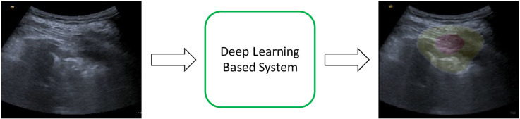
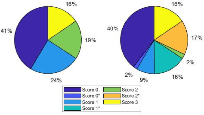
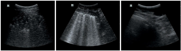
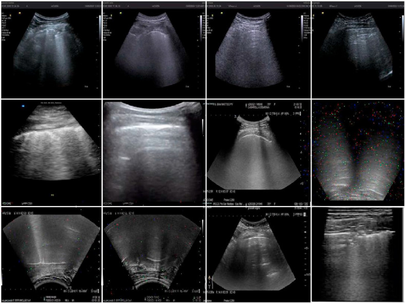
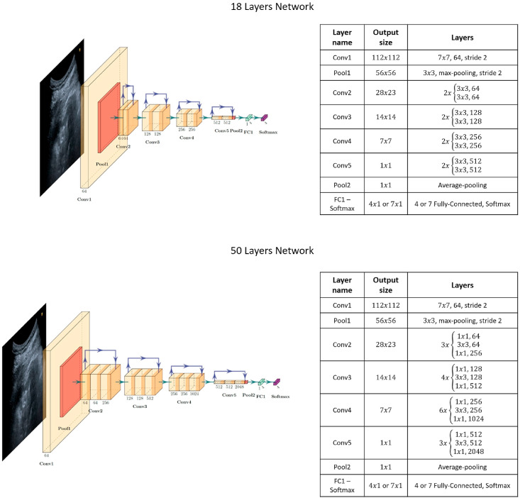
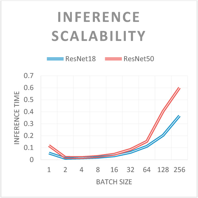
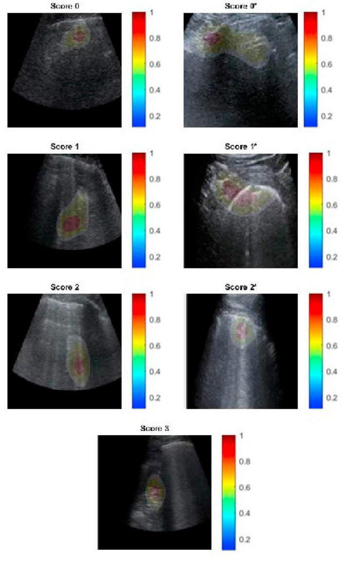
The Covid-19 European outbreak in February 2020 has challenged the world's health systems, eliciting an urgent need for effective and highly reliable diagnostic instruments to help medical personnel. Deep learning (DL) has been demonstrated to be useful for diagnosis using both computed tomography (CT) scans and chest X-rays (CXR), whereby the former typically yields more accurate results. However, the pivoting function of a CT scan during the pandemic presents several drawbacks, including high cost and cross-contamination problems. Radiation-free lung ultrasound (LUS) imaging, which requires high expertise and is thus being underutilised, has demonstrated a strong correlation with CT scan results and a high reliability in pneumonia detection even in the early stages. In this study, we developed a system based on modern DL methodologies in close collaboration with Fondazione IRCCS Policlinico San Matteo's Emergency Department (ED) of Pavia. Using a reliable dataset comprising ultrasound clips originating from linear and convex probes in 2908 frames from 450 hospitalised patients, we conducted an investigation into detecting Covid-19 patterns and ranking them considering two severity scales. This study differs from other research projects by its novel approach involving four and seven classes. Patients admitted to the ED underwent 12 LUS examinations in different chest parts, each evaluated according to standardised severity scales. We adopted residual convolutional neural networks (CNNs), transfer learning, and data augmentation techniques. Hence, employing methodological hyperparameter tuning, we produced state-of-the-art results meeting F1 score levels, averaged over the number of classes considered, exceeding 98%, and thereby manifesting stable measurements over precision and recall.
Keywords: Deep learning; LUS Score; Lung ultrasound; SARS-CoV-2.
Copyright © 2021 Elsevier Ltd. All rights reserved.
Conflict of interest statement
We declare that neither the manuscript nor any parts of its content are currently under consideration or published in another journal and that there is no conflict of interest in submitting our paper to your journal.
Figures
Similar articles
-
Perceptive SARS-CoV-2 End-To-End Ultrasound Video Classification through X3D and Key-Frames Selection.Bioengineering (Basel). 2023 Feb 21;10(3):282. doi: 10.3390/bioengineering10030282. Bioengineering (Basel). 2023. PMID: 36978673 Free PMC article.
-
Comparative study of lung ultrasound and chest computed tomography scan in the assessment of severity of confirmed COVID-19 pneumonia.Intensive Care Med. 2020 Sep;46(9):1707-1713. doi: 10.1007/s00134-020-06186-0. Epub 2020 Jul 29. Intensive Care Med. 2020. PMID: 32728966 Free PMC article.
-
COVID-19 detection in CT and CXR images using deep learning models.Biogerontology. 2022 Feb;23(1):65-84. doi: 10.1007/s10522-021-09946-7. Epub 2022 Jan 22. Biogerontology. 2022. PMID: 35064446 Free PMC article.
-
Development and integration of VGG and dense transfer-learning systems supported with diverse lung images for discovery of the Coronavirus identity.Inform Med Unlocked. 2022;32:101004. doi: 10.1016/j.imu.2022.101004. Epub 2022 Jul 8. Inform Med Unlocked. 2022. PMID: 35822170 Free PMC article. Review.
-
Are Lung Ultrasound Findings in COVID-19 Pneumonia Typical or Specific?Praxis (Bern 1994). 2021 Jun;110(8):421-425. doi: 10.1024/1661-8157/a003696. Praxis (Bern 1994). 2021. PMID: 34107756 Review.
Cited by
-
Review of Machine Learning in Lung Ultrasound in COVID-19 Pandemic.J Imaging. 2022 Mar 5;8(3):65. doi: 10.3390/jimaging8030065. J Imaging. 2022. PMID: 35324620 Free PMC article. Review.
-
Thoracic Ultrasound in Others Scenarios: An Expanding Tool.Open Respir Arch. 2025 Feb 27;6(Suppl 2):100420. doi: 10.1016/j.opresp.2025.100420. eCollection 2024 Oct. Open Respir Arch. 2025. PMID: 40226769 Free PMC article. Review.
-
Using Ultrasound Image Augmentation and Ensemble Predictions to Prevent Machine-Learning Model Overfitting.Diagnostics (Basel). 2023 Jan 23;13(3):417. doi: 10.3390/diagnostics13030417. Diagnostics (Basel). 2023. PMID: 36766522 Free PMC article.
-
Spinal tissue identification using a Forward-oriented endoscopic ultrasound technique.Biomed Eng Lett. 2024 Oct 26;15(1):193-201. doi: 10.1007/s13534-024-00440-w. eCollection 2025 Jan. Biomed Eng Lett. 2024. PMID: 39781062
-
Cross-domain lung opacity detection via adversarial learning and box fusion.Sci Rep. 2024 Dec 28;14(1):31353. doi: 10.1038/s41598-024-82719-7. Sci Rep. 2024. PMID: 39732945 Free PMC article.
References
MeSH terms
LinkOut - more resources
Full Text Sources
Other Literature Sources
Medical
Miscellaneous
