. 2021 Nov 1:284:119881.
doi: 10.1016/j.lfs.2021.119881. Epub 2021 Aug 10.

In Vivo protection from SARS-CoV-2 infection by ATN-161 in k18-hACE2 transgenic mice

Affiliations

In Vivo protection from SARS-CoV-2 infection by ATN-161 in k18-hACE2 transgenic mice

Narayanappa Amruta et al. Life Sci. .

Abstract

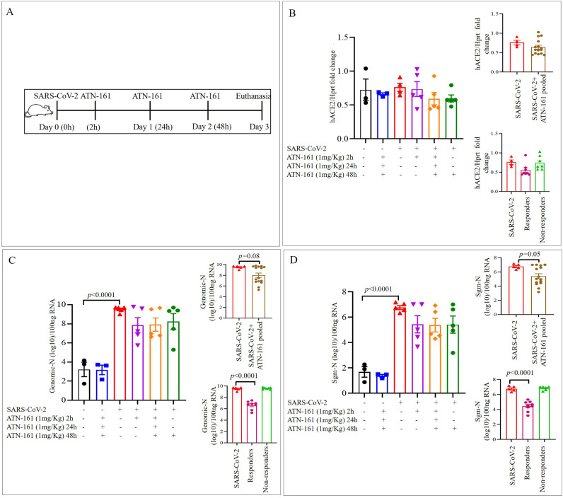
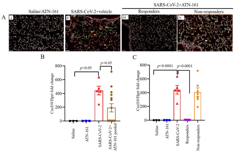
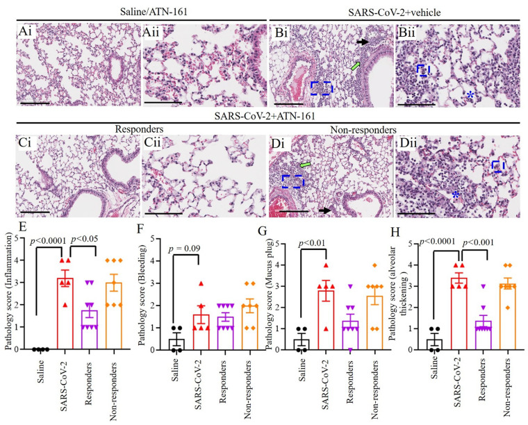
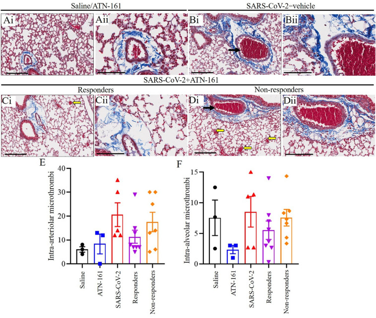
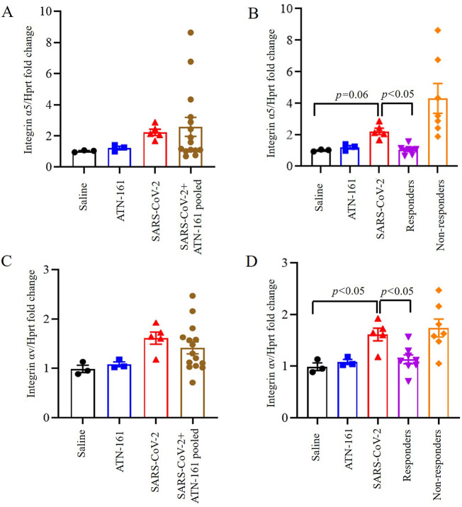
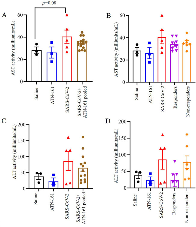
Severe acute respiratory syndrome coronavirus 2 (SARS-CoV-2) is an infectious disease that has spread worldwide. Current treatments are limited in both availability and efficacy, such that improving our understanding of the factors that facilitate infection is urgently needed to more effectively treat infected individuals and to curb the pandemic. We and others have previously demonstrated the significance of interactions between the SARS-CoV-2 spike protein, integrin α5β1, and human ACE2 to facilitate viral entry into host cells in vitro. We previously found that inhibition of integrin α5β1 by the clinically validated small peptide ATN-161 inhibits these spike protein interactions and cell infection in vitro. In continuation with our previous findings, here we have further evaluated the therapeutic potential of ATN-161 on SARS-CoV-2 infection in k18-hACE2 transgenic (SARS-CoV-2 susceptible) mice in vivo. We discovered that treatment with single or repeated intravenous doses of ATN-161 (1 mg/kg) within 48 h after intranasal inoculation with SARS-CoV-2 lead to a reduction of lung viral load, viral immunofluorescence, and improved lung histology in a majority of mice 72 h post-infection. Furthermore, ATN-161 reduced SARS-CoV-2-induced increased expression of lung integrin α5 and αv (an α5-related integrin that has also been implicated in SARS-CoV-2 interactions) as well as the C-X-C motif chemokine ligand 10 (Cxcl10), further supporting the potential involvement of these integrins, and the anti-inflammatory potential of ATN-161, respectively, in SARS-CoV-2 infection. To the best of our knowledge, this is the first study demonstrating the potential therapeutic efficacy of targeting integrin α5β1 in SARS-CoV-2 infection in vivo and supports the development of ATN-161 as a novel SARS-CoV-2 therapy.

Keywords: ATN-161; Cxcl10; Integrins; SARS-CoV-2; hACE2.


Conflict of interest statement

The authors have declared that no conflict of interest exists.

Figures

Fig. 1
Fig. 1
ATN-161 reduces the Genomic-N and Sub genomic N (Sgm—N) viral load in SARS-CoV-2 infected K18-hACE2 mice lung tissue Schematic overview of experimental timeline for K18-hACE2 mice (A) 10-week old male K18-hACE2 transgenic mice administered saline (black bar) or ATN-161 (1 mg/kg, blue bar) intravenously (via retro-orbital). Mice were inoculated via the intranasal route with Severe acute respiratory syndrome coronavirus 2 (SARS-CoV-2) (2 × 105 TCID50) + Saline (i.v. by retroorbital rout) (Red bar). ATN-161 (1 mg/Kg) treatment was administered at 3 different time periods, SARS-CoV-2 + ATN-161-2 h post infection (purple bar), SARS-CoV-2 + ATN-161-daily (2 h, 24 h and 48 h) administration (orange bar), and SARS-CoV-2 + ATN-161-48 h administration (green bar) post SARS-CoV-2 intranasal inoculation. Panels B, C, and D top inserts with brown bar represents SARS-CoV-2 + ATN-161 all treatment groups pooled data; Panels B, C, and D bottom insert with pink bar represents responding mice and light green bar represents nonresponding with SARS-CoV-2 + ATN-161 (either 2, daily or 48 h) treatment. 3 days post infection (3 dpi) the mice were euthanized, and RNA isolated from the left lungs by Trizol method for qRT-PCR (B) hACE2 expression in lungs of K18-hACE2 mice (C) viral genomic-N (Total-N) and (D) sub genomic-N mRNA (sgm-—N). Experimental groups are divided into 6 groups. Saline n = 3; ATN-161 n = 3; SARS-CoV-2 (5 mice for vehicle, 5 mice for each 3 ATN-161 groups). Data are presented as mean ± SEM. P values represent saline vs SARS-CoV-2 + vehicle and SARS-CoV-2 + vehicle vs SARS-CoV-2 + ATN-161 treatment groups. (For interpretation of the references to colour in this figure legend, the reader is referred to the web version of this article.)
Fig. 2
Fig. 2
ATN-161 reduces the expression of virus in SARS-CoV-2 infected K18-hACE2 mice lung tissue. Immunohistochemistry staining for SARS-CoV-2 (Ai-iv, orange arrowheads indicates SARS-CoV-2 viral stain (green) positive cells). SARS-CoV-2 viral staining representative image from saline/ATN-161 (i); SARS-CoV-2 + vehicle (ii); Responders (iii) and Non-responders (iv) with SARS-CoV-2 + ATN-161 administration either 2, daily or 48 h administration, post SARS-CoV-2 intranasal inoculation. The tissue analysis at 3 dpi using the protocol described in Fig. 1. All K-18 hACE2 mice infected with SARS-CoV-2 + Saline or non-responders from SARS-CoV-2 + ATN-161 administration group with have multifocal regions of SARS-CoV-2–positive cells (ii, iv), whereas treatment with ATN-161, 2 h and daily administered groups show 3 mice are completely negative for SARS-CoV-2 protein and 2 mice are negative (responders) in 48 h ATN-161 group out of 5 mice challenged for SARS-CoV-2. (B–C) Cxcl10 mRNA expression in lung. Scale bars, 50 μm. Green = SARS-CoV-2; White = nuclei/DAPI; Red = empty/autofluorescence. Data are presented as mean ± SEM. P values represent saline vs SARS-CoV-2 + vehicle and SARS-CoV-2 + vehicle vs SARS-CoV-2 + ATN-161 pooled, SARS-CoV-2 + ATN-161 responders, and SARS-CoV-2 + ATN-161 non-responders. (For interpretation of the references to colour in this figure legend, the reader is referred to the web version of this article.)
Fig. 3
Fig. 3
Histopathological Analysis of SARS-CoV-2 infected K18-hACE2 mice lung tissue (Ai–Dii) Hematoxylin and eosin staining of lung sections from saline/ATN-161 (Ai–Aii); SARS-CoV-2 + vehicle (Bi–ii); Responders (Ci–ii) and Non-responders (Di–ii) with SARS-CoV-2 + ATN-161(either 2 h, daily, or 48 h) administration post SARS-CoV-2 intranasal inoculation with tissue analysis at 3 dpi using the protocol described in Fig. 1. Histopathological observations indicated that multifocal lesion, moderate interstitial pneumonia and alveolar septal thickening (Bii, Dii, blue frames), infiltration of lymphocytes (Inflammation) (Bi, Di, green arrow), mucus plug and fibrin exudation (Bi, Di, black arrow), bleeding and pulmonary edema (Bii, Dii, asterisk). (E–H) Semi quantitative analysis of histopathological findings is based on subjective scoring as described in methods. Control n = 3; ATN-161 n = 3; SARS-CoV-2 (5 mice for vehicle, 5 mice for each 3 ATN-161 groups). Scale bar: 200 μm (Ai, Bi, Ci, and Di) and 100 μm (Aii, Bii, Cii, and Dii). Data are presented as mean ± SEM. P values represent saline vs SARS-CoV-2 + vehicle and SARS-CoV-2 + vehicle vs SARS-CoV-2 + ATN-161 responders, and SARS-CoV-2 + ATN-161 non-responders. (For interpretation of the references to colour in this figure legend, the reader is referred to the web version of this article.)
Fig. 4
Fig. 4
Histopathological Analysis of SARS-CoV-2 infected K18-hACE2 mice lung tissue. Representative images of Masson's Trichrome-stained sections (Ai–Dii) of lung sections from Saline/ATN-161 (Ai-Aii); SARS-CoV-2 + vehicle (Bi–ii); Responders (Ci–ii) and Non-responders (Di–ii) with SARS-CoV-2 + ATN-161(either 2 h, daily, or 48 h) administration post SARS-CoV-2 intranasal inoculation with tissue analysis at 3 dpi using the protocol described in Fig. 1. Histopathological observations indicated that showing multiple intra-arteriolar microthrombi (black arrows), intra-alveolar microthrombi (yellow arrow). However, the groups did not differ on the extent of fibrosis histopathology (collagen area). Semi quantitative analysis of histopathological findings is based on subjective scoring as described in methods (E–F). Control n = 3; ATN-161 n = 3; SARS-CoV-2 (5 mice for vehicle, 5 mice for each 3 ATN-161 groups). Scale bar: 200 μm (Ai, Bi, Ci, and Di) and 100 μm (Aii, Bii, Cii, and Dii). (For interpretation of the references to colour in this figure legend, the reader is referred to the web version of this article.)
Fig. 5
Fig. 5
Induced expression of integrin α5, and integrin αv in the lungs of SARS-CoV-2 infected K18-hACE2 mice and is inhibited by ATN-161 treatment. Integrin α5 (A–B), and Integrin αv (C–D) mRNA expression in lung at different time periods of ATN-161 by intravenous injection (i.v) after infection with SARS-CoV-2 (TCID50 = 2 × 105 /0.05 mL) was determined by qRT-PCR. Data are presented as mean ± SEM. P values represent saline vs SARS-CoV-2 + vehicle and SARS-CoV-2 + vehicle vs SARS-CoV-2 + ATN-161 pooled, SARS-CoV-2 + ATN-161 responders, and SARS-CoV-2 + ATN-161 non-responders.
Fig. 6
Fig. 6
Effect of ATN-161 on hepatic enzyme levels in the serum of SARS-CoV-2 infected K18-hACE2 mice. (A–B) aspartate aminotransferase (AST); (C–D) Alanine aminotransferase (ALT). Data are presented as mean ± SEM. P values represent saline vs SARS-CoV-2 + vehicle and SARS-CoV-2 + vehicle vs SARS-CoV-2 + ATN-161 pooled, SARS-CoV-2 + ATN-161 responders, and SARS-CoV-2 + ATN-161 non-responders.

References

    1. Zhou F., Yu T., Du R., Fan G., Liu Y., Liu Z., et al. Clinical course and risk factors for mortality of adult inpatients with COVID-19 in Wuhan, China: a retrospective cohort study. Lancet. 2020;395(10229):1054–1062. doi: 10.1016/S0140-6736(20)30566-3. - DOI - PMC - PubMed
    1. Carvacho I., Piesche M. RGD-binding integrins and TGF-beta in SARS-CoV-2 infections - novel targets to treat COVID-19 patients? Clin. Transl. Immunol. 2021;10(3) doi: 10.1002/cti2.1240. - DOI - PMC - PubMed
    1. Control CfD . 2020. Prevention. Interim Clinical Guidance for Management of Patients With Confirmed Coronavirus Disease (COVID-19)
    1. Perra N. Non-pharmaceutical interventions during the COVID-19 pandemic: a review. Phys. Rep. 2021 doi: 10.1016/j.physrep.2021.02.001. - DOI - PMC - PubMed
    1. Group CC-W. Liu Y., Morgenstern C., Kelly J., Lowe R., Jit M. The impact of non-pharmaceutical interventions on SARS-CoV-2 transmission across 130 countries and territories. BMC Med. 2021;19(1):40. doi: 10.1186/s12916-020-01872-8. - DOI - PMC - PubMed

MeSH terms