. 2021 Aug 13;12(1):4909.
doi: 10.1038/s41467-021-24881-4.

Structures of tmRNA and SmpB as they transit through the ribosome

Affiliations

Structures of tmRNA and SmpB as they transit through the ribosome

Charlotte Guyomar et al. Nat Commun. .

Abstract

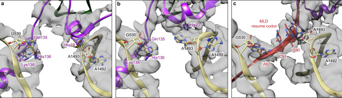
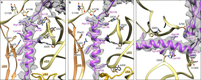
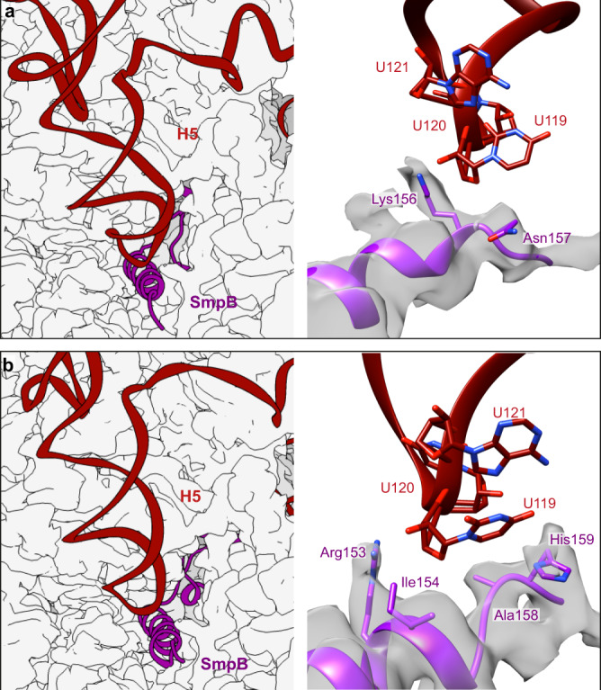
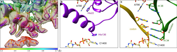
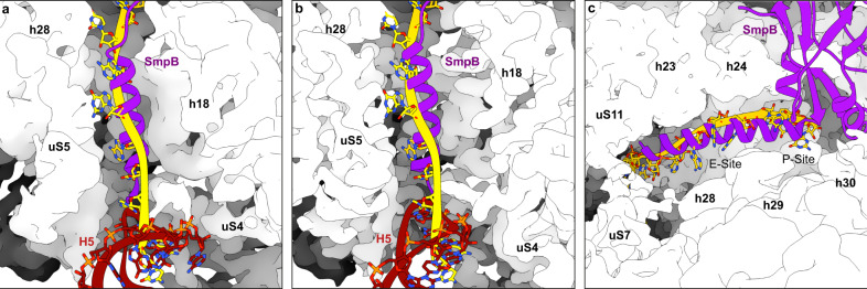
In bacteria, trans-translation is the main rescue system, freeing ribosomes stalled on defective messenger RNAs. This mechanism is driven by small protein B (SmpB) and transfer-messenger RNA (tmRNA), a hybrid RNA known to have both a tRNA-like and an mRNA-like domain. Here we present four cryo-EM structures of the ribosome during trans-translation at resolutions from 3.0 to 3.4 Å. These include the high-resolution structure of the whole pre-accommodated state, as well as structures of the accommodated state, the translocated state, and a translocation intermediate. Together, they shed light on the movements of the tmRNA-SmpB complex in the ribosome, from its delivery by the elongation factor EF-Tu to its passage through the ribosomal A and P sites after the opening of the B1 bridges. Additionally, we describe the interactions between the tmRNA-SmpB complex and the ribosome. These explain why the process does not interfere with canonical translation.


Conflict of interest statement

The authors declare no competing interests.

Figures

Fig. 1
Fig. 1. High-resolution structures of four consecutive trans-translation states.
Shown are the a pre-accommodation (PRE-ACC), b accommodation (ACC), c translocation (TRANS) and d intermediate post-translocation (TRANS*) complexes. Top, schematic representations of the complexes showing the ribosomal 50S (blue) and 30S (khaki) subunits, non-stop mRNA (yellow), P-site tRNAphe (green), elongation factor EF-Tu (pink), SmpB (purple) and tmRNA (red). Also indicated are the ribosome’s A, P and E sites, and the tmRNA structural domains: the H2 helix, H5 stem-loop, pseudoknot (PK) ring, mRNA-like domain (MLD) and tRNA-like domain (TLD). Middle, electron density maps contoured at 2.5 σ, where sigma refers to the variance in the map. Bottom, the same density maps rotated by 180° around the Y-axis and 45° around the X-axis. eh Cross-sections of the 30S subunits in the same states as (ad), respectively, showing the SmpB C-terminal tail, non-stop mRNA and the MLD in the mRNA channel.
Fig. 2
Fig. 2. The H5 and PK2 domains of tmRNA interact with the uS2, uS3, uS4 and uS5 proteins on the small ribosomal subunit.
Shown are the ac pre-accommodation state, df accommodation state, and gi translocation state. Left column, overview of the interactions between tmRNA (red), SmpB (purple), and the ribosomal proteins uS2 (tan), uS3 (khaki) uS4 (gold) and uS5 (sandy brown). To highlight the motion of the H5 stem-loop during translocation, all structures are aligned on uS3. Middle, close-up of the interactions between the PK2 pseudoknot and uS3’s KH2 RNA-binding domain. Right, close-up of the interactions between the H5 stem-loop and SmpB, uS3, uS4 and uS5. Residues and nucleotides within 4 Å of each other are indicated, and the cryo-electron density map is displayed.
Fig. 3
Fig. 3. Detailed view of the decoding centre during trans-translation.
Close-up of the interactions between SmpB (purple) and the conserved nucleotides G530, A1492 and A1493 of the 16S rRNA (khaki) in the a pre-accommodation and b accommodation states. c Interactions between tmRNA’s resume codon (red) and the same nucleotides during the translocation state. Residues and nucleotides within 4 Å of each other are indicated, and the cryo-electron density map is displayed.
Fig. 4
Fig. 4. Interactions between SmpB’s C-terminal tail and the 30S small ribosomal subunit.
Each panel details the contacts that stabilize the C-terminal tail of SmpB in the mRNA channel during the a pre-accommodation, b accommodation, and c translocation states. SmpB is purple, 16S rRNA is khaki, and the ribosomal proteins uS4 and uS5 are gold and sandy brown, respectively. Residues and nucleotides within 4 Å of each other are indicated, and the cryo-electron density map of SmpB is displayed.
Fig. 5
Fig. 5. Detailed view of the interactions between the C-terminal tail of SmpB and the H5 stem-loop of tmRNA.
Left: position of the H5 stem-loop at the entrance of the mRNA channel. Right: focus on the residues involved in the interactions between SmpB and H5 during the a pre-accommodation and b accommodation states. SmpB is purple, tmRNA is red and the surface of the ribosomal small subunit is light grey. Residues and nucleotides within 4 Å of each other are indicated, and the cryo-electron density map of SmpB is displayed.
Fig. 6
Fig. 6. The structure of translocated tmRNA reveals how the right resume codon is selected.
a Left, focus on the interactions between SmpB (purple) and the four nucleotides just upstream of the tmRNA (red) resume codon. The cryo-electron density map is displayed and coloured according to the local resolutions as computed with ResMap. Right, same but without the map. For clarity, cartoon representation is used for SmpB and only the four residues involved in the codon selection are shown. b An A84U/U85G double mutation in tmRNA maintains a high level of trans-translation but also promotes -1 frameshifting. c Mutation of tmRNA’s highly conserved A86 nucleotide into a pyrimidine nucleotide lowers the stacking interaction with SmpB Tyr55 and results in a +1 frameshift,. d An SmpB triple mutant (Y24C, E107V and V129A) can partially reverse the effect of the tmRNA A86C mutation, allowing for both +1 frameshifting and in-frame re-registration. The boxes show part of the MLD sequence, with the wild-type sequence repeated at the top for reference. The first two codons are grey, the mutated nucleotides are green and the five nucleotides presented in the figure are in capital letters. Where appropriate, the sequences of the mutants are shifted to highlight the frameshifts caused by the mutations.
Fig. 7
Fig. 7. SmpB mimics the interactions of a tRNA anticodon loop in the P site.
a Focus on the interactions between SmpB and the 16S rRNA P-site in the translocation state. The cryo-electron density map is displayed and coloured according to the local resolutions as computed with ResMap. b Same as (a), but without the map. For clarity, cartoon representation is used for SmpB and only His22 and His126 are shown. c Comparison with a P-site tRNA (PDB code 7K00). The SmpB C-terminal tail occupies the mRNA path, replacing the codon–anticodon interaction. SmpB also reproduces the anticodon loop interactions with the 16S rRNA. The stacking interaction between His22 and A790 replaces the interaction usually observed with the sugar of tRNA’s nucleotide (nt) 38. Meanwhile, His136 replaces the anticodon’s nucleotide 34, stacking on the C1400 rRNA nucleotide. SmpB is purple, tRNA is green, mRNA is yellow and 16S rRNA is khaki.
Fig. 8
Fig. 8. Transit of tmRNA through the B1a, B1b, and B1c bridges during trans-translation.
Close-up view of the opening and closing of bridges B1a, B1b and B1c during a pre-accommodation, b accommodation, c translocation and d intermediate translocation (TRANS*) states. The 50S ribosomal subunit is blue, 30S is khaki, elongation factor EF-Tu is pink, tmRNA is red, SmpB is purple and tRNAphe is green. The tmRNA helix H2 and the 50S helix H38 are also labelled. Black arrows indicate the degree of rotation and tilt of the 30S head measured with respect to its body, as per Nguyen and Whitford. Orange arrows highlight the opening of bridges B1b and B1c. All maps are contoured at 2.5 σ, where sigma represents variance in the map.
Fig. 9
Fig. 9. Close-up of the mRNA path during trans-translation.
In the a pre-accommodation and b accommodation states, the C-terminal tail of SmpB (purple) occupies the mRNA channel, while the H5 stem-loop of tmRNA (red) closes the entrance. Both domains would clash with a canonical mRNA (yellow, PDB code 6ZTJ), forcing the selection of stalled ribosomes. c After translocation, the C-terminal tail is rotated by 62° and deeply inserted into the mRNA exit tunnel. There again, it clashes with the mRNA (PDB code 7JT1), resulting in the ejection of the non-stop mRNA from the ribosome. The cryo-electron density map of the 30S small subunit is shown as a white surface.

References

    1. Giudice E, Gillet R. The task force that rescues stalled ribosomes in bacteria. Trends Biochem Sci. 2013;38:403–411. doi: 10.1016/j.tibs.2013.06.002. - DOI - PubMed
    1. Keiler KC, Feaga HA. Resolving nonstop translation complexes is a matter of life or death. J. Bacteriol. 2014;196:2123–2130. doi: 10.1128/JB.01490-14. - DOI - PMC - PubMed
    1. Buskirk, A. R. & Green, R. Ribosome pausing, arrest and rescue in bacteria and eukaryotes. Philos Trans R Soc Lond B Biol Sci. 372, 20160183 (2017). - PMC - PubMed
    1. Bessho Y, et al. Structural basis for functional mimicry of long-variable-arm tRNA by transfer-messenger RNA. Proc. Natl Acad. Sci. U.S.A. 2007;104:8293–8298. doi: 10.1073/pnas.0700402104. - DOI - PMC - PubMed
    1. Komine Y, Kitabatake M, Yokogawa T, Nishikawa K, Inokuchi H. A tRNA-like structure is present in 10Sa RNA, a small stable RNA from Escherichia coli. Proc. Natl Acad. Sci. U.S.A. 1994;91:9223–9227. doi: 10.1073/pnas.91.20.9223. - DOI - PMC - PubMed

Publication types

MeSH terms