. 2021 Aug 13;12(1):4908.
doi: 10.1038/s41467-021-25082-9.

Nuclear export and translation of circular repeat-containing intronic RNA in C9ORF72-ALS/FTD

Affiliations

Nuclear export and translation of circular repeat-containing intronic RNA in C9ORF72-ALS/FTD

Shaopeng Wang et al. Nat Commun. .

Abstract

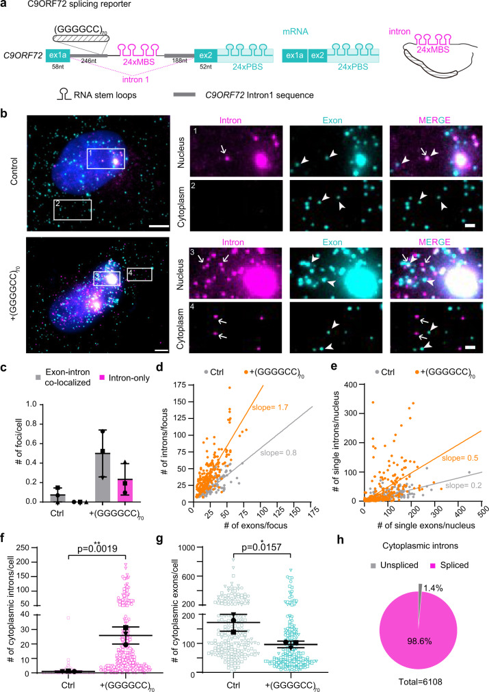
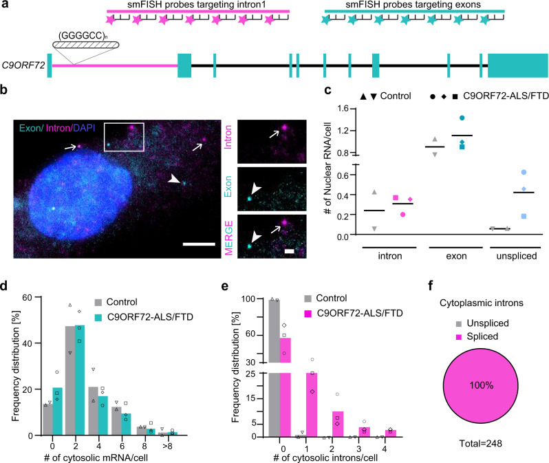
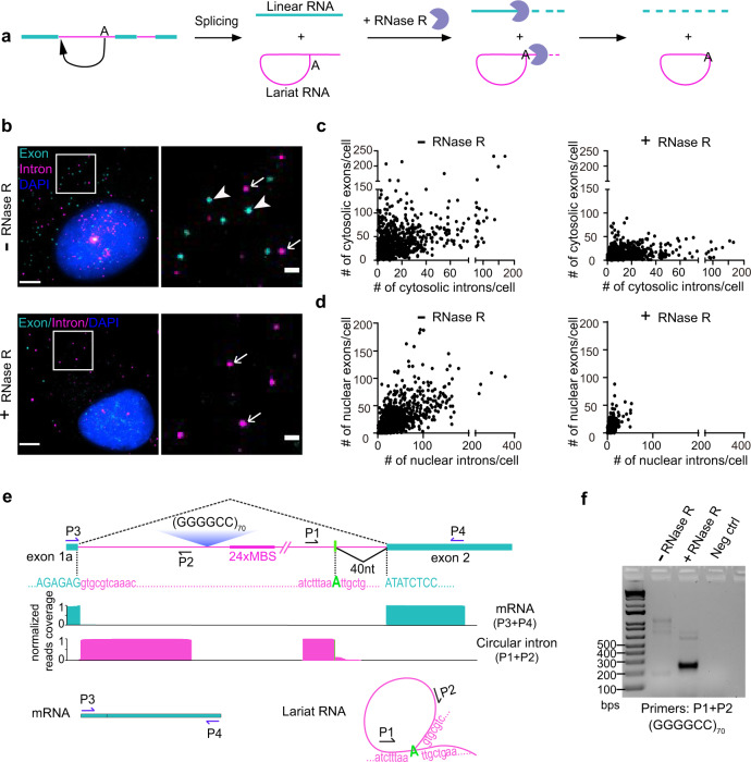
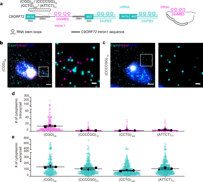
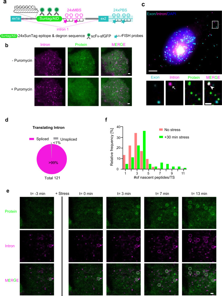
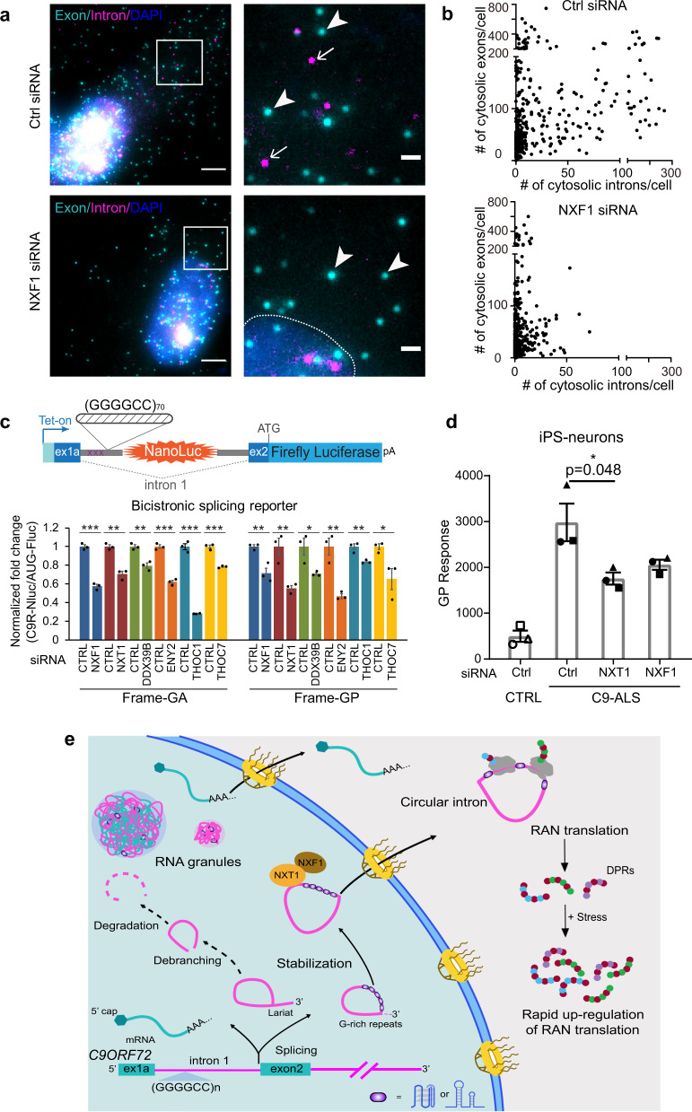
C9ORF72 hexanucleotide GGGGCC repeat expansion is the most common genetic cause of amyotrophic lateral sclerosis (ALS) and frontotemporal dementia (FTD). Repeat-containing RNA mediates toxicity through nuclear granules and dipeptide repeat (DPR) proteins produced by repeat-associated non-AUG translation. However, it remains unclear how the intron-localized repeats are exported and translated in the cytoplasm. We use single molecule imaging approach to examine the molecular identity and spatiotemporal dynamics of the repeat RNA. We demonstrate that the spliced intron with G-rich repeats is stabilized in a circular form due to defective lariat debranching. The spliced circular intron, instead of pre-mRNA, serves as the translation template. The NXF1-NXT1 pathway plays an important role in the nuclear export of the circular intron and modulates toxic DPR production. This study reveals an uncharacterized disease-causing RNA species mediated by repeat expansion and demonstrates the importance of RNA spatial localization to understand disease etiology.


Conflict of interest statement

The authors declare no competing interests.

Figures

Fig. 1
Fig. 1. C9ORF72 repeat-containing introns form nuclear RNA granules and are exported into cytoplasm.
a Schematic of the C9ORF72 splicing reporter construct. MBS/PBS: MS2/PP7 binding sites; ex1a, ex2: exon 1a and 2 of C9ORF72 gene. Two sets of RNA FISH probes were designed to target either 24×MBS or 24×PBS with different fluorescent dyes. The mature mRNA only shows PBS signal, spliced intron only has MBS signal, and the unspliced pre-mRNA contains both MBS and PBS signals. b Representative images of two-color smFISH experiment to study the spatial distribution of RNA species in control or +(GGGGCC)70 reporter cells. The boxes 1–4 were enlarged on the right. Magenta: intron (MBS); cyan: exon (PBS); blue: DAPI; arrow: single-intron molecule; arrowhead: single exon molecule. Scale bar: 5 μm and 1 μm for zoom in, respectively. Quantification shown in panels cg. c Number of RNA granules per nucleus. Repeat-containing introns formed large granules colocalized with exons (gray) as well as intron only (pink) granules. Cells were treated with transcription inhibitor actinomycin D (1 μg/mL for 20 min) to exclude the effect of transcription. Data are mean ± SD from three biological replicates. The quantified cell number: Ctrl (88, 82, 41) and (GGGGCC)70 (72, 43, 73). d Scatter plot of intron vs exon numbers in each RNA granule. Each dot represented one granule (from three biological replicates). The lines were linear fit to the scatter plot. The slope reflects the ratio of intron vs exon molecules in each granule. e Scatter plot of the numbers of single introns vs exons in each nucleus. Each dot represented a single nucleus. The lines were linear fit to the scatter plot. The slope reflects the ratio of intron vs exon molecules in each nucleus. f, g Quantification of cytoplasmic intron (f) and exon (g) number per cell. Each symbol represented a single cell and the three shapes represented three biological replicates. The mean of each replicate (larger black shapes) was used to calculate the average (horizontal bar) and SD (error bars) in each group, as well as for statistic comparison between groups. *P < 0.05, **P < 0.01, two-tailed t-test. The cell number in dg: Ctrl (60, 43, 93) and (GGGGCC)70 (93, 95, 49). h Percentage of cytoplasmic introns colocalized (unspliced) or uncolocalized (spliced) with exons in +(GGGGCC)70 cells. The exported GGGGCC repeat-containing RNAs are predominantly spliced introns. Source data are provided as a Source Data File.
Fig. 2
Fig. 2. The distribution and molecular identity of endogenous C9ORF72 RNAs in patient fibroblast cells.
a Schematic of smFISH probes targeting endogenous C9ORF72 RNAs. A set of 76 probes targets intron 1 (magenta) and a 77-probe set targets all the exons (cyan). b Representative two-color smFISH images of endogenous C9ORF72 RNAs in C9ORF72-ALS patient-derived fibroblasts. arrow: single-intron molecule; arrowhead: single exon molecule. Scale bar: 5 μm, and 1 μm for zoom in. c Quantification of nuclear introns, mRNAs and pre-mRNAs per nucleus. Datapoints of different shapes represent individual fibroblast line derived from two healthy controls and three C9ORF72-ALS patients. The horizontal bar indicated the mean value. d, e The percentage of cells containing different numbers of cytosolic exons (d) and introns (e) were quantified and calculated from each line with three technical replicates. Datapoints of different shapes represent individual fibroblast line derived from two healthy controls and three C9ORF72-ALS patients. The quantified cell number: Ctrl-1 (81), Ctrl-2 (165), C9-2 (135), C9-5 (131), and C9-6 (93). f Cytoplasmic introns were not colocalized with exons in C9ORF72-ALS patient fibroblasts, supporting the nuclear export of spliced intron. Source data are provided as a Source Data File.
Fig. 3
Fig. 3. The cytoplasmic repeat-containing introns are exported in the circular form.
a Schematic of splicing and circular RNA enrichment by RNase R treatment. RNase R only degrades linear RNA. The tail of the lariat can be degraded, but the circular part will be preserved. b Representative images of two-color smFISH in +(GGGGCC)70 reporter cells treated with RNase R (bottom) or just buffer (top). Magenta: intron (MBS); cyan: exon (PBS); blue: DAPI; arrow: intron; arrowhead: exon. Scale bar: 5 μm, and 1 μm for zoom in. Quantification shown in panels c and d. c, d Scatter plot of intron vs exon numbers in the cytoplasm (c) or nucleus (d) of each cell without RNase R (left) or with RNase R treatment (right). Each dot represented one cell. Total three technical replicates were carried out: 287, 289, 263 cells quantified for RNase R treatment and 222, 369, 342 cells for control (without RNase R treatment). e Deep sequencing identified circular boundaries of intron RNAs. The major splicing junction in mRNAs and branch site in circular RNAs were annotated. The libraries were prepared using the indicated primers (P1–P2 for circular intron; P3–P4 for mRNA), and the whole fragments were sequenced by 1 × 300nt MiSeq. The reads spanning the exon–exon junctions in mRNAs were aligned, shown in cyan. The reads spanning the junction between branch site and 5’-end of the intron in circular RNAs were aligned and shown in magenta. f RT-PCR of circular intron across the branch site boundary from +(GGGGCC)70 reporter cells with or without RNase R treatment. The same amount of RNAs before and after RNase R treatment were used to synthesize the cDNA. PCR products using the divergent primers P1+P2 were only evident in RNase R-treated samples. Total three independent biological replicates were examined with similar results. Source data are provided as a Source Data File.
Fig. 4
Fig. 4. G-rich repeats stabilize the spliced intron and mediate the nuclear export.
a Schematic of splicing reporter constructs with four other repeat sequences in the context of C9ORF72 gene. The GGGGCCexp was replaced with the indicated repeats in the reporter of Fig. 1a. MBS/PBS: MS2/PP7 binding sites; ex1a, ex2: exon 1a and 2 of C9ORF72 gene. From these reporters, the mature mRNA only shows PBS signal, spliced intron only has MBS signal, and the unspliced pre-mRNA contains both MBS and PBS signals. b, c Representative images of two-color smFISH of C9ORF72 splicing reporter cells containing intronic (CGG)98 (b) and (CCCCGG)70 (c) repeats. Scale bar: 5 μm, and 1 μm for zoom in. d, e Quantification of cytoplasmic intron (d) and exon (e) numbers per cell in the splicing reporter cell lines with four different repeat expansions. Each symbol represented a single cell and the three shapes represented three biological replicates (n = 75, 80, 73 cells in (CGG)98; n = 162, 146, 103 cells in (CCCCGG)70; n = 147, 171, 119 cells in (CCTG)240; n = 180,114,151 cells in (ATTCT)73). The mean of each replicate (larger black shapes) was used to calculate the average (horizontal bar) and SD (error bars) in each reporter line. Source data are provided as a Source Data File.
Fig. 5
Fig. 5. Translation of circular repeat-containing intron.
a Diagram of single-molecule translation imaging reporter for C9ORF72 repeat-containing intron. The SINAPS reporter lacking the ATG start codon was inserted after the (GGGGCC)70 repeats in the GR frame in the splicing construct. The SunTag fluorescence (green, scFv-sfGFP) on the RNA represents the RAN translation signal. The auxin-inducible degron (AID) at the end of the reporter was used to degrade the mature proteins and decrease the background signals. b Live cell imaging illustrates that the fluorescence puncta are translation sites. U-2 OS cells stably expressing membrane tethered stdMCP protein, were transiently transfected with intronic translation reporter in a. Cells were treated with translation inhibitor puromycin (100 μg/mL) at time 0 and imaged at 10 min. Translation was shut down efficiently. Green (protein): SunTag-scFv-sfGFP; magenta (intron): stdMCP-Halotag-CAAX-JF646. Scale bar: 2 μm. Total three independent biological replicates were examined with similar results. c Representative smFISH & IF images to measure RNA and translation. Green: protein; magenta: intron; cyan: exon. Arrow: intron; arrowhead: translation site. Scale bar: 5 μm, and 1 μm for zoom in. The protein signals colocalized with the spliced intron (magenta) indicated the translation of the spliced intron. Quantification shown in panel f (also see Supplementary Movie 2). d Percentage of translating introns colocalized (unspliced) or uncolocalized (spliced) with exons. From a total of 121 translation introns, only 1 showed colocalization with exon signals. e Time-lapse images illustrating the upregulation of RAN translation by stress stimuli. U-2 OS cells stably expressing membrane tethered stdMCP protein were transiently transfected with translation reporter for C9ORF72 repeat-containing intron. Cells were treated with 2 mM sodium arsenite at time 0 and imaged every 30 s for 30 min. Green (protein): SunTag-scFv-sfGFP; magenta (intron): stdMCP-Halotag-CAAX-JF646. Scale bar: 2 μm. Total three independent biological replicates were examined with similar results (also see Supplementary Movie 3). f Histogram of translation site intensities measured by the number of nascent peptides on each translation site, which increased upon stress stimuli. Data are quantified from three biological replicates: control (530, 480, 504 cells) and under stress (550, 512, 430 cells). Source data are provided as a Source Data File.
Fig. 6
Fig. 6. The NXF1-NXT1 pathway regulates the circular intronic repeat RNA export.
a Two-color smFISH in +(GGGGCC)70 splicing reporter cells transfected with nontargeting control (top) or NXF1-targeting (bottom) siRNA. Magenta: intron (MBS); cyan: exon (PBS); blue: DAPI. Scale bar: 5 μm. The boxes were zoomed and shown on the right, scale bar: 1 μm. Arrow: intron; arrowhead: exon. Dash line: nuclear boundary. Quantification shown in panel b. b Scatter plot of cytosolic intron vs exon numbers in each cell with either nontargeting control siRNA (top) or NXF1-targeting siRNA (bottom) transfection. Each dot represented one cell (total 418 cells for control and 490 for NXF1 siRNA, from three biological replicates). c HeLa Flp-In bicistronic splicing reporter cells (C9R-NLuc is located in the C9ORF72 intron 1, flanked by C9 exon 1 and 2, pA: poly-A tail) were induced to express translation reporters by doxycycline after two days of siRNA transfection, and luciferase activities were measured after another 24 h. NLuc signals were normalized to FLuc in each sample and the relative expression was compared to nontargeting siRNA control. *P < 0.05, **P < 0.01, ***P < 0.001, two-tailed t-test. Data are mean ± SEM. from three biological replicates. The exact P values for frame-GA from left to right are 0.0001, 0.0012, 0.0036, 0.0001, 0.00002, 0.0003. The exact P values for frame-GP from left to right are 0.009, 0.0093, 0.033, 0.0046, 0.0085, 0.0417. d C9ORF72-ALS iPSNs were transfected with NXT1, NXF1 or nontargeting siRNA at day 16 post-differentiation. Poly-GP was measured by ELISA after another 16 days of differentiation and maturation. Different shapes of datapoints represent independent cell lines. Data are presented as mean ± SEM. from three control and three C9ORF72-ALS cell lines. *P < 0.05 by One-way ANOVA followed by Dunn’s post hoc. e Working model: G-rich repeats stabilize introns in circular form and mediate the nuclear export. In C9ORF72-ALS/FTD, the circular intron with GGGGCCexp is the template for RAN translation in the cytoplasm, which can be upregulated by stress stimuli. The NXF1-NXT1 pathway plays an important role in the nuclear export of the circular intron, mediated by the GGGGCCexp. Source data are provided as a Source Data File.

Similar articles

Cited by

References

    1. Paulson H. Repeat expansion diseases. Handb. Clin. Neurol. 2018;147:105–123. doi: 10.1016/B978-0-444-63233-3.00009-9. - DOI - PMC - PubMed
    1. DeJesus-Hernandez M, et al. Expanded GGGGCC hexanucleotide repeat in noncoding region of C9ORF72 causes chromosome 9p-linked FTD and ALS. Neuron. 2011;72:245–256. doi: 10.1016/j.neuron.2011.09.011. - DOI - PMC - PubMed
    1. Renton AE, et al. A hexanucleotide repeat expansion in C9ORF72 is the cause of chromosome 9p21-linked ALS-FTD. Neuron. 2011;72:257–268. doi: 10.1016/j.neuron.2011.09.010. - DOI - PMC - PubMed
    1. Haeusler AR, Donnelly CJ, Rothstein JD. The expanding biology of the C9orf72 nucleotide repeat expansion in neurodegenerative disease. Nat. Rev. Neurosci. 2016;17:383–395. doi: 10.1038/nrn.2016.38. - DOI - PMC - PubMed
    1. Kumar V, Hasan GM, Hassan MI. Unraveling the role of RNA mediated toxicity of C9orf72 repeats in C9-FTD/ALS. Front. Neurosci. 2017;11:711. doi: 10.3389/fnins.2017.00711. - DOI - PMC - PubMed

Publication types

MeSH terms