. 2021 Aug 13;12(1):4926.
doi: 10.1038/s41467-021-25227-w.

The missing linker between SUN5 and PMFBP1 in sperm head-tail coupling apparatus

Affiliations

The missing linker between SUN5 and PMFBP1 in sperm head-tail coupling apparatus

Ying Zhang et al. Nat Commun. .

Abstract

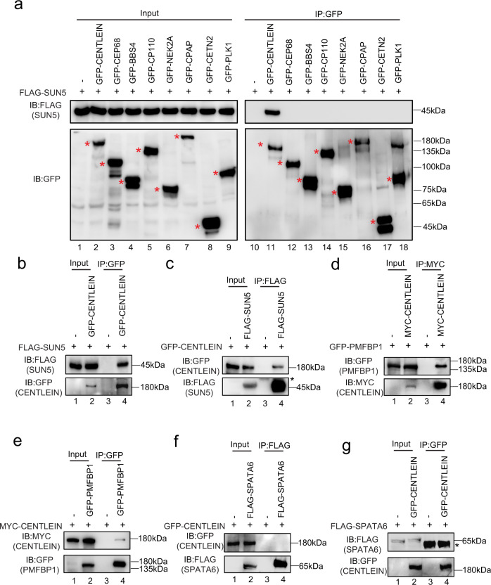
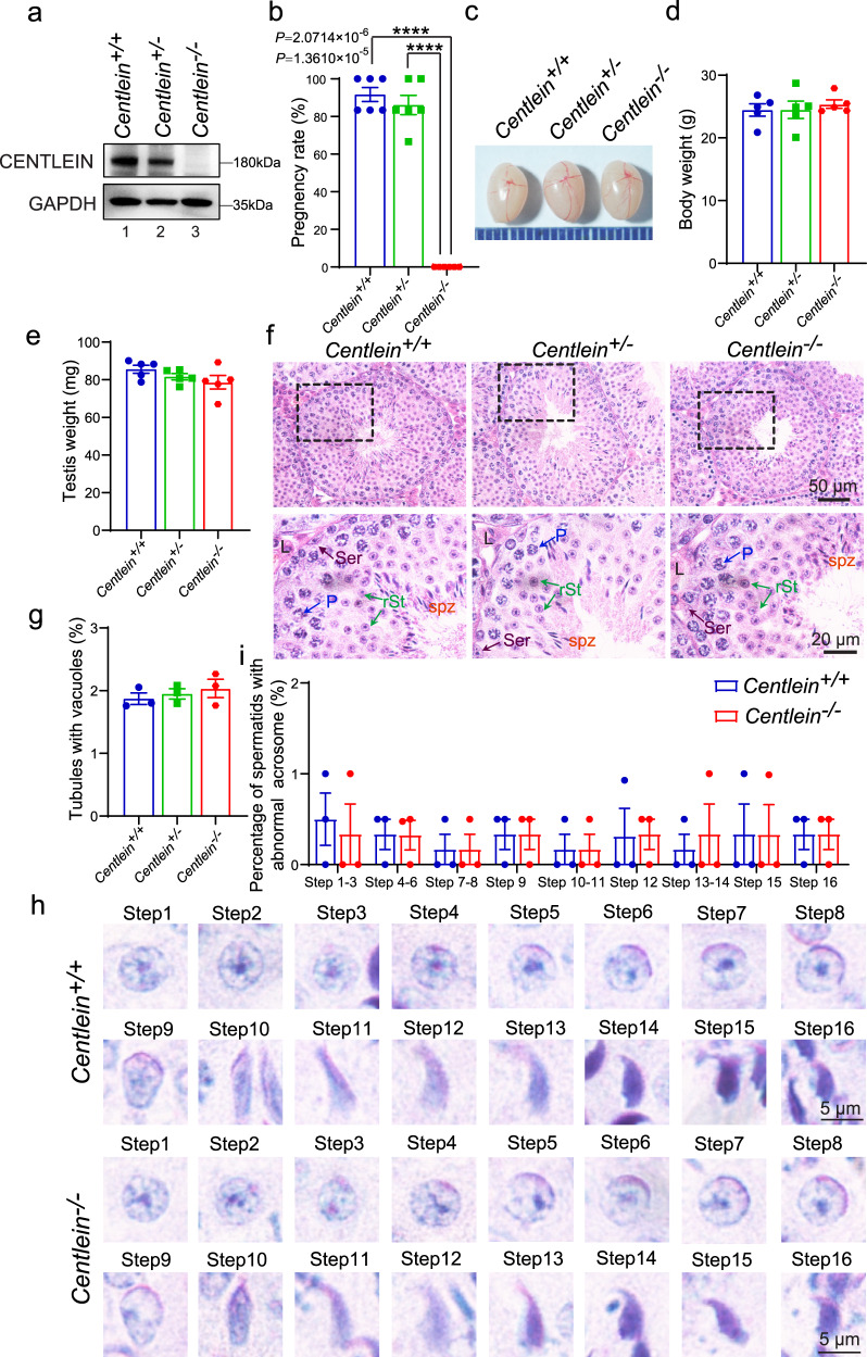
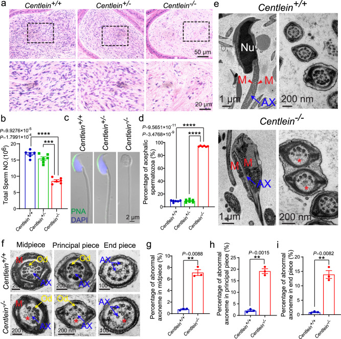
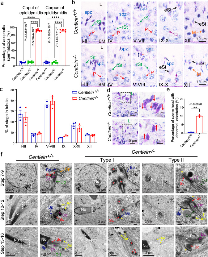
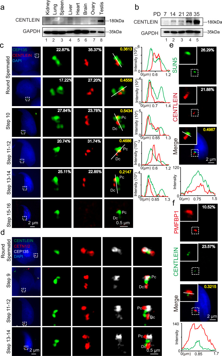
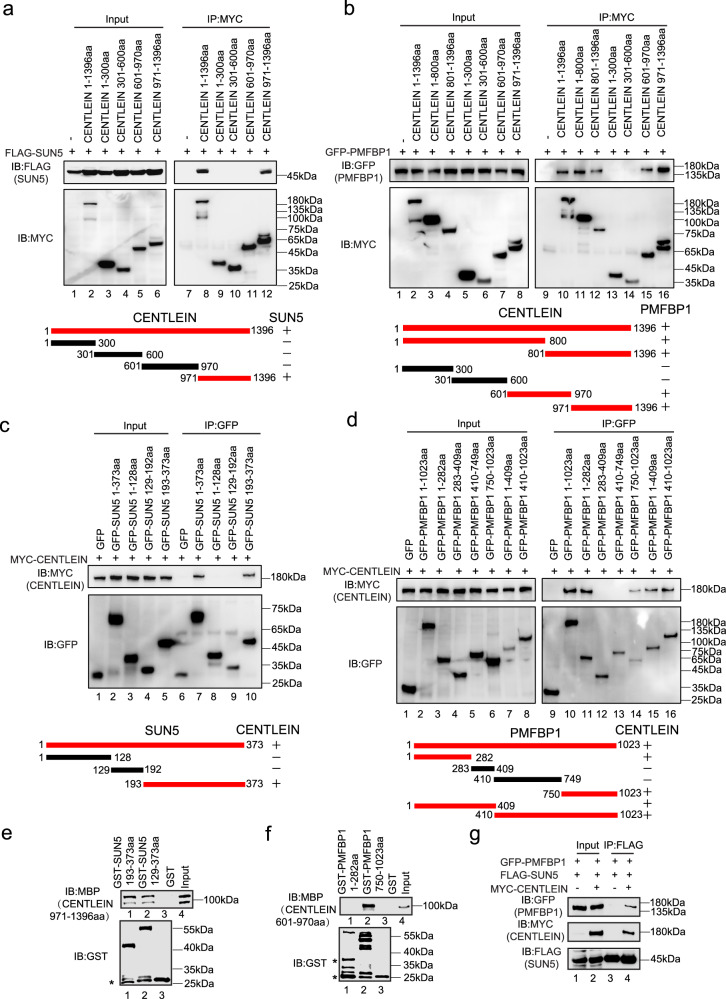
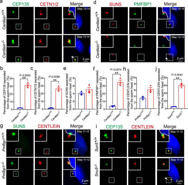
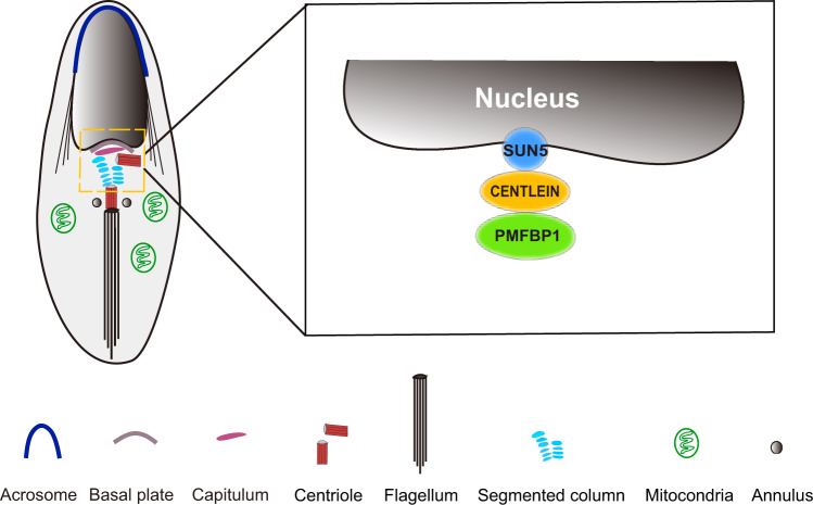
The sperm head-to-tail coupling apparatus (HTCA) ensures sperm head-tail integrity while defective HTCA causes acephalic spermatozoa, rendering males infertile. Here, we show that CENTLEIN is indispensable for HTCA integrity and function, and that inactivation of CENTLEIN in mice leads to sperm decapitation and male sterility. We demonstrate that CENTLEIN directly interacts with both SUN5 and PMFBP1, two proteins localized in the HTCA and related with acephalic spermatozoa syndrome. We find that the absence of Centlein sets SUN5 and PMFBP1 apart, the former close to the sperm head and the latter in the decapitated tail. We show that lack of Sun5 results in CENTLEIN and PMFBP1 left in the decapitated tail, while disruption of Pmfbp1 results in SUN5 and CENTLEIN left on the detached sperm head. These results demonstrate that CENTLEIN cooperating with SUN5 and PMFBP1 participates in the HTCA assembly and integration of sperm head to the tail, indicating that impairments of CENTLEIN might be associated with acephalic spermatozoa syndrome in humans.


Conflict of interest statement

The authors declare no competing interests.

Figures

Fig. 1
Fig. 1. CENTLEIN interacts with both SUN5 and PMFBP1.
a A candidate-based approach by identification of SUN5-binding centrosomal proteins. FLAG-SUN5 and one of the GFP-tagged plasmid, including the empty vector, GFP-CENTLEIN, GFP-CEP68, GFP-BBS4, GFP-CP110, GFP-NEK2A, GFP-CPAP, GFP-CETN2, and GFP-PLK1, were co-transfected into HEK293T cells. Twenty-four hours after transfection, cells were collected for immunoprecipitation (IP) with anti-GFP antibody and analyzed with FLAG and GFP antibodies, respectively. Red asterisks indicate GFP-tagged proteins. b, c CENTLEIN could bind with SUN5. Empty vector, GFP-CENTLEIN and FLAG-SUN5 were co-transfected into HEK293T cells. Twenty-four hours after transfection, cells were collected for immunoprecipitation (IP) with anti-GFP antibody (b) or anti-FLAG antibody (c) and analyzed with FLAG and GFP antibodies, respectively. Asterisk indicates IgG heavy chains. d, e CENTLEIN could interact with PMFBP1. Empty vector, MYC-CENTLEIN, and GFP-PMFBP1 were co-transfected into HEK293T cells. Twenty-four hours after transfection, cells were collected for immunoprecipitation (IP) with anti-MYC antibody (d) or anti-GFP antibody (e) and analyzed with MYC and GFP antibodies, respectively. f, g CENTLEIN could not interact with SPATA6. Empty vector, GFP-CENTLEIN, and FLAG-SPATA6 were co-transfected into HEK293T cells. Twenty-four hours after transfection, cells were collected for immunoprecipitation (IP) with anti-FLAG antibody (f) or anti-GFP antibody (g) and analyzed with FLAG and GFP antibodies, respectively. Asterisk indicates IgG heavy chains. The experiment was repeated three times independently with similar results (ag).
Fig. 2
Fig. 2. The disruption of Centlein in mice leads to male infertility.
a The CENTLEIN protein was completely absent in the Centlein−/− testis. Immunoblotting of CENTLEIN was performed in the Centlein+/+, Centlein+/, and Centlein−/− testes. GAPDH served as a loading control. Biologically independent mice were examined in three separate experiments with similar results. b Centlein-deficient male mice were completely infertile. The fertility assessment experiments were performed in Centlein+/+, Centlein+/, and Centlein−/− male mice (n = 6 independent experiments). Data are presented as mean ± SEM. A two-tailed Student’s t test was performed, ****P < 0.0001. c The size of the testes was not altered in the Centlein+/+, Centlein+/, and Centlein−/− mice. d Quantification ratio of body weight in Centlein+/+, Centlein+/, and Centlein−/− male mice (n = 5 independent experiments). Data are presented as mean ± SEM. e Quantification ratio of testis weight in Centlein+/+, Centlein+/, and Centlein−/− male mice (n = 5 independent experiments). Data are presented as mean ± SEM. f The histomorphology of Centlein-deficient seminiferous tubules was similar to the control groups as shown by H&E staining. L: Leydig cells, Ser (brown): Sertoli cells, P (blue): pachytene spermatocytes, rSt (green): round spermatid, spz (red): spermatozoa. g Quantification ratio of seminiferous tubules with vacuoles in the Centlein+/+, Centlein+/−, and Centlein−/− testes (n = 3 independent experiments). Data are presented as mean ± SEM. h Acrosome and nucleus morphology in different steps of spermatid development was normal in Centlein-deficient mice. The periodic acid-Schiff (PAS) and hematoxylin staining was performed in Centlein+/+ and Centlein−/− mouse. i Quantification ratio of spermatids with abnormal acrosome in the Centlein+/+ and Centlein−/− testes (n = 3 independent experiments). Data are presented as mean ± SEM. Source data are provided as a Source data file. Blue dots indicate Centlein+/+mice, green dots indicate Centlein+/ mice, and red dots indicate Centlein−/− mice.
Fig. 3
Fig. 3. Ablation of Centlein leads to acephalic spermatozoa.
a Fewer sperm heads are present in Centlein−/− epididymis. The H&E staining of the caudal epididymis of Centlein+/+, Centlein+/−, and Centlein−/− mice are shown. The spermatozoa within the Centlein-deficient caudal epididymis appear to be stained less with hematoxylin compared with those in the Centlein+/+ and Centlein+/− caudal epididymis. Biologically independent mice for each genotype were examined in three separate experiments with similar results. b The sperm counts in the caudal epididymis of Centlein−/− mice was significantly reduced compared with Centlein+/+ and Centlein+/− mice (n = 6 independent experiments). Data are presented as mean ± SEM. A two-tailed Student’s t test was performed, ***P  < 0.001, ****P  0.0001. c The Centlein-null spermatozoa are headless. Single-sperm PNA (green) staining was performed using Centlein+/+, Centlein+/−, and Centlein−/− spermatozoa. Nuclei were stained with DAPI (blue). d Proportion of decapitated tails in Centlein+/+, Centlein+/−, and Centlein−/− caudal epididymis (n = 6 independent experiments). Data are presented as mean ± SEM. A two-tailed Student’s t test was performed, ****P < 0.0001. e Ultrastructure of Centlein+/+ and Centlein−/− spermatozoa from caudal epididymis showing that the Centlein-null spermatozoa had no sperm head and contained a residual droplet of cytoplasm at the top of the flagellum with misarranged mitochondria inside. Nu: nuclear, M (red): mitochondrion, AX (blue): axoneme. The red asterisk indicates the missing microtubule doublets of axoneme in Centlein-null spermatozoa. Biologically independent mice were examined in three separate experiments with similar results. f Ultrastructure of the midpiece, principal piece, and end piece of Centlein+/+ and Centlein−/− spermatozoa from caudal epididymis. M (red): mitochondrion, AX (blue): axoneme, Od (yellow): outer dense fibers. The red asterisk indicates the missing microtubule doublets of axoneme in Centlein-null spermatozoa. gi Quantification ratio of abnormal axoneme in midpiece (g), principal piece (h), and the end piece (i) of Centlein+/+ and Centlein−/− spermatozoa (n = 3 independent experiments). Data are presented as mean ± SEM. A two-tailed Student’s t test was performed, **P < 0.01. Source data are provided as a Source Data file. Blue dots indicate Centlein+/+mice, green dots indicate Centlein+/− mice, and red dots indicate Centlein−/− mice.
Fig. 4
Fig. 4. Centlein-deficient spermatids display impaired head-to-tail anchorage.
a The proportion of decapitated tails in Centlein+/+, Centlein+/−, and Centlein−/− corpus and caput epididymis (n = 6 independent experiments). Data are presented as mean ± SEM. A two-tailed Student’s t test was performed, ****P < 0.0001. b PAS and hematoxylin staining were performed in Centlein+/+ and Centlein−/− mouse. The mature sperm head could still be detected at stages IX–X in Centlein-deficient testes. The arrows indicate the orientation of the sperm heads. L: lumen, BM: basement membrane, P (red): pachytene spermatocyte, D (purple): diplonema spermatocyte, rST (green): round spermatid, eST (black): elongating spermatid, M (orange): meiotic spermatocyte, spz (blue): spermatozoa. Biologically independent mice were examined in three separate experiments with similar results. c The proportion of different stages of tubule cross-sections in Centlein+/+ and Centlein−/− mice (n = 3 independent experiments). d Centlein-null spermatids have lost their orientation toward the basement membrane during spermiation in stage V–VIII seminiferous epithelia. The arrows indicate the orientation of the sperm heads. L: lumen, BM: basement membrane. e Quantification ratio of spermatids with abnormal orientation in Centlein+/+ and Centlein−/− testes (n = 3 independent experiments). Data are presented as mean ± SEM. A two-tailed Student’s t test was performed, **P < 0.01. f The coupling apparatus could not be tightly attached to the sperm head in Centlein-null mice. TEM analyses of the stepwise development of the coupling apparatus were performed in Centlein+/+ and Centlein−/− testes. The red asterisk indicates destroyed coupling apparatus. The yellow asterisk indicates the gap between the nuclear envelope and coupling apparatus. Nu: nuclear, Ac: acrosome, Bp (blue): basal plate, Cp (orange): capitulum, Sc (red): segmented column, Pc (white): proximal centriole, Dc (purple): distal centriole, An (yellow): annulus, Od (green): outer dense fibers. Biologically independent mice were examined in three separate experiments with similar results. Source data are provided as a Source data file. Blue dots indicate Centlein+/+mice, green dots indicate Centlein+/− mice, and red dots indicate Centlein−/− mice.
Fig. 5
Fig. 5. CENTLEIN is localized at the sperm head–tail coupling apparatus.
a CENTLEIN was predominately expressed in the testis. Immunoblotting of CENTLEIN was performed in the kidney, lung, spleen, liver, heart, brain, ovary, and testis. GAPDH served as the loading control. b The onset of CENTLEIN expression in P14 testes. Immunoblotting of CENTLEIN was performed in the testis at different postnatal days. GAPDH served as the loading control. c The localization of CENTLEIN at different developmental stages. Super-resolution microscopic images of CENTLEIN (red) and CEP135 (green) in testicular germ cells. Nuclei were stained with DAPI (blue). The pixel overlaps of CENTLEIN and CEP135 were quantified using the IMARIS software. Pearson’s correlation coefficients were determined for the correlation of voxel intensity between CENTLEIN (red) and CEP135 (green) channels and are displayed in yellow. The Pearson’s coefficients are 0.3613, 0.4558, 0.5434, 0.4686, and 0.2147. Line-scan analysis (white lines) using the ZEN software (Right). Pc proximal centriole, Dc distal centriole. d Super-resolution microscopic images of CENTLEIN (green), CETN1/2 (red), and CEP135 (white) in testicular germ cells. Nuclei were stained with DAPI (blue). Pc proximal centriole, Dc distal centriole. e The immunofluorescence analysis CENTLEIN (red) and SUN5 (green) was performed in testicular germ cells. Nuclei were stained with DAPI (blue). The pixel overlaps of CENTLEIN and SUN5 were quantified using the IMARIS software. Pearson’s correlation coefficient was determined for the correlation of voxel intensity between CENTLEIN (red) and SUN5 (green) channels and are displayed in yellow. The Pearson’s coefficient is 0.4987. Line-scan analysis (white line) using the LAS X software (Lower). f The immunofluorescence analysis for CENTLEIN (green) and PMFBP1 (red) was performed in testicular germ cells. Nuclei were stained with DAPI (blue). The pixel overlaps of CENTLEIN and PFMBP1 were quantified using the IMARIS software. Pearson’s correlation coefficients were determined for the correlation of voxel intensity between CENTLEIN (green) and PMFBP1 (red) channels and are displayed in yellow. The Pearson’s coefficient is 0.3215. Line-scan analysis (white line) using the LAS X software (Lower). The experiment was repeated three times independently with similar results (af).
Fig. 6
Fig. 6. CENTLEIN directly interacts with SUN5 and PMFBP1.
a Amino acids 971–1396 of CENTLEIN are necessary to bind to SUN5. HEK293T cells were co-transfected with FLAG-SUN5 and the indicated fragments of MYC-CENTLEIN, immunoprecipitated with anti-MYC antibody, and then immunoblotted with FLAG and MYC antibodies, respectively. +, red, interaction; −, black, no interaction. b Amino acids 601–970 and 971–1396 of CENTLEIN are necessary to bind to PMFBP1. HEK293T cells were co-transfected with GFP-PMFBP1 and the indicated fragments of MYC-CENTLEIN, immunoprecipitated with anti-MYC antibody, and then immunoblotted with MYC and GFP antibodies, respectively. +, red, interaction; −, black, no interaction. c The SUN domain of SUN5 is necessary to bind to CENTLEIN. HEK293T cells were co-transfected with MYC-CENTLEIN and the indicated fragments of GFP-SUN5, immunoprecipitated with anti-GFP antibody, and then immunoblotted with GFP and MYC antibodies, respectively. +, red, interaction; −, black, no interaction. d Amino acids 1–282 and 750–1023 of PMFBP1 are necessary to bind to CENTLEIN. HEK293T cells were co-transfected with MYC-CENTLEIN and the indicated fragments of GFP-PMFBP1, immunoprecipitated with anti-GFP antibody, and then immunoblotted with MYC and GFP antibodies, respectively. +, red, interaction; −, black, no interaction. e The SUN domain of SUN5 directly bind the 971–1396aa region of CENTLEIN. GST-SUN5 129–373aa and GST-SUN5 193–373aa were purified and used to pull down MBP-CENTLEIN 971–1396aa; GST was used as a control. Asterisks in the bottom panel indicate GST products cleaved from the fused proteins. f Amino acids 750–1023 of PMFBP1 directly bind the 601–970aa region of CENTLEIN. GST-PMFBP1 1–282aa and GST-PMFBP1 750–1023aa were purified and used to pull down MBP-CENTLEIN 601–970aa; GST was used as a control. Asterisks in the bottom panel indicate GST products cleaved from the fused proteins. g CENTLEIN acts as a molecular linker between SUN5 and PMFBP1. GFP-PMFBP1, FLAG-SUN5, and empty vector or MYC-CENTLEIN plasmids were co-transfected into HEK293T cells. Twenty-four hours after transfection, cells were collected for immunoprecipitation (IP) with anti-FLAG antibody and analyzed with FLAG, MYC, and GFP antibodies, respectively. The experiment was repeated three times independently with similar results (ag).
Fig. 7
Fig. 7. CENTLEIN cooperates with SUN5 and PMFBP1 to connect the sperm tail to its head.
a Immunofluorescence analysis for CEP135 (green) and CETN1/2 (red) was performed in Centlein+/+ and Centlein−/− testicular germ cells. Nuclei were stained with DAPI (blue). The arrow head indicates detached CEP135 and CETN1/2 from sperm head. b, c Quantification ratio of CEP135 (b) and CETN1/2 (c) separated from the sperm head >1.5 μm in Centlein+/+ and Centlein−/− mice (n = 3 independent experiments). Blue dots indicate Centlein+/+ mice and red dots indicate Centlein−/− mice. d Ablation of Centlein impairs the localization of PMFBP1 to the coupling apparatus. Immunofluorescence analysis for PMFBP1 (green) and SUN5 (red) was performed in Centlein+/+ and Centlein−/− testicular germ cells. Nuclei were stained with DAPI (blue). The arrow head indicates detached PMFBP1 from sperm head. e, f Quantification ratio of SUN5 (e) and PMFBP1 (f) separated from the sperm head >1.5 μm in Centlein+/+ and Centlein−/− mice (n = 3 independent experiments). Blue dots indicate Centlein+/+mice and red dots indicate Centlein−/− mice. g Disruption of Pmfbp1 has no influence on the localization of CENTLEIN to the coupling apparatus. Immunofluorescence analysis for CENTLEIN (red) and SUN5 (green) was performed in Pmfbp1+/+ and Pmfbp1−/− testicular germ cells. Nuclei were stained with DAPI (blue). h Quantification ratio of CENTLEIN separated from the sperm head >1.5 μm in Pmfbp1+/+ and Pmfbp1−/− mice (n = 3 independent experiments). Blue dots indicate Pmfbp1+/+mice and red dots indicate Pmfbp1−/− mice. i Lack of Sun5 perturbs the localization of CENTLEIN to the coupling apparatus. Immunofluorescence analysis for CENTLEIN (red) and CEP135 (green) was performed in Sun5+/+ and Sun5−/− testicular germ cells. Nuclei were stained with DAPI (blue). The arrow head indicates detached CENTLEIN and CEP135 from the sperm head. j Quantification ratio of CENTLEIN separated from the sperm head >1.5 μm in Sun5+/+ and Sun5−/− mice (n = 3 independent experiments). Blue dots indicate Sun5+/+ mice and red dots indicate Sun5−/− mice. Data in b, c, e, f, h, j are presented as mean ± SEM. A two-tailed Student’s t test was performed, **P < 0.01.
Fig. 8
Fig. 8. A proposed model for the role of CENTLEIN in integration of sperm head to the tail.
CENTLEIN works as a linker between SUN5 and PMFBP1 to maintain the integrity of HTCA.

References

    1. Ray PF, et al. Genetic abnormalities leading to qualitative defects of sperm morphology or function. Clin. Genet. 2017;91:217–232. doi: 10.1111/cge.12905. - DOI - PubMed
    1. Chemes HE, et al. Lack of a head in human-spermatozoa from sterile patients - a syndrome associated with impaired fertilization. Fertil. Steril. 1987;47:310–316. doi: 10.1016/S0015-0282(16)50011-9. - DOI - PubMed
    1. Perotti ME, Giarola A, Gioria M. Ultrastructural study of the decapitated sperm defect in an infertile man. J. Reprod. Fertil. 1981;63:543. doi: 10.1530/jrf.0.0630543. - DOI - PubMed
    1. Chemes HE, et al. Acephalic spermatozoa and abnormal development of the head-neck attachment: a human syndrome of genetic origin. Hum. Reprod. 1999;14:1811–1818. doi: 10.1093/humrep/14.7.1811. - DOI - PubMed
    1. Baccetti B, et al. Morphogenesis of the decapitated and decaudated sperm defect in 2 brothers. Gamete Res. 1989;23:181–188. doi: 10.1002/mrd.1120230205. - DOI - PubMed

Publication types

MeSH terms