. 2021 Aug 13;11(1):16478.
doi: 10.1038/s41598-021-94769-2.

Soft robotic constrictor for in vitro modeling of dynamic tissue compression

Affiliations

Soft robotic constrictor for in vitro modeling of dynamic tissue compression

Jungwook Paek et al. Sci Rep. .

Abstract

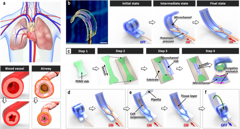
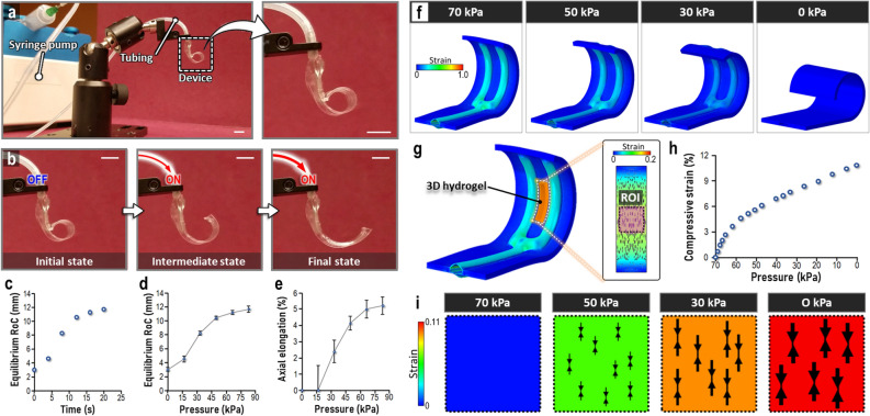
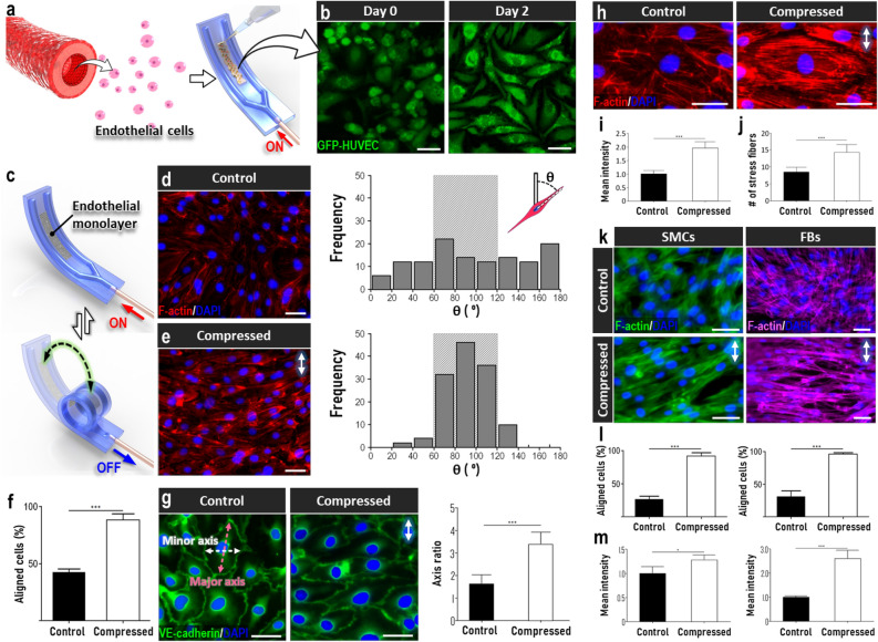
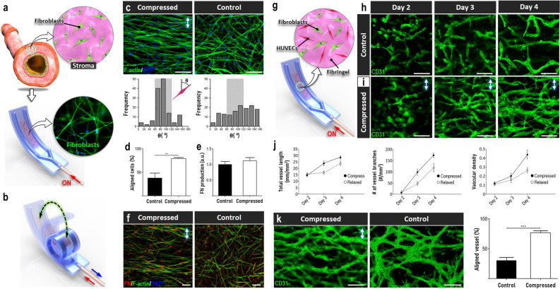
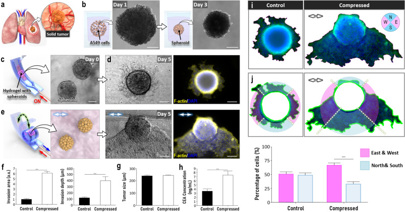
Here we present a microengineered soft-robotic in vitro platform developed by integrating a pneumatically regulated novel elastomeric actuator with primary culture of human cells. This system is capable of generating dynamic bending motion akin to the constriction of tubular organs that can exert controlled compressive forces on cultured living cells. Using this platform, we demonstrate cyclic compression of primary human endothelial cells, fibroblasts, and smooth muscle cells to show physiological changes in their morphology due to applied forces. Moreover, we present mechanically actuatable organotypic models to examine the effects of compressive forces on three-dimensional multicellular constructs designed to emulate complex tissues such as solid tumors and vascular networks. Our work provides a preliminary demonstration of how soft-robotics technology can be leveraged for in vitro modeling of complex physiological tissue microenvironment, and may enable the development of new research tools for mechanobiology and related areas.


Conflict of interest statement

D.H. is a co-founder of Vivodyne Inc. and holds equity in Vivodyne Inc. and Emulate Inc. J.P., J.S., E.B., Y.M., C.O., and V.S. declare no potential conflict of interest.

Figures

Figure 1
Figure 1
A soft-robotic constrictor for dynamic compressive mechanical loading of living tissue. (a) Circumferential tissue compression occurs during the constriction of tubular organs such as blood vessels and airways. (b) Normally coiled soft robotic cell culture platform and its pneumatically driven unrolling motion. Scale bar, 5 mm. (c) Fabrication of the soft robotic device through irreversible bonding of a thin stretched PDMS strip to a microchannel-containing PDMS slab (Steps 1, 2, and 3) and subsequent release of the bonded assembly (Step 4). (d,e) Sequential steps for establishing cell culture on the microengineered device. Red arrows labeled “ON” at the bottom of schematics represent application of pneumatic pressure to the microchannels. (f) Compression of cultured tissue layer through removal of air pressure from the microchannels (shown with a blue arrow labeled “OFF”) and the resultant bending of the culture substrate.
Figure 2
Figure 2
Pneumatic actuation of soft robotic constrictor. (a) Experimental setup for controlled pneumatical actuation of the soft robotic device. Scale bars, 5 mm. (b) Photographs of the uncoiling device due to applied air pressure. Scale bars, 5 mm. (c) Quantification of equilibrium RoC during the application of pneumatic pressure. Plots of equilibrium RoC (d) and axial elongation (e) at different levels of applied pneumatic pressure. (f) Finite element analysis-based computation prediction of substrate geometry and strain during pneumatic actuation of the soft robot. The heatmap shows maximum nominal principal strain. (g) Inclusion of 3D hydrogel for finite element modeling of 3D tissue. (h,i) In silico prediction of changes in mean compressive strain in the ROI with decreasing pneumatic pressure. The size of arrow pairs in (i is proportional to the level of compressive strain. The heatmap shows maximum nominal principal compressive strain. Data show mean ± SD with n = 3.
Figure 3
Figure 3
Responses of mechanosensitive cells to dynamic compression. (a,b) Monolayer culture of green fluorescence protein (GFP)-expressing HUVECs in the uncoiled device. Scale bar, 50 µm. (c) Cyclic pneumatic actuation of the endothelium-containing device. (d–f) Alignment of compressed endothelial cells due to applied compressive force as quantitatively assessed by the distribution of orientation angle (d,e) and the percentage of aligned cells (f). Scale bars, 50 µm. Any given cell was evaluated to be aligned when its orientation angle defined by the angle between its major axis and the vertical axis was between 60° and 120° (represented by the shaded boxes on the plots). (g) Compression-induced cell elongation quantified by the ratio of major to minor axes of cell body. Scale bars, 50 µm. (h) Fluorescence visualization of altered intracellular architecture of HUVECs due to compression. The architectural changes were quantified by the mean fluorescence intensity of F-actin staining (i) and the number of stress fibers (j). Scale bars, 50 µm. (k) Mechanosensitive responses of primary human smooth muscle cells (SMCs) and lung fibroblasts (FBs) as evidenced by their reorientation in the perpendicular direction to applied compressive force. Scale bar, 50 µm. (l,m) Quantification of cell alignment (l) and F-actin fluorescence intensity (m). Arrows in this figure show the direction of applied cyclic compression. ***P < 0.001, **P < 0.01, *P < 0.05. Data show mean ± SD with n = 3.
Figure 4
Figure 4
Dynamic soft robotic compression of cells and tissues in 3D culture. (a,b) In vitro modeling of stromal tissue in the lung using a fibroblast-laden collagen hydrogel construct formed in the soft robotic device. (c,d) Alignment of cultured fibroblasts due to cyclic compression. Any given cell was evaluated to be aligned when its orientation angle defined by the angle between its major axis and the vertical axis was between 60° and 120°, (represented by the shaded boxes on the plots). Scale bars, 100 µm. (e,f) The production of FN by fibroblasts was not influenced by compressive mechanical loading. Scale bars, 100 µm. (g) Co-culture of primary human lung fibroblasts and HUVECs for in vitro modeling of vasculogenesis. (h,i) Self-assembly of 3D vascular networks over a period of 6 days in the absence (h) or presence of compression (i). Green shows CD31 staining. Scale bars, 50 µm. (j) Quantification of total vessel length, the number of vascular junctions, and vascular density. (k) Formation of de novo vessels aligned perpendicularly to applied compression. Scale bars, 50 µm. Arrows in this figure indicate force direction. ***P < 0.001, **P < 0.01, *P < 0.05. Data show mean ± SD with n = 3.
Figure 5
Figure 5
In vitro modeling of solid tumor in the soft robotic constrictor. (a,b) A549 spheroids formed in low-attachment wells are used to model malignant tumors in the lung. Scale bars, 100 µm. (c) Micrograph of tumor spheroids embedded in type I collagen hydrogel in the device. Scale bar, 100 µm. (d) Phase contrast (left) and fluorescence (right) images of lung tumor after 5 days of static culture. Scale bars, 50 µm. (e) Micrographs of dynamically compressed lung tumor at Day 5. Scale bars, 100 µm. (f) Quantification of invasion area and depth. (g) Comparison of tumor size in the presence or absence of compression. (h) ELISA-based quantification of CEA secreted by lung tumors. (i) Delineation of the outline of tumor and invading cancer cells using fluorescence micrographs shown in (d,e) E, W, S, and N in the circular direction map represent east, west, south, and north, respectively. (j) Radial segmentation of tumor regions and quantification of directional cancer cell migration. Arrows in this figure show the direction of applied compressive force. ***P < 0.001, **P < 0.01, *P < 0.05. Data show mean ± SD with n = 3.

Similar articles

Cited by

References

    1. Wang JH, Thampatty BP. An introductory review of cell mechanobiology. Biomech. Model. Mechanobiol. 2006;5:1–16. doi: 10.1007/s10237-005-0012-z. - DOI - PubMed
    1. Bates JH. Systems physiology of the airways in health and obstructive pulmonary disease. Wiley Interdiscip. Rev. Syst. Biol. Med. 2016;8(5):423. doi: 10.1002/wsbm.1347. - DOI - PMC - PubMed
    1. Viola H, Chang J, Grunwell JR, Hecker L, Tirouvanziam R, Grotberg JB, Takayama S. Microphysiological systems modeling acute respiratory distress syndrome that capture mechanical force-induced injury-inflammation-repair. APL Bioeng. 2019;3:41503. doi: 10.1063/1.5111549. - DOI - PMC - PubMed
    1. Tschumperlin DJ, Dai G, Maly IV, Kikuchi T, Laiho LH, McVittie AK, Haley KJ, Lilly CM, So PT, Lauffenburger DA, Kamm RD, Drazen JM. Mechanotransduction through growth-factor shedding into the extracellular space. Nature. 2004;429:83. doi: 10.1038/nature02543. - DOI - PMC - PubMed
    1. Choe MM, Sporn PH, Swartz MA. Extracellular matrix remodeling by dynamic strain in a three-dimensional tissue-engineered human airway wall model. Am. J. Respir. Cell Mol. Biol. 2006;35:30. doi: 10.1165/rcmb.2005-0443OC. - DOI - PMC - PubMed

Publication types