. 2021 Nov;58(11):5703-5721.
doi: 10.1007/s12035-021-02508-5. Epub 2021 Aug 14.

Dolutegravir Inhibition of Matrix Metalloproteinases Affects Mouse Neurodevelopment

Affiliations

Dolutegravir Inhibition of Matrix Metalloproteinases Affects Mouse Neurodevelopment

Aditya N Bade et al. Mol Neurobiol. 2021 Nov.

Abstract

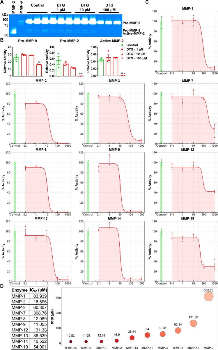
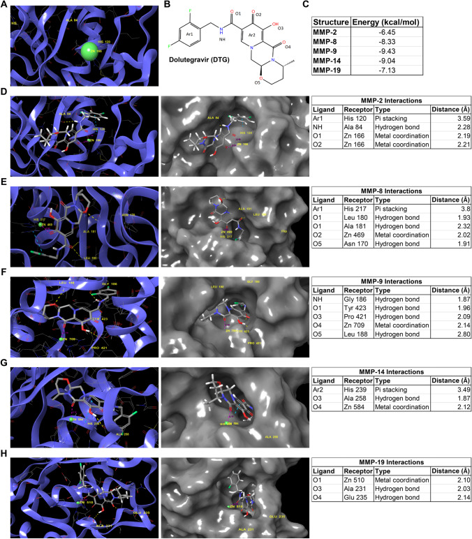
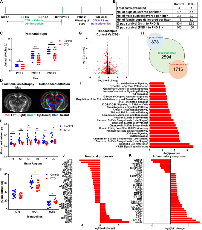
Dolutegravir (DTG) is a first-line antiretroviral drug (ARV) used in combination therapy for the treatment of human immunodeficiency virus type-1 (HIV-1) infection. The drug is effective, safe, and well tolerated. Nonetheless, concerns have recently emerged for its usage in pregnant women or those of child-bearing age. Notably, DTG-based ARV regimens have been linked to birth defects seen as a consequence of periconceptional usages. To this end, uncovering an underlying mechanism for DTG-associated adverse fetal development outcomes has gained clinical and basic research interest. We now report that DTG inhibits matrix metalloproteinases (MMPs) activities that could affect fetal neurodevelopment. DTG is a broad-spectrum MMPs inhibitor and binds to Zn++ at the enzyme's catalytic domain. Studies performed in pregnant mice show that DTG readily reaches the fetal central nervous system during gestation and inhibits MMP activity. Postnatal screenings of brain health in mice pups identified neuroinflammation and neuronal impairment. These abnormalities persist as a consequence of in utero DTG exposure. We conclude that DTG inhibition of MMPs activities during gestation has the potential to affect prenatal and postnatal neurodevelopment.

Keywords: Antiretroviral drug; Dolutegravir; HIV-1; Matrix metalloproteinases; Neurodevelopment; Pregnancy outcomes.


Conflict of interest statement

Drs. Benson Edagwa and Howard E. Gendelman are co-founders of Exavir Therapeutics, Inc., a biotechnology company focused on the development of long-acting antiretroviral medicines. The remaining authors declare that they have no competing interests.

Figures

Fig. 1
Fig. 1
DTG inhibition of MMPs. A Gelatin zymogram. Activity of MMP-2 and MMP-9 was evaluated in serum-free medium of THP-1 cells following treatment with DTG (1, 10, or 100 µM). Vehicle-treated cells were used as control. Human recombinant enzymes were used as standards. B Relative activity of MMP-9 or MMP-2 was measured and normalized with respective standard. A one-way ANOVA followed by Dunnett’s test was used to compare activity of individual MMP between each treatment concentration and untreated control (**p < 0.01, ***p < 0.001). C Fluorometric substrate assay. Inhibition profile of DTG (0.1, 1, 10, 100, and 1000 µM) was determined against ten human recombinant MMP enzymes. D IC50 values for DTG against each MMP were calculated from dose–response curve (fluorometric substrate assay) using four-parameter hill-slope model. For both assays, data are expressed as the mean ± SEM, N = 3 biological replicates. Experiments were repeated independently three times with equivalent results
Fig. 2
Fig. 2
Molecular interaction between DTG and MMP. A A 3D representative image of MMP-2 catalytic domain containing Zn++ (green ball) is shown. B Chemical structure of DTG with ligand labeling used for molecular docking. C Calculated binding energies for each MMP with DTG using Schrodinger’s software suite. DH 3D representations of molecular docking complex of DTG with D MMP-2, E MMP-8, F MMP-9, G MMP-14, and H MMP-19. 3D representations of molecular docking complexes are shown in ribbon (blue color; left side of the figure) and surface interaction (gray color; middle of the figure) formats. Interactions with Zn++ (green ball) and other amino acids (yellow color) are identified. Interactions with Zn++ are shown by pink dotted line. Hydrogen bond interaction with amino acids is shown by yellow dotted line. Pi staking is shown by blue dotted line. Interaction details for each complex are provided in the tabular format (right side of the figure)
Fig. 3
Fig. 3
DTG PK and BD during and after pregnancy. A Schematic presentation of the experimental timeline. B DTG concentrations in plasma of dams. DTG levels were measured during prenatal and postnatal period to evaluate DTG levels in the mother’s blood. C DTG concentrations in placental tissue at GD 16.5. D DTG concentrations in brain tissue of embryos or pups. Whole brains from embryos and pups were processed for DTG levels at GD 16.5 and after birth at PND 4. BD Each sample (plasma or tissue) represents distinct litter. For drug concentration quantitation in all samples, data are expressed as mean ± SEM, N = minimum 4 animals at each time point
Fig. 4
Fig. 4
In utero DTG exposure effects on MMP activity in the fetal CNS. A Schematic presentation of timeline of the experiment. B Weights of dams from GD 0.5 to GD 15.5. No differences between DTG and control groups were observed. C Embryo phenotype assessment. D Representative images of embryos after vehicle (control) or DTG treatment at GD 16.5. From DTG group, one embryo out of 103 viable embryos showed exencephaly. E Fluorometric substrate assay. Broad-spectrum MMP activity in a whole brain tissue homogenate of embryos at GD 16.5 was evaluated. Data are expressed as the mean ± SEM, N = 9 animals (control) and N = 17 animals (DTG). t test (two-tailed) with Welch’s correction was used to compare the broad-spectrum MMP activity between DTG and control groups (*p < 0.05). F Protein levels of MMP-2 in embryo brain tissue. G Protein levels of MMP-9 embryo brain tissue. H Protein levels of MMP-14 embryo brain tissue. F, G Each lane is representative of six animals from each group. I Quantitation of protein levels in DTG compared to control using ImageJ software. Data are expressed as the mean ± SEM, N = 6 animals/group. t test (two-tailed) with Welch’s correction was used to compare protein levels of individual MMP between DTG and control (#p < 0.1)
Fig. 5
Fig. 5
Postnatal neurodevelopment following in utero DTG exposure. A Schematic presentation of timeline. B Pregnancy and postnatal outcomes. C Postnatal animal (pups) weights. D Representative diffusion tensor imaging (DTI) image of PND 28 pup from DTG group. Red: left–right; green: up-down; blue: in–out. E DTI: fractional anisotropy (FA) measurements in six different brain regions: hippocampus (HI), cortex (CT), striatum (ST), thalamus (TH), hypothalamus (HY), and cerebellum (CE). F Magnetic resonance spectroscopy (1H MRS): metabolite concentration measurements in hippocampus. Total creatine (tCre), N-acetylaspartate (NAA), and total choline (tCho) were measured. C, E, F Data are expressed as mean ± SEM, N = minimum 8 animals/group for each parameter evaluation. Student’s t test (two-tailed) was used to compare animal weights at each time point, FA (DTI) in each brain region or each metabolite concentration (1H MRS), between DTG and control (#p < 0.1, *p < 0.05, **p < 0.01, ***p < 0.001). GK Comparative transcriptomic analysis of total RNA from hippocampus region between DTG and control groups. N = 4 animals/group. G Volcano plot; p ≤ 0.05; red color: log2FoldChange ≥ 0.5 or ≤  − 0.5. H Total number of upregulated and downregulated genes. I Major affected pathways were determined by using Ingenuity pathway analysis (IPA). J, K Differentially expressed genes associated with neuronal processes and neuroinflammation

References

    1. World Health Organization (2016) Consolidated guidelines on the use of antiretroviral drugs for treating and preventing HIV infection: recommendations for a public health approach—second edition. https://www.who.int/hiv/pub/arv/chapter4.pdf?ua=1. Accessed February 27 2021 - PubMed
    1. Crowell CS, Williams PL, Yildirim C, Van Dyke RB, Smith R, Chadwick EG, Seage GR, 3rd, Diperna A, Hazra R, Pediatric HIVACS. Safety of in-utero antiretroviral exposure: neurologic outcomes in children who are HIV-exposed but uninfected. AIDS. 2020;34(9):1377–1387. doi: 10.1097/QAD.0000000000002550. - DOI - PMC - PubMed
    1. Ramokolo V, Goga AE, Slogrove AL, Powis KM. Unmasking the vulnerabilities of uninfected children exposed to HIV. BMJ. 2019;366:l4479. doi: 10.1136/bmj.l4479. - DOI - PMC - PubMed
    1. Evans C, Jones CE, Prendergast AJ. HIV-exposed, uninfected infants: new global challenges in the era of paediatric HIV elimination. Lancet Infect Dis. 2016;16(6):e92–e107. doi: 10.1016/S1473-3099(16)00055-4. - DOI - PubMed
    1. Department of Health and Human Services (DHHS), Panel on Antiretroviral Guidelines for Adults and Adolescents. Guidelines for the use of antiretroviral agents in adults and adolescents living with HIV. https://clinicalinfo.hiv.gov/sites/default/files/guidelines/documents/Ad.... Accessed June 11 2021

MeSH terms