. 2021 Sep;297(3):101079.
doi: 10.1016/j.jbc.2021.101079. Epub 2021 Aug 13.

Phosphorylation of slit diaphragm proteins NEPHRIN and NEPH1 upon binding of HGF promotes podocyte repair

Affiliations

Phosphorylation of slit diaphragm proteins NEPHRIN and NEPH1 upon binding of HGF promotes podocyte repair

Ashish K Solanki et al. J Biol Chem. 2021 Sep.

Abstract

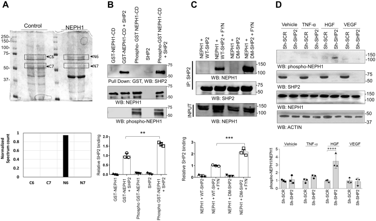
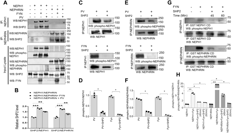
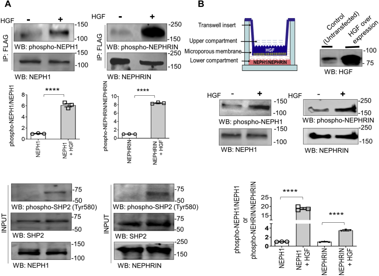
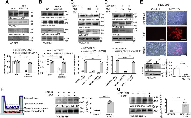
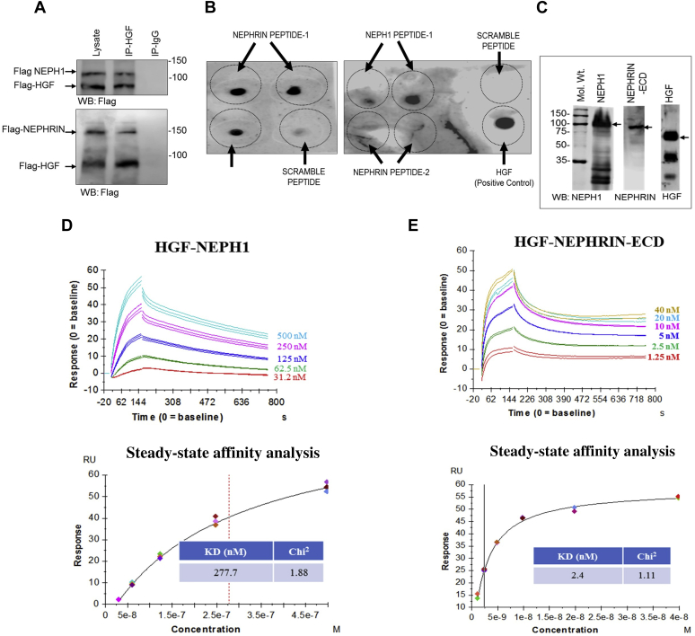
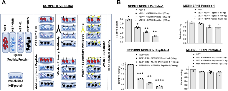
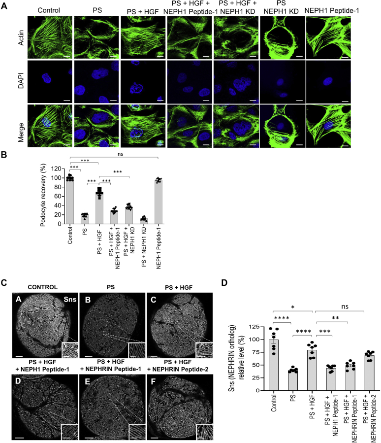
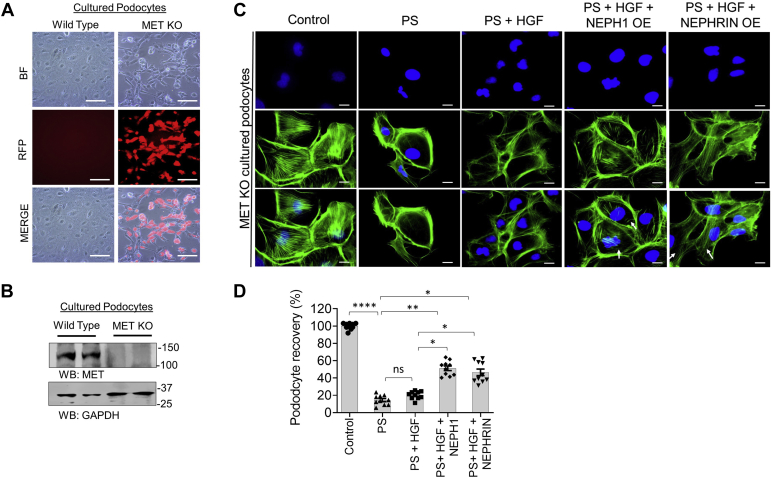
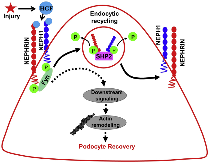
Phosphorylation (activation) and dephosphorylation (deactivation) of the slit diaphragm proteins NEPHRIN and NEPH1 are critical for maintaining the kidney epithelial podocyte actin cytoskeleton and, therefore, proper glomerular filtration. However, the mechanisms underlying these events remain largely unknown. Here we show that NEPHRIN and NEPH1 are novel receptor proteins for hepatocyte growth factor (HGF) and can be phosphorylated independently of the mesenchymal epithelial transition receptor in a ligand-dependent fashion through engagement of their extracellular domains by HGF. Furthermore, we demonstrate SH2 domain-containing protein tyrosine phosphatase-2-dependent dephosphorylation of these proteins. To establish HGF as a ligand, purified baculovirus-expressed NEPHRIN and NEPH1 recombinant proteins were used in surface plasma resonance binding experiments. We report high-affinity interactions of NEPHRIN and NEPH1 with HGF, although NEPHRIN binding was 20-fold higher than that of NEPH1. In addition, using molecular modeling we constructed peptides that were used to map specific HGF-binding regions in the extracellular domains of NEPHRIN and NEPH1. Finally, using an in vitro model of cultured podocytes and an ex vivo model of Drosophila nephrocytes, as well as chemically induced injury models, we demonstrated that HGF-induced phosphorylation of NEPHRIN and NEPH1 is centrally involved in podocyte repair. Taken together, this is the first study demonstrating a receptor-based function for NEPHRIN and NEPH1. This has important biological and clinical implications for the repair of injured podocytes and the maintenance of podocyte integrity.

Keywords: HGF; SHP-2; phosphorylation–dephosphorylation; podocytes.


Conflict of interest statement

Conflict of interest The authors declare that they have no conflicts of interest with the contents of this article.

Figures

Figure 1
Figure 1
The phosphatase SHP-2 binds NEPH1.A, immunoprecipitation of endogenous NEPH1 from podocytes followed by mass spectrometry analysis identified SHP-2 (N6) as a novel NEPH1 interacting protein. The plot shows the normalized spectrum values of four different bands excised between 75 and 50 kDa (highlighted as C6 and C7 from IgG controls and N6 and N7 from NEPH1 antibody) derived by using Scaffold software (Proteome Software, Inc). B, direct binding was evaluated by mixing purified phosphorylated GST-NEPH1-Cytoplasmic Domain (NEPH1-CD, produced in TKB1 cells) with purified recombinant SHP-2, which showed increased binding of SHP-2 with phosphorylated GST-NEPH1-CD. C, NEPH1 was coexpressed with SHP-2 or the substrate trapping SHP-2 mutant (DM SHP-2) with or without the phosphorylating agent FYN. To evaluate NEPH1 binding, SHP-2 was immunoprecipitated from the cell lysate and Western blotted (WB) with NEPH1 antibody. This demonstrated that NEPH1 bound SHP-2 in the presence of FYN. Significantly enhanced binding was observed with DM SHP-2 indicating that NEPH1 is a SHP-2 substrate. D, control podocytes and podocytes with stable SHP-2 knockdown were treated with growth factors (TNF-α, HGF, and VEGF), and the phosphorylation of endogenous NEPH1 was evaluated using phospho-NEPH1 antibody. NEPH1 phosphorylation was significantly increased only following HGF stimulation of SHP-2 KD podocytes. Experiments BD were performed in triplicate, repeated three times with similar results, and representative images of the results are presented in the figure. Data are presented as mean ± SEM, and p-values were calculated using the Sidak's multiple comparisons test (two-way ANOVA). ∗∗p ≤ 0.01, ∗∗∗p ≤ 0.001, ∗∗∗∗p ≤ 0.0001. SCR, scrambled.
Figure 2
Figure 2
SHP-2 binds NEPH1 and NEPHRIN and acts as a phosphatase.A and B, NEPH1 or NEPHRIN were phosphorylated by either coexpressing with FYN or treating with pervanadate and were immunoprecipitated with their respective antibodies. Western blotting of the immunoprecipitated complexes showed increased SHP-2 binding to phosphorylated NEPHRIN and NEPH1. Data are presented as mean ± SEM, and p-values were calculated using the Kruskal–Wallis one-way analysis of variance. CF, HEK293 cells expressing NEPHRIN or NEPH1 were treated with PV followed by immunoprecipitation of NEPH1 and NEPHRIN (set 1). Simultaneously, NEPH1 and NEPHRIN were overexpressed in HEK293 cells and the proteins were immunoprecipitated using their respective antibodies and incubated with purified FYN (500 ng) (set 2). Recombinant SHP-2 protein was then added to the immunoprecipitated complexes (both sets) and the extent of dephosphorylation was measured by their respective phosphoantibodies. Data are presented as mean ± SEM, and p-values were calculated using the Mann–Whitney (nonparametric) test, one-tailed. G and H, purified recombinant GST-NEPH1 and GST-NEPHRIN cytoplasmic domain (CD) proteins were phosphorylated by incubating with recombinant active FYN immobilized on nickel beads. Post phosphorylation, FYN beads were removed and the phosphorylated GST-NEPH1 cytoplasmic domain (GST-NEPH1 CD) and GST-NEPHRIN CD proteins were incubated with purified recombinant SHP-2 for the indicated times and Western blot was performed with the respective phosphoantibodies, which confirmed SHP-2-mediated dephosphorylation. Data are presented as mean ± SEM, and p-values were calculated using the Kruskal–Wallis one-way analysis of variance. ∗p ≤ 0.05, ∗∗p ≤ 0.005, ∗∗∗p ≤ 0.0005. All experiments were performed in triplicate and repeated three times with similar results, and representative images of the results are presented in the figure.
Figure 3
Figure 3
HGF is a novel inducer of NEPHRIN and NEPH1 phosphorylation.A, NEPHRIN-FLAG and NEPH1-FLAG were coexpressed with HGF in HEK293 cells, and phosphorylation was analyzed using their respective phosphoantibodies. SHP2 was also phosphorylated in the presence of HGF. Data are presented as mean ± SEM, and p-values were calculated using a two-tailed Student’s t test. B, HGF-expressing HEK293 cells were grown on the Transwell filter and stable NEPH1- and NEPHRIN-expressing HEK293 cells were cultured on the plastic at the bottom of the well. NEPHRIN and NEPH1 phosphorylation was measured by lysing the cells in the well, and Western blot analysis was performed with their respective phosphoantibodies. Data are presented as mean ± SEM, and p-values were calculated using a two-tailed Student’s t test. ∗∗∗∗p ≤ 0.001. All experiments were performed in triplicate and repeated three times with similar results, and representative images of the results are presented in the figure.
Figure 4
Figure 4
The MET receptor is not required for HGF-induced NEPHRIN and NEPH1 phosphorylation. Recombinant HGF (20 ng/ml) was added to HEK293 cells overexpressing (A) NEPH1-FLAG or (B) NEPHRIN-FLAG in the presence or absence of 100 nM Crizotinib, a MET inhibitor. Phosphorylation of NEPH1 and NEPHRIN was unchanged by the presence of Crizotinib. Data are presented as mean ± SEM, and p-values were calculated using the Kruskal–Wallis one-way analysis of variance. C, D, NEPH1-FLAG or NEPHRIN-FLAG were overexpressed in HEK293 cells with stable shRNA-mediated knockdown (KD) of the MET receptor. Recombinant HGF (20 ng/ml) was added to these cells, and NEPHRIN and NEPH1 phosphorylation in the cell lysate showed no change in phosphorylation following MET knockdown (KD). Data are presented as mean ± SEM, and p-values were calculated using the Kruskal–Wallis one-way analysis of variance. E, using CRISPR-Cas9, stable MET knockout HEK293 cells were generated and transfected with NEPHRIN- and NEPH1-expressing plasmids. Red fluorescent protein (RFP) is a marker for transfection and confirms the stable knockout of MET post puromycin selection. Data are presented as mean ± SEM, and p-values were calculated using a two-tailed Student’s t test. The scale bar represents 25 μm. F and G, using HGF-overexpressing cells grown on the Transwell filter and HEK293 MET knockout cells expressing NEPH1 or NEPHRIN growing on the plastic at the bottom of well (first panel, schematic), we demonstrate that NEPH1 and NEPHRIN phosphorylation occurs following complete loss of the MET receptor. Data are presented as mean ± SEM, and p-values were calculated using a two-tailed Student’s t test. ns, nonsignificant, ∗p ≤ 0.05, ∗∗p ≤ 0.01, ∗∗∗∗p ≤ 0.0001. All experiments were performed in triplicate and repeated three times with similar results, and representative images of the results are presented in the figure.
Figure 5
Figure 5
HGF interacts with NEPHRIN and NEPH1.A, NEPHRIN-FLAG and NEPH1-FLAG were coexpressed with HGF-FLAG in HEK293 cells, respectively. HGF was immunoprecipitated using the HGF antibody, and the immunoprecipitated complexes were analyzed for NEPHRIN-FLAG and NEPH1-FLAG binding by Western blot using the FLAG antibody. B, dot blot assay: Synthetic peptides corresponding to the HGF-binding regions of NEPHRIN and NEPH1 were spotted onto nitrocellulose membranes and probed with recombinant HGF. HGF binding was seen with the peptides NEPH1 Peptide-1 and NEPHRIN Peptide-1 but not with NEPHRIN Peptide-2 or control (scrambled) peptides. CE, baculovirus-expressed NEPHRIN- extracellular domain (ECD) and NEPH1 (C) were mixed with HGF in the indicated amounts and subjected to surface plasmon resonance. Surface plasmon resonance analysis showed the concentration-dependent binding of NEPH1 (D) and NEPHRIN (E) with HGF. The steady-state affinity analysis and the calculated respective KD values have been shown for NEPH1 (D, lower panel) and NEPHRIN (E, lower panel).
Figure 6
Figure 6
The HGF-binding site for MET does not overlap with the HGF-binding sites for NEPH1 and NEPHRIN.A, schematic for the competitive ELISA. HGF was immobilized on the wells of an ELISA plate, and its binding to NEPH1, NEPHRIN, and MET alone and in combination with NEPH1 peptide-1 and NEPHRIN peptide-1 was analyzed. B, quantification of the data. All comparisons are with NEPH1 or NEPHRIN alone. Data are presented as mean ± SEM, and p-values were calculated using a two-tailed Student’s t test. ∗p ≤ 0.05, ∗∗p ≤ 0.01, ∗∗∗p ≤ 0.001, ∗∗∗∗p < 0.0001.
Figure 7
Figure 7
HGF treatment repairs podocytes/nephrocytes in a NEPH1- and NEPHRIN-dependent fashion.A and B, cultured human podocytes were treated with protamine sulfate (PS), and actin cytoskeleton (green) disorganization was visualized by phalloidin staining. To induce recovery, HGF (50 ng/ml) was added to the PS-treated podocytes. The addition of NEPH1 inhibitory peptide blocked HGF-induced recovery. KD of NEPH1 also prevented HGF-induced recovery. Ten cells per experimental condition were evaluated from three experimental replicates. The scale bar represents 25 μm. Data are presented as mean ± SEM, and p-values were calculated using Tukey's multiple comparisons test (one-way ANOVA). C and D, Sns (Drosophila ortholog of NEPHRIN) staining of Drosophila nephrocytes treated with PS in the presence or absence of HGF and NEPHRIN or NEPH1 peptides. Decreased Sns staining was noted in PS-treated nephrocytes, which was rescued by treatment with HGF. Addition of HGF-interacting NEPH1 Peptide-1 and NEPHRIN Peptide-1, but not NEPHRIN Peptide-2, blocked the rescue by HGF. The scale bar represents 5 μm; scale bar for insets represents 1 μm. For quantification, seven nephrocytes from three flies for each condition were analyzed. Data are presented as mean ± SEM, and p-values were calculated using the Tukey's multiple comparisons test (one-way ANOVA). ns, nonsignificant, ∗p ≤ 0.05, ∗∗p ≤ 0.01, ∗∗∗p ≤ 0.001, ∗∗∗∗p ≤ 0.0001.
Figure 8
Figure 8
NEPH1/NEPHRIN augments HGF-mediated recovery in protamine sulfate (PS)-induced injury in MET knockout podocytes.A and B, using CRIPR-Cas9 MET knockout podocytes were generated. Red fluorescent protein (RFP) is a marker for transfection and confirms the stable knock out of MET post puromycin selection. Western blot for the MET knockout and control podocyte cell lysates. C and D, MET knockout podocytes were transfected with NEPHRIN- and NEPH1-expressing plasmids and then treated with PS and the actin cytoskeletal (green) disorganization was visualized by phalloidin staining. To induce recovery, HGF (50 ng/ml) was added to the PS-treated podocytes. Podocytes transfected with NEPHRIN and NEPH1 showed significantly improved recovery with HGF. Ten cells per experimental condition were evaluated from three experimental trials. The scale bar represents 25 μm. Data are presented as mean ± SEM, and p-values were calculated using the Kruskal–Wallis test one-way analysis of variance. ns, nonsignificant, ∗p ≤ 0.05, ∗∗ p ≤ 0.01, ∗∗∗p ≤ 0.001.
Figure 9
Figure 9
Schematic of the phosphorylation (activation) and dephosphorylation (deactivation) mechanisms for NEPHRIN and NEPH1 in podocytes. In response to injury, HGF initiates the recovery process by phosphorylating NEPHRIN and NEPH1, which induces actin cytoskeleton reorganization leading to podocyte repair. To maintain homeostasis, subsequent dephosphorylation is mediated in a SHP2-dependent manner.

References

    1. Benzing T. Signaling at the slit diaphragm. J. Am. Soc. Nephrol. 2004;15:1382–1391. - PubMed
    1. Nihalani D., Solanki A.K., Arif E., Srivastava P., Rahman B., Zuo X., Dang Y., Fogelgren B., Fermin D., Gillies C.E., Sampson M.G., Lipschutz J.H. Disruption of the exocyst induces podocyte loss and dysfunction. J. Biol. Chem. 2019;294:10104–10119. - PMC - PubMed
    1. Patrakka J., Tryggvason K. Nephrin--a unique structural and signaling protein of the kidney filter. Trends Mol. Med. 2007;13:396–403. - PubMed
    1. Donoviel D.B., Freed D.D., Vogel H., Potter D.G., Hawkins E., Barrish J.P., Mathur B.N., Turner C.A., Geske R., Montgomery C.A., Starbuck M., Brandt M., Gupta A., Ramirez-Solis R., Zambrowicz B.P. Proteinuria and perinatal lethality in mice lacking NEPH1, a novel protein with homology to NEPHRIN. Mol. Cell. Biol. 2001;21:4829–4836. - PMC - PubMed
    1. Rantanen M., Palmén T., Pätäri A., Ahola H., Lehtonen S., Aström E., Floss T., Vauti F., Wurst W., Ruiz P., Kerjaschki D., Holthöfer H. Nephrin TRAP mice lack slit diaphragms and show fibrotic glomeruli and cystic tubular lesions. J. Am. Soc. Nephrol. 2002;13:1586–1594. - PubMed

Publication types