. 2021:20:100133.
doi: 10.1016/j.mcpro.2021.100133. Epub 2021 Aug 12.

Optimized Liquid and Gas Phase Fractionation Increases HLA-Peptidome Coverage for Primary Cell and Tissue Samples

Affiliations

Optimized Liquid and Gas Phase Fractionation Increases HLA-Peptidome Coverage for Primary Cell and Tissue Samples

Susan Klaeger et al. Mol Cell Proteomics. 2021.

Abstract

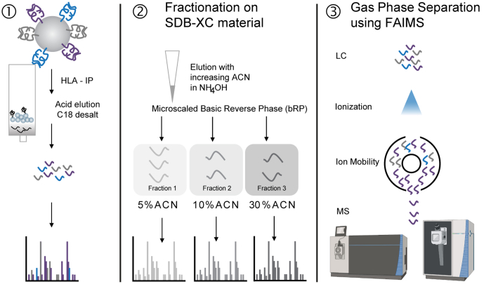
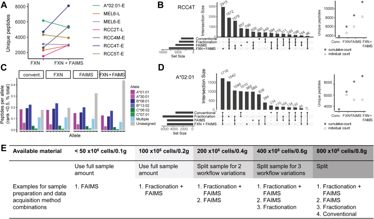
MS is the most effective method to directly identify peptides presented on human leukocyte antigen (HLA) molecules. However, current standard approaches often use 500 million or more cells as input to achieve high coverage of the immunopeptidome, and therefore, these methods are not compatible with the often limited amounts of tissue available from clinical tumor samples. Here, we evaluated microscaled basic reversed-phase fractionation to separate HLA peptide samples offline followed by ion mobility coupled to LC-MS/MS for analysis. The combination of these two separation methods enabled identification of 20% to 50% more peptides compared with samples analyzed without either prior fractionation or use of ion mobility alone. We demonstrate coverage of HLA immunopeptidomes with up to 8107 distinct peptides starting with as few as 100 million cells. The increased sensitivity obtained using our methods can provide data useful to improve HLA-binding prediction algorithms as well as to enable detection of clinically relevant epitopes such as neoantigens.

Keywords: FAIMS; HLA; basic reversed-phase fractionation; immunopeptidomics; ion mobility.


Conflict of interest statement

Conflict of interest D. A. B. reported nonfinancial support from Bristol-Myers Squibb, honoraria from LM Education/Exchange Services, and personal fees from Octane Global, Defined Health, Dedham Group, Adept Field Solutions, Slingshot Insights, Blueprint Partnerships, Charles River Associates, Trinity Group, and Insight Strategy, outside the submitted work. P. A. O. has received research funding from and has advised Neon Therapeutics, Bristol-Meyers Squibb, Merck, CytomX, Pfizer, Novartis, Celldex, Amgen, Array, AstraZeneca/MedImmune, Armo BioSciences, and Roche/Genentech, outside the submitted work. D. B. K. has previously advised Neon Therapeutics and has received consulting fees from Neon Therapeutics. D. B. K. owns equity in Agenus, Armata Pharmaceuticals, Breakbio, BioMarin Pharmaceutical, Bristol Myers Squibb, Celldex Therapeutics, Chinook Therapeutics, Editas Medicine, Exelixis, Gilead Sciences, IMV, Lexicon Pharmaceuticals, Moderna, and Regeneron Pharmaceuticals. BeiGene, a Chinese biotech company, supports unrelated research at TIGL. C. J. W. holds equity in BioNTech, Inc and receives research funding from Pharmacyclics, Inc. S. A. C. is a member of the scientific advisory boards of Kymera, PTM BioLabs, and Seer and an ad hoc scientific advisor to Pfizer and Biogen. All the other authors declare no competing interests.

Figures

None
Graphical abstract
Fig. 1
Fig. 1
Schematic overview of HLA sample preparation approaches. (1) Prior standard workflow in which HLA-I peptides are acid eluted and desalted before LC–MS/MS analysis with repeat injection. (2) Offline basic reversed-phase separation of eluted peptides, further separated into three fractions using SDB-XC material using increasing ACN concentrations in ammonium hydroxide (5%, 10%, and 30% ACN in 0.1% NH4OH, pH 10). (3) Separation of ions in the gas phase using FAIMS coupled to the mass spectrometer. ACN, acetonitrile; FAIMS, high field asymmetric waveform ion mobility spectrometry; HLA, human leukocyte antigen.
Fig. 2
Fig. 2
Basic reversed-phase separation increases peptide yield.A, the number of unique peptides identified by LC–MS/MS obtained without off-line fractionation (empty squares, “conventional” on x-axis) and using fractionation (filled black squares) across monoallelic and multiallelic samples. Unfilled bars correspond to peptides identified in both approaches; blue-filled bars are peptides unique to unfractionated analyses, whereas purple-filled bars are peptides unique to fractionated samples. Numbers in bars indicate the percent of total peptides per approach. B, motif of 9-mer peptides identified in the unfractionated (top) and fractionated (bottom) acquisition of the monoallelic HLA-A∗02:01 sample. C, nonmetric multidimensional scaling (NMDS) plot (see Experimental procedures section) showing clusters of peptides identified in both (gray), unfractionated (blue), and fractionated (purple) samples of A∗02:01 cells. D, UpSet plot showing peptide spectrum matches (PSMs) identified in the three fractions of the A∗02:01 peptidome. Horizontal bars at bottom left indicate the number of total PSMs per fraction; dots and lines under the vertical bars identify peptides identified in only one (single dot) or multiple fractions (dots with lines). E, Same as D but for RCC1T. F, allele assignment of peptides identified in the individual fractions of RCC1T (top left, fraction of assigned peptides at HLAthena rank <0.5) as well as in conventional and fractionated RCC1T (bottom left, fraction of peptides at HLAthena rank <0.5). Top right box plot shows peptide pI of peptides per fraction, allele motifs, and median pI of all peptides assigned to each allele are depicted at the bottom. HLA, human leukocyte antigen.
Fig. 3
Fig. 3
Ion mobility separation increases peptide yield.A, the number of HLA-I eluted peptide identifications obtained using FAIMS (filled black squares) or without FAIMS (empty squares) across monoallelic and multiallelic samples. Unfilled bars correspond to peptides identified in both approaches; blue filled bars are peptides unique to no-FAIMS (conventional), whereas red-filled bars are peptides unique to samples analyzed with FAIMS. Numbers in bars indicate the percent of peptides per approach. B, motif of 9-mer peptides identified in without FAIMS (top) and with FAIMS (bottom) acquisition of the monoallelic HLA-B∗53:01 sample on the Orbitrap Exploris. C, NMDS plot of peptides identified in both (gray), conventional (blue), and with FAIMS (red) samples of B∗53:01. D, precursor isolation purity of peptides in FAIMS (red) and conventional (blue) acquisition. E, number of identifications in all samples across the chromatographic gradient colored by detection in normal acquisition (no FAIMS, blue), + FAIMS acquisition (red), and both acquisition modes (gray, bin size = 30). FAIMS, high field asymmetric waveform ion mobility spectrometry; HLA, human leukocyte antigen.
Fig. 4
Fig. 4
Combination of fractionation and FAIMS for greater coverage of the immunopeptidome.A, number of unique 8–11-mer identifications in fractionated samples acquired with and without FAIMS. “L” and “E” indicate the instrument, Fusion Lumos or Orbitrap Exploris, used for acquisition. B, peptides identified across all modes of acquisition of RCC4T. Horizontal bars at bottom left indicate the number of total unique peptides per acquisition; dots and lines under the vertical bars identify peptides identified in only one (single dot) or multiple acquisitions (dots with lines). Each acquisition (empty circle) adds unique peptide identifications to the total cumulative peptidome (filled circle). C, allele assignment of peptides identified in RCC4T (HLAthena, rank <0.5) across four different acquisitions. D, same as B but for HLA-A∗02:01 sample. E, the table with recommended sample preparation and data acquisition strategies based on input available. FAIMS, high field asymmetric waveform ion mobility spectrometry.
Fig. 5
Fig. 5
Optimized data acquisition strategies facilitate neoantigen discovery.A, neoantigens identified in tumor-derived cell lines MEL 1 and MEL 6 acquired with fractionation ± FAIMS, their protein of origin, detection method, transcript abundance level (TPM), and the most likely allele the peptide would bind to as predicted by HLAthena using MSi (peptide sequence–only model) and MSiCE (cleavability and expression integrative model). B, mirror plots for the MS spectra of two neoantigen sequences (LLHTELERFL and NSKKKWFLF) in the discovery experiment (top) and the corresponding synthetic peptide spectrum (bottom, red: y-ion, blue: b-ion, and green: internal ion). C, targeted MS experiment for mutated peptide YIHGRGWAL using a heavy peptide. After optimization, transitions of y6- and y7-ion were acquired of the heavy peptide alone (left, blank) and of the heavy peptide spiked into a peptidome sample of MEL6. Endogenous peptide detection is shown on the top, and detection of the heavy peptide is shown at the bottom. D, CD137 signal of neoantigen-specific TCRs in different target backgrounds (PMA/ionomycin = positive control; MEL= tumor cell line, PBMCs from the corresponding patient, APCs pulsed with DMSO, mutant, or WT peptide). Percent upregulation is shown for each panel. APC, antigen-presenting cell; DMSO, dimethyl sulfoxide; FAIMS, high field asymmetric waveform ion mobility spectrometry; MEL, melanoma; PBMC, peripheral blood mononuclear cell; PMA, phorbol myristate acetate; TCR, T-cell receptor.

References

    1. Bilusic M., Madan R.A. Therapeutic cancer vaccines: the latest advancement in targeted therapy. Am. J. Ther. 2012;19:e172–e181. - PMC - PubMed
    1. Keskin D.B., Anandappa A.J., Sun J., Tirosh I., Mathewson N.D., Li S., Oliveira G., Giobbie-Hurder A., Felt K., Gjini E., Shukla S.A., Hu Z., Li L., Le P.M., Allesøe R.L., et al. Neoantigen vaccine generates intratumoral T cell responses in phase Ib glioblastoma trial. Nature. 2019;565:234–239. - PMC - PubMed
    1. Ott P.A., Hu Z., Keskin D.B., Shukla S.A., Sun J., Bozym D.J., Zhang W., Luoma A., Giobbie-Hurder A., Peter L., Chen C., Olive O., Carter T.A., Li S., Lieb D.J., et al. An immunogenic personal neoantigen vaccine for patients with melanoma. Nature. 2017;547:217–221. - PMC - PubMed
    1. Sahin U., Derhovanessian E., Miller M., Kloke B.P., Simon P., Löwer M., Bukur V., Tadmor A.D., Luxemburger U., Schrörs B., Omokoko T., Vormehr M., Albrecht C., Paruzynski A., Kuhn A.N., et al. Personalized RNA mutanome vaccines mobilize poly-specific therapeutic immunity against cancer. Nature. 2017;547:222–226. - PubMed
    1. Abelin J.G., Keskin D.B., Sarkizova S., Hartigan C.R., Zhang W., Sidney J., Stevens J., Lane W., Zhang G.L., Eisenhaure T.M., Clauser K.R., Hacohen N., Rooney M.S., Carr S.A., Wu C.J. Mass spectrometry profiling of HLA-associated peptidomes in mono-allelic cells enables more accurate epitope prediction. Immunity. 2017;46:315–326. - PMC - PubMed

Publication types