Review
. 2021 Oct 7;42(38):3904-3916.
doi: 10.1093/eurheartj/ehab544.

Artificial intelligence in the diagnosis and management of arrhythmias

Affiliations
Review

Artificial intelligence in the diagnosis and management of arrhythmias

Venkat D Nagarajan et al. Eur Heart J. .

Abstract

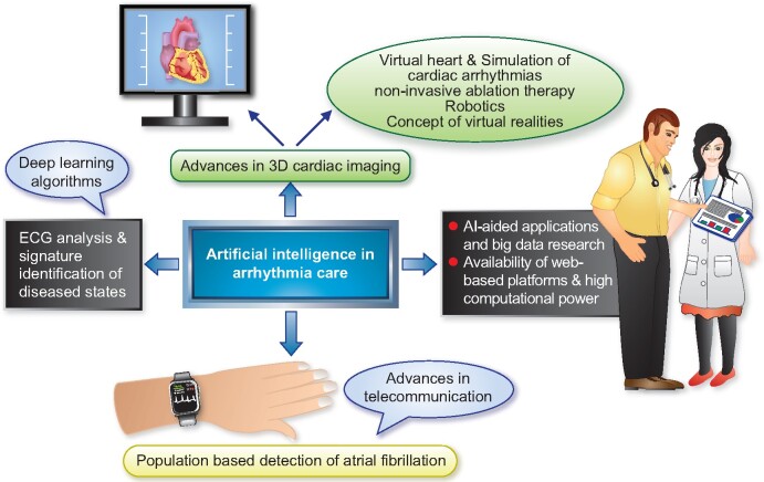
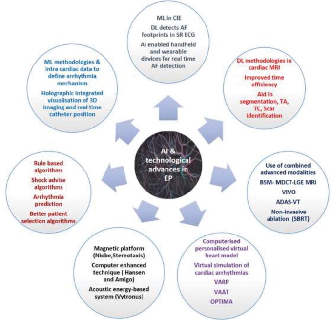
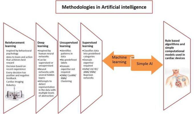
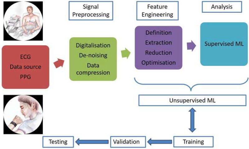
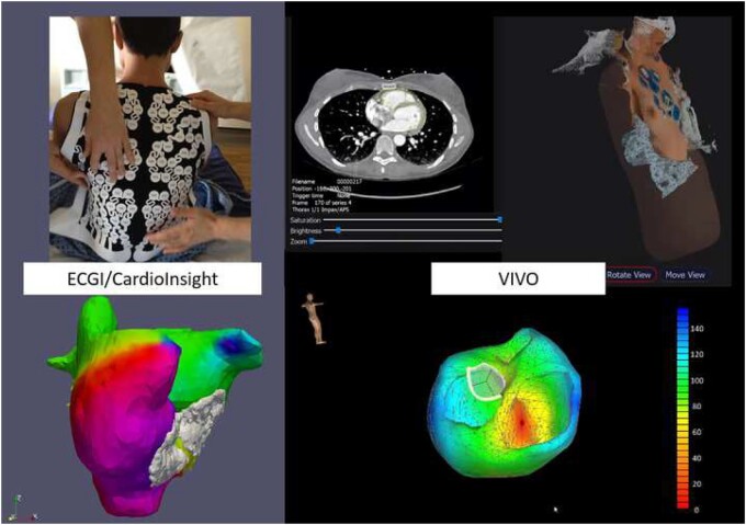
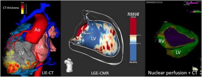
The field of cardiac electrophysiology (EP) had adopted simple artificial intelligence (AI) methodologies for decades. Recent renewed interest in deep learning techniques has opened new frontiers in electrocardiography analysis including signature identification of diseased states. Artificial intelligence advances coupled with simultaneous rapid growth in computational power, sensor technology, and availability of web-based platforms have seen the rapid growth of AI-aided applications and big data research. Changing lifestyles with an expansion of the concept of internet of things and advancements in telecommunication technology have opened doors to population-based detection of atrial fibrillation in ways, which were previously unimaginable. Artificial intelligence-aided advances in 3D cardiac imaging heralded the concept of virtual hearts and the simulation of cardiac arrhythmias. Robotics, completely non-invasive ablation therapy, and the concept of extended realities show promise to revolutionize the future of EP. In this review, we discuss the impact of AI and recent technological advances in all aspects of arrhythmia care.

Keywords: Ablation; Atrial fibrillation; Electrophysiology; Machine learning; Artificial intelligence.


Figures

None
Artificial intelligence-enhanced arrhythmia care.
Figure 1
Figure 1
An illustration highlighting the impact of artificial intelligence and recent technological advancements on all aspects of patient care in the field of cardiac electrophysiology. ADAS, Automatic Detection of Arrhythmic Substrate; AF, atrial fibrillation; BSM, body surface mapping; CIE, computerized interpretation of electrocardiography; DL, deep learning; EAM, electro anatomical mapping; EP, electrophysiology; LGE, late gadolinium enhancement; MDCT, multidetector computed tomography; ML, machine learning; MRI, magnetic resonance imaging; OPTIMA, optimal target identification via modelling of arrhythmogenesis; SBRT, stereotactic body radiotherapy; SR, sinus rhythm; TA, texture analysis; TC, tissue characterization; VAAT, virtual heart arrhythmia ablation targeting; VARP, virtual heart arrhythmia risk predictor approach; VIVO, view into ventricular onset; VT, ventricular tachycardia.
Figure 2
Figure 2
Artificial intelligence methodologies with their individual characteristics. AI, artificial intelligence; ANN, artificial neural network; CovNN, convolutional neural network; DNN, deep neural network; KNN, K-nearest neighbours; LR, logistic regression; RF, random forest; RNN, recurrent neural network; SVM, support vector machine.
Figure 3
Figure 3
Schematic representation of steps involved in cardiac impulse analysis from the data acquisition to analysis by machine learning algorithm. ECG, electrocardiogram; ML, machine learning; PPG, photo plethysmography.
Figure 4
Figure 4
Left panel: Example of non-invasive simultaneous mapping of atrial fibrillation of both the right and left atrium using the electrocardiogram imaging technology. Several mechanisms occur in various areas of the atria simultaneously and thereby maintain atrial fibrillation. Right panel: Example of non-invasive simultaneous mapping of ventricular ectopy using the view into ventricular onset technology.
Figure 5
Figure 5
Left panel: 3D image information from computed tomography and myocardial thickness in a patient with coronary artery disease and apical scar after myocardial infarction. Middle panel: Image information of late gadolinium enhancement from cardiac magnetic resonance imaging of the left ventricle with identification of the potentially arrhythmogenic channels within the scar responsible for ventricular re-entrant tachycardia. Right panel: Example of perfusion information from functional nuclear imaging superimposed on a contrast computed tomography scan in a patient with arrhythmogenic right ventricular disease. Ao, aorta; CMR, cardiac magnetic resonance; LA, left atrium; LGE, late gadolinium enhancement; LV, left ventricle; RV, right ventricle.

References

    1. Turing A. Computing machinery and intelligence. Mind 1950;LIX:433–460.
    1. Moore J. The Dartmouth College Artificial Intelligence Conference: the next fifty years. AI Mag 2006;27:87–91.
    1. Marques E, Filho DS, Fernandes FDA, Lacerda C, Soares DA, Seixas L, Augusto A, Sarmet MD, Gismondi RA, Mesquita ET, Mesquita CT. Artificial intelligence in cardiology: concepts, tools and challenges—“the horse is the one who runs, you must be the jockey”. Arq Bras Cardiol 2020;114:718–725. - PMC - PubMed
    1. Martin-Isla C, Campello VM, Izquierdo C, Raisi-Estabragh Z, Baeßler B, Petersen SE, Lekadir K. Image-based cardiac diagnosis with machine learning: a review. Front Cardiovasc Med 2020;7:1. - PMC - PubMed
    1. Schläpfer J, Wellens HJ. Computer-interpreted electrocardiograms benefits and limitations. J Am Coll Cardiol 2017;70:1183–1192. - PubMed