. 2021 Dec;45(12):3592-3602.
doi: 10.1007/s00268-021-06288-w. Epub 2021 Aug 15.

Immunohistochemical Inflammation in Histologically Normal Appendices in Patients with Right Iliac Fossa Pain

Affiliations

Immunohistochemical Inflammation in Histologically Normal Appendices in Patients with Right Iliac Fossa Pain

Emmanouil Psaltis et al. World J Surg. 2021 Dec.

Abstract

Background: Histologically normal appendices resected for right iliac fossa pain in children demonstrate immunohistochemical markers of inflammation. We aimed to establish if subclinical inflammation was present in histologically normal appendices resected from adults with right iliac fossa pain.

Methods: Immunohistochemistry was performed on formalin-fixed paraffin-embedded appendices for tumour necrosis factor (TNF)-α, interleukin (IL)-6, IL-2R and serotonin in four groups: Group I (n = 120): uncomplicated appendicitis, Group II (n = 118): complicated appendicitis (perforation or gangrene), Group III (n = 104): histologically normal appendices resected for right iliac fossa pain and Group IV (n = 106) appendices resected at elective colectomy. Expression was quantified using the H-scoring system.

Results: Median, interquartile range expression of TNF-α was increased in Groups I (5.9, 3.1-9.8), II (6.8, 3.6-12.1) and III (9.8, 6.2-15.2) when compared with Group IV (3.0, 1.4-4.7, p < 0.01). Epithelial expression of IL-6 in Groups II (44.0, 8.0-97.0) and III (71.0, 18.5-130.0) was increased when compared with Group IV (9.5, 1.0-60.2, p < 0.01). Expression of mucosal IL-2R in Groups I (47.4, 34.8-69.0), II (37.8, 25.4-60.4) and III (18.4, 10.1-34.7) was increased when compared with Group IV (2.8, 1.2-5.7, p < 0.01). Serotonin content in Groups I (3.0, 0-30.0) and II (0, 0-8.5) was decreased when compared with Groups III (49.7, 16.7-107.5) and IV (43.5, 9.5-115.8, p < 0.01).

Conclusion: Histologically normal appendices resected from symptomatic patients exhibited increased proinflammatory cytokine expression on immunohistochemistry suggesting the presence of an inflammatory process not detected on conventional microscopy.


Conflict of interest statement

None of the authors has a conflict of interest to report.

Figures

Fig. 1
Fig. 1
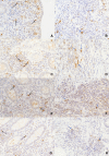
Expression of TNF-α (brown cytoplasmic staining) in monocytes (black arrows) using immunohistochemical staining at × 400 magnification. Expression of TNF-α in monocytes of patients with uncomplicated acute appendicitis (a); complicated acute appendicitis (b); histologically normal appendices (c); and patients from the control group (d). The vast majority of monocytes that expressed TNF-α were macrophages
Fig. 2
Fig. 2
Expression of IL-6 (brown cytoplasmic staining) in epithelial (black arrows) and inflammatory (blue arrows) cells using immunohistochemical staining at × 400 magnification. Expression of IL-6 in epithelial (a) and inflammatory (b) cells in patients with uncomplicated acute appendicitis; epithelial (c) and inflammatory (d) cells in patients with complicated acute appendicitis; epithelial (e) and inflammatory (f) cells in patients with histologically normal appendices; and epithelial (g) and inflammatory (h) cells in patients from the control group. IL-6 expressed by both mononuclear and polymorphonuclear cells
Fig. 3
Fig. 3
Expression of IL-2R (brown membrane staining) in the mucosa (black arrows) and submucosa (blue arrows) using immunohistochemical staining at × 400 magnification. Expression of IL-2R in the mucosa (a) and submucosa (b) of patients with uncomplicated acute appendicitis; mucosa (c) and submucosa (d) of patients with complicated acute appendicitis; mucosa (e) and submucosa (f) of patients with histologically normal appendices; and mucosa (g) and submucosa (h) of patients from the control group. Il-2R was expressed by lymphocytes
Fig. 4
Fig. 4
Serotonin contents (brown cytoplasmic staining) of enterochromaffin (ECC) and subepithelial neuroendocrine cells (SNC) using immunohistochemical staining at × 400 magnification. Serotonin contents of ECCs (a) and SNCs (b) in patients with uncomplicated acute appendicitis; ECCs (c) and SNCs (d) in patients with complicated acute appendicitis; ECCs (e) and SNCs (f) in patients with histologically normal appendices; and ECCs (g) and SNCs (h) in patients from the control group. ECCs (black arrows) appeared as small polygonal cells located in the crypts between the intestinal villi. SNCs (blue arrows) appeared as solitary or small clusters close to the crypts

References

    1. Wang Y, Reen DJ, Puri P. Is a histologically normal appendix following emergency appendicectomy alway normal? Lancet. 1996;347:1076–1079. doi: 10.1016/S0140-6736(96)90279-2. - DOI - PubMed
    1. Nemeth L, Reen DJ, O'Briain DS, McDermott M, Puri P. Evidence of an inflammatory pathologic condition in "normal" appendices following emergency appendectomy. Arch Pathol Lab Med. 2001;125:759–764. doi: 10.5858/2001-125-0759-EOAIPC. - DOI - PubMed
    1. Murphy CG, Glickman JN, Tomczak K, Wang YY, Beggs AH, Shannon MW, et al. Acute appendicitis is characterized by a uniform and highly selective pattern of inflammatory gene expression. Mucosal Immunol. 2008;1:297–308. doi: 10.1038/mi.2008.13. - DOI - PMC - PubMed
    1. Olsen T, Goll R, Cui G, Husebekk A, Vonen B, Birketvedt GS, et al. Tissue levels of tumor necrosis factor-alpha correlates with grade of inflammation in untreated ulcerative colitis. Scand J Gastroenterol. 2007;42:1312–1320. doi: 10.1080/00365520701409035. - DOI - PubMed
    1. Charalambous MP, Maihofner C, Bhambra U, Lightfoot T, Gooderham NJ, Colorectal Cancer Study Group Upregulation of cyclooxygenase-2 is accompanied by increased expression of nuclear factor-kappa B and I kappa B kinase-alpha in human colorectal cancer epithelial cells. Br J Cancer. 2003;88:1598–1604. doi: 10.1038/sj.bjc.6600927. - DOI - PMC - PubMed

Publication types