

Diffusion-weighted magnetic resonance imaging for differentiating between benign and malignant thoracic lymph nodes: a meta-analysis
- PMID: 34393288
- PMCID: PMC8354191
- DOI: 10.1590/0100-3984.2020.0084
Diffusion-weighted magnetic resonance imaging for differentiating between benign and malignant thoracic lymph nodes: a meta-analysis
Abstract
Objective: To establish the diagnostic performance of diffusion-weighted magnetic resonance imaging (DWI) in discriminating malignant from non-malignant thoracic lymph nodes.
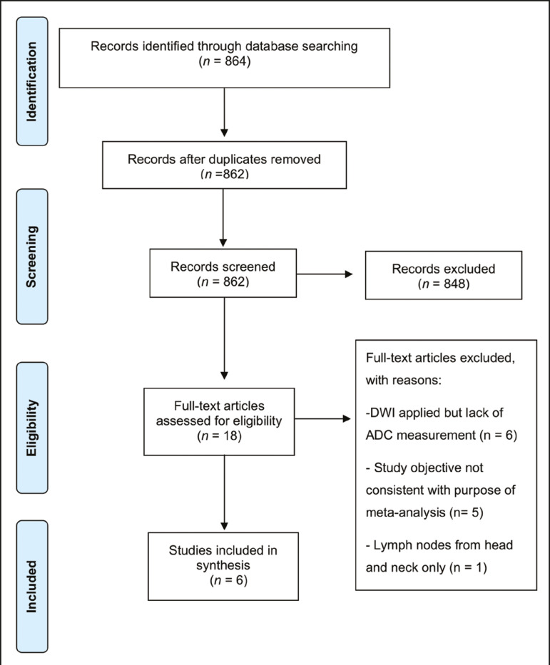
Materials and methods: This was a meta-analysis involving systematic searches of the MEDLINE, EMBASE, and Web of Science databases up through April 2020. Studies reporting thoracic DWI and lymph node evaluation were included. The pooled sensitivity, specificity, diagnostic odds ratio, positive predictive value, negative predictive value, and area under the receiver operating characteristic curve (AUC) were calculated.
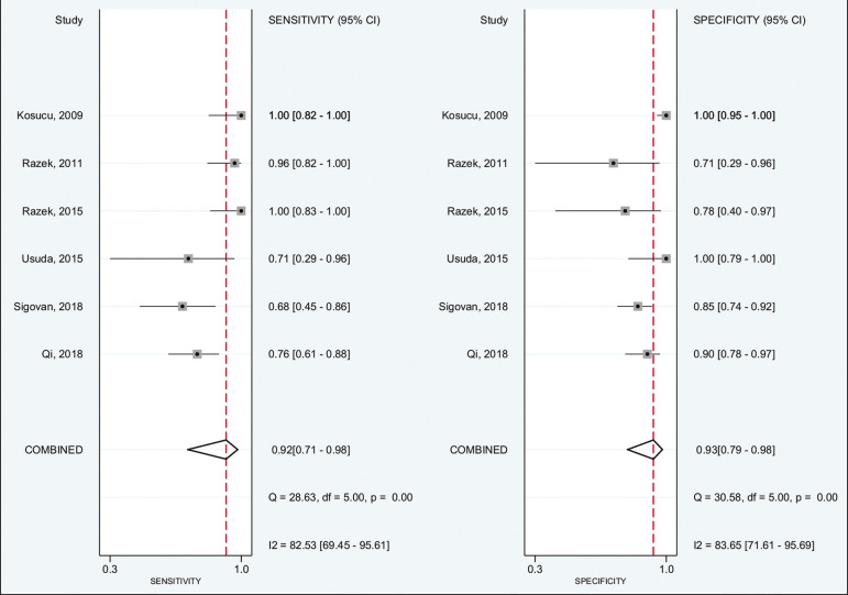
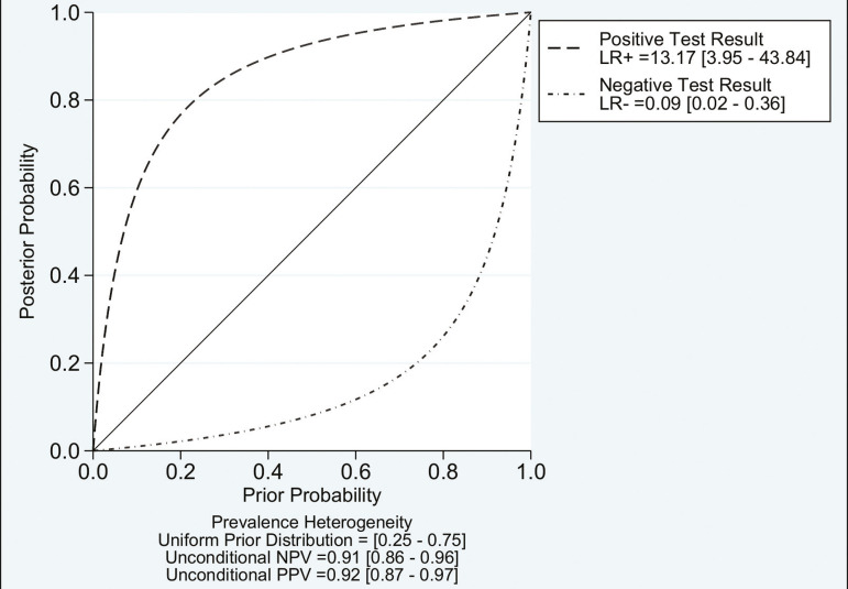
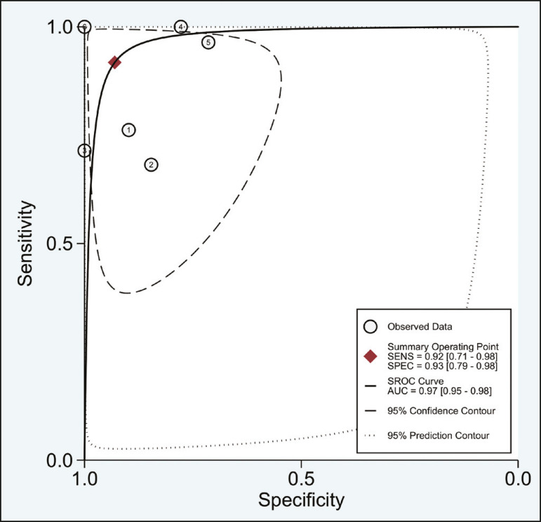
Results: We evaluated six studies, involving a collective total of 356 mediastinal lymph nodes in 214 patients. Thoracic DWI had a pooled sensitivity and specificity of 92% (95% confidence interval [95% CI]: 71-98%) and 93% (95% CI: 79-98%), respectively. The positive and negative likelihood ratios were 13.2 (95% CI: 4.0-43.8) and 0.09 (95% CI: 0.02-0.36), respectively. The diagnostic odds ratio was 149 (95% CI: 18-1,243), and the AUC was 0.97 (95% CI: 0.95-0.98).
Conclusion: DWI is a reproducible technique and has demonstrated high accuracy for differentiating between malignant and benign states in thoracic lymph nodes.
Objetivo: Uma meta-análise foi realizada para estabelecer o desempenho diagnóstico da ressonância magnética com imagem ponderada em difusão (DWI) na discriminação de linfonodos torácicos malignos de benignos.
Materiais e métodos: MEDLINE, EMBASE e Web of Science foram sistematicamente pesquisados até abril de 2020. Foram incluídos estudos que relatavam o uso de DWI na avaliação de linfonodos torácicos. Sensibilidade, especificidade, razão de chances de diagnóstico, valores preditivos positivos e negativos e área sob a curva (AUC) foram calculados.
Resultados: Foram encontrados 356 linfonodos mediastinais de 214 pacientes nos seis estudos incluídos. DWI produziu sensibilidade e especificidade combinadas de 92% (intervalo de confiança 95% [IC 95%]: 71-98%) e 93% (IC 95%: 79-98%), respectivamente. A razão de verossimilhança positiva foi de 13,2 (IC 95%: 4,0-43,8), a razão de verossimilhança negativa foi de 0,09 (IC 95%: 0,02-0,36); A razão de chances de diagnóstico foi de 149 (IC 95%: 18-1.243). A DWI teve uma AUC de 0,97 (IC 95%: 0,95-0,98).
Conclusão: DWI é uma técnica reprodutível que demonstrou alta acurácia na diferenciação de estados malignos e benignos nos linfonodos torácicos.
Keywords: Diffusion magnetic resonance imaging; Lymph nodes/diagnostic imaging; Lymphadenopathy; Magnetic resonance imaging; Meta-analysis; Thoracic neoplasms/diagnosis.
Figures

Similar articles
-
Diagnostic value of diffusion-weighted magnetic resonance imaging: differentiation of benign and malignant lymph nodes in different regions of the body.Clin Imaging. 2015 Sep-Oct;39(5):856-62. doi: 10.1016/j.clinimag.2015.05.006. Epub 2015 May 22. Clin Imaging. 2015. PMID: 26091745
-
The Diagnostic Value of Diffusion-Weighted Imaging in Differentiating Metastatic Lymph Nodes of Head and Neck Squamous Cell Carcinoma: A Systematic Review and Meta-Analysis.AJNR Am J Neuroradiol. 2018 Oct;39(10):1889-1895. doi: 10.3174/ajnr.A5813. Epub 2018 Sep 13. AJNR Am J Neuroradiol. 2018. PMID: 30213809 Free PMC article.
-
Evaluation of the diagnostic performance of apparent diffusion coefficient (ADC) values on diffusion-weighted magnetic resonance imaging (DWI) in differentiating between benign and metastatic lymph nodes in cases of cholangiocarcinoma.Abdom Radiol (NY). 2019 Feb;44(2):473-481. doi: 10.1007/s00261-018-1742-6. Abdom Radiol (NY). 2019. PMID: 30151713
-
Diagnostic Performance of Diffusion-Weighted Imaging for Differentiating Malignant From Benign Intraductal Papillary Mucinous Neoplasms of the Pancreas: A Systematic Review and Meta-Analysis.Front Oncol. 2021 Jul 5;11:637681. doi: 10.3389/fonc.2021.637681. eCollection 2021. Front Oncol. 2021. PMID: 34290974 Free PMC article.
-
Diffusion-weighted magnetic resonance imaging for the detection of metastatic lymph nodes in patients with lung cancer: A meta-analysis.Mol Clin Oncol. 2017 Mar;6(3):344-354. doi: 10.3892/mco.2017.1153. Epub 2017 Feb 6. Mol Clin Oncol. 2017. PMID: 28451411 Free PMC article.
Cited by
-
Diagnostic modalities in the mediastinum and the role of bronchoscopy in mediastinal assessment: a narrative review.Mediastinum. 2024 Dec 6;8:51. doi: 10.21037/med-24-32. eCollection 2024. Mediastinum. 2024. PMID: 39781205 Free PMC article. Review.
-
Comparative analysis of F-18 FDG PET/CT images between scrub typhus and systemic lupus erythematosus.Sci Rep. 2024 Jul 3;14(1):15264. doi: 10.1038/s41598-024-65256-1. Sci Rep. 2024. PMID: 38961124 Free PMC article.
References
-
- Mennini ML, Catalano C, Del Monte M, et al. Computed tomography and magnetic resonance imaging of the thoracic lymphatic system. Thorac Surg Clin. 2012;22:155–160. - PubMed
-
- Passlick B. Detection of disseminated lung cancer cells in lymph nodes by monoclonal antibodies: impact on staging and prognosis. Ann Ital Chir. 1999;70:839–840. - PubMed
-
- Gupta S, Seaberg K, Wallace MJ, et al. Imaging-guided percutaneous biopsy of mediastinal lesions: different approaches and anatomic considerations. Radiographics. 2005;25:763–786. - PubMed
-
- Goldberg SN, Raptopoulos V, Boiselle PM, et al. Mediastinal lymphadenopathy: diagnostic yield of transbronchial mediastinal lymph node biopsy with CT fluoroscopic guidance-initial experience. Radiology. 2000;216:764–767. - PubMed
-
- Torabi M, Aquino SL, Harisinghani MG. Current concepts in lymph node imaging. J Nucl Med. 2004;45:1509–1518. - PubMed
LinkOut - more resources
Full Text Sources
Miscellaneous