. 2021 Aug 6:14:3767-3780.
doi: 10.2147/JIR.S322960. eCollection 2021.

Clinical Strains of Pseudomonas aeruginosa Secrete LasB Elastase to Induce Hemorrhagic Diffuse Alveolar Damage in Mice

Affiliations

Clinical Strains of Pseudomonas aeruginosa Secrete LasB Elastase to Induce Hemorrhagic Diffuse Alveolar Damage in Mice

Yajie Zhu et al. J Inflamm Res. .

Abstract

Background: Acute lung injury and acute respiratory distress syndrome (ALI/ARDS) are most often caused by bacterial pneumonia and characterized by severe dyspnea and high mortality. Knowledge about the lung injury effects of current clinical bacterial strains is lacking. The aim of this study was to investigate the ability of representative pathogenic bacteria isolated from patients to cause ALI/ARDS in mice and identify the major virulence factor.

Methods: Seven major bacterial species were isolated from clinical sputum and unilaterally instilled into the mouse airway. A histology study was performed to determine the lung injury effect. Virulence genes were examined by PCR. Sequence types of P. aeruginosa strains were identified by MLST. LC-MS/MS was used to analysis the bacterial exoproducts proteome. LasB was purified through a DEAE-cellulose column, and its toxicity was tested both in vitro and in vivo.

Results: Staphylococcus aureus, Streptococcus pneumoniae, Streptococcus agalactiae, Acinetobacter baumannii, Klebsiella pneumoniae, Pseudomonas aeruginosa and Escherichia coli were randomly separated and tested 3 times. Among them, gram-negative bacteria have much more potential to cause acute lung injury than gram-positive bacteria. However, P. aeruginosa is the only pathogen that induces diffuse alveolar damage, hemorrhage and hyaline membranes in the lungs of mice. The lung injury effect is associated with the excreted LasB elastase. Purified LasB recapitulated lung injury similar to P. aeruginosa infection in vivo. We found that this was due to the powerful degradation effect of LasB on the extracellular matrix of the lung and key proteins in the coagulation cascade without inducing obvious cellular apoptosis. We also report for the first time that LasB could induce DIC-like coagulopathy in vitro.

Conclusion: P. aeruginosa strains are most capable of inducing ALI/ARDS in mice among major clinical pathogenic bacteria tested, and this ability is specifically attributed to their LasB production.

Keywords: ALI/ARDS; LasB elastase; Pseudomonas aeruginosa; unilateral lung injury.


Conflict of interest statement

The authors declare no conflicts of interest in this work.

Figures
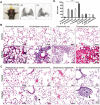
Figure 1
Figure 1
Histological assessment of acute lung injury induced by major bacterial species separated from clinical sputum (A) Unilateral endotracheal intubation. Unilateral lung consolidation appeared 4 hours after LPS instillation (3D CT reconstruction). (B) Hematoxylin and eosin (HE) stain of lung sections inoculated with gram-negative bacteria. Alveolar injury was observed in all groups (n=9 in each group). K. pneumoniae: neutrophils were accumulated in the alveolar wall and the alveolar space was filled with bacteria; A. baumannii: patchy neutrophil infiltration; E. coli: diffuse neutrophil infiltration; P. aeruginosa: hemorrhage, diffuse alveolar damage, hyaline membranes (arrows), vessel congestion, alveolar wall thickening and neutrophil infiltration were all observed. (C) HE stain of lung sections of gram-positive bacterial infection. Neutrophil infiltration was found limited in the periarterial space (arrowheads) without causing alveolar damage, especially in hypervirulent (hv) Streptococcus pneumonia group (n=9 in each group). (D) Score of 7 major bacterial species induced ALI 24h post-inoculation in mice, compared with normal C57/BL6 mice (n=6 per group). ***P < 0.0005.
Figure 2
Figure 2
ALI/ARDS induced by different strains of P. aeruginosa in mice (A) Kaplan-Meier survival curves for mice infected with different strains (n=10 in each group). (B) ALI score of mice infected with different strains (n≥4). Mice were euthanized at 12 h post infection. A, ATCC27853; ***P < 0.0005. (C) Rate of occurrence of bilateral infiltration 12h after unilateral inoculation (n=10 in each group). (D) Representative histology findings of the most and least virulent clinical P. aeruginosa strain Pa 4 and Pa 3. Severe DAD and bilateral infiltration occurred in Pa 4 infected lungs. The bronchial lumen (*) was inundated with red blood cells. DAD, hyaline membranes (arrowheads) and neutrophil accumulation even occurred in the uninoculated control lung in Pa 4 infected mice; In Pa 3 infected group, less hemorrhage but more obvious bacterial proliferation and infiltration found in bilateral lungs (arrows). HE stain; (E) pink frothy sputum and bloody sputum observed from the nose and mouth of mice. (F) Rate of occurrence of pink frothy/bloody sputum in different mice groups (n=10 per group).
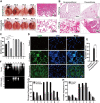
Figure 3
Figure 3
P. aeruginosa exoproducts induce hemorrhagic ALI in mice. (A) Representative gross picture of P. aeruginosa exoproducts injured mice lung. Unilateral lung injury was largely inhibited by PIR (protease inhibitors cocktail with EDTA added). Typical DAD, hyaline membranes and bleeding were induced by Pa 4 exoproducts. (B) Histological difference of mice died with pneumothorax or not 3 h after high concentration of exoproducts instillation. Severe lung tissue compression appeared in mice died of pneumothorax (n=14), compared with emphysematous alveolar destruction in survived mice (no pneumothorax, n=10). (C) Modified ALI score of different P. aeruginosa strain exoproducts challenged lungs. n=3. (D) TUNEL stain of P. aeruginosa bacteria/exoproducts challenged lung tissue. Cell nuclei (Blue), TUNEL-positive cells (Green). (E) Apoptosis rate of bacteria/exoproducts challenged lung cells. (F) Gelatin zymography of exoproducts. (G) Fibrinogen degradation assay of exoproducts. (H) Thrombin degradation assay of exoproducts. *P < 0.05; **P < 0.005; ***P < 0.0005.
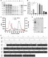
Figure 4
Figure 4
Proteomic analysis of exoproducts and purification of the virulent protease LasB (A) SDS-PAGE of exoproducts from 8 P. aeruginosa strains. (B) Relative LasB expression of 8 P. aeruginosa strains. *P < 0.05; **P < 0.005; (C) elution profile of LasB protease on DEAE 52 cellulose column. (D) Verification of LasB purity by SDS-PAGE. 10, purified LasB (Fraction 10); 15, impurities (Fraction 15); 35, impurities (Fraction 35); CE, crude enzyme solution. (E) Amino acid sequence alignment of Las B from Pa 4, Pa 2 and PAO1.
Figure 5
Figure 5
LasB elastase provokes hemorrhagic DAD, degrades extracellular matrix and causes hemostasis disorder (A) HE stain of mice lung unilaterally instilled with 3μg LasB elastase. DAD, hemorrhage, hyaline membranes, neutrophil infiltration and multiple bullae (arrowheads) occurred in LasB treated left lung; L, left; R, right. (B) ALI score of LasB injured lungs and the effect of different LasB inhibitors (n=3 in each group). EDTA: 25 mM; Ilomastat: 1mM. (C) Gelatin zymography of purified LasB and the inhibition effect of EDTA. (D) Degradation of fibrinogen and thrombin with or without inhibitors in vitro. Fib, fibrinogen; 1: LasB + fibrinogen; 2: LasB + ilomastat + fibrinogen; 3: LasB + TLCK + fibrinogen; 4: LasB + TLCK + fibrinogen; Thr, thrombin; 5: LasB + thrombin; 6: LasB + ilomastat + thrombin; 7: LasB + TLCK + thrombin; 8: LasB + TLCK + thrombin; (E) the effect of LasB on overall coagulation function. Blood from healthy donors was incubated with LasB for 35 min, room temperature. (F) The impact of LasB on coagulation function in vivo. Hemostasis of SD rats were measured 1 h after injection of LasB elastase through tail vein. (G) TUNEL stain of LasB injured lung tissue. No apparent cellular apoptosis was found. Cell nuclei (Blue), TUNEL-positive cells (Green). (H) Apoptosis rate of LasB injured lung cells. (I) Cytotoxicity assay of LasB, Pa 4 bacteria or exoproducts to THP-1 cells. LasB 1: 100 μg/mL; LasB 2: 200 μg/mL. *P < 0.05; **P < 0.005; ***P < 0.0005.

References

    1. Fan E, Brodie D, Slutsky AS. Acute respiratory distress syndrome: advances in diagnosis and treatment. JAMA. 2018;319(7):698–710. doi:10.1001/jama.2017.21907 - DOI - PubMed
    1. Silva PL, Pelosi P, Rocco PRM. Personalized pharmacological therapy for ARDS: a light at the end of the tunnel. Expert Opin Investig Drugs. 2020;29(1):49–61. doi:10.1080/13543784.2020.1699531 - DOI - PubMed
    1. Bellani G, Laffey JG, Pham T, et al. Epidemiology, patterns of care, and mortality for patients with acute respiratory distress syndrome in intensive care units in 50 countries. JAMA. 2016;315(8):788–800. doi:10.1001/jama.2016.0291 - DOI - PubMed
    1. Tanner JR, Kingsley RA. Evolution of salmonella within hosts. Trends Microbiol. 2018;26(12):986–998. doi:10.1016/j.tim.2018.06.001 - DOI - PMC - PubMed
    1. Defraine V, Fauvart M, Michiels J. Fighting bacterial persistence: current and emerging anti-persister strategies and therapeutics. Drug Resist Updat. 2018;38:12–26. doi:10.1016/j.drup.2018.03.002 - DOI - PubMed