. 2021 Jul 30:15:692232.
doi: 10.3389/fncel.2021.692232. eCollection 2021.

An Autism-Associated de novo Mutation in GluN2B Destabilizes Growing Dendrites by Promoting Retraction and Pruning

Affiliations

An Autism-Associated de novo Mutation in GluN2B Destabilizes Growing Dendrites by Promoting Retraction and Pruning

Jacob A Bahry et al. Front Cell Neurosci. .

Erratum in

Abstract

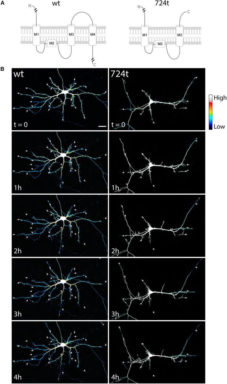
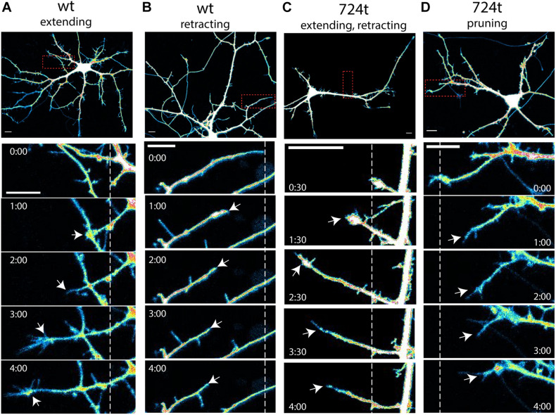
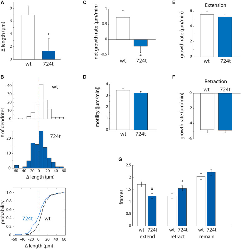
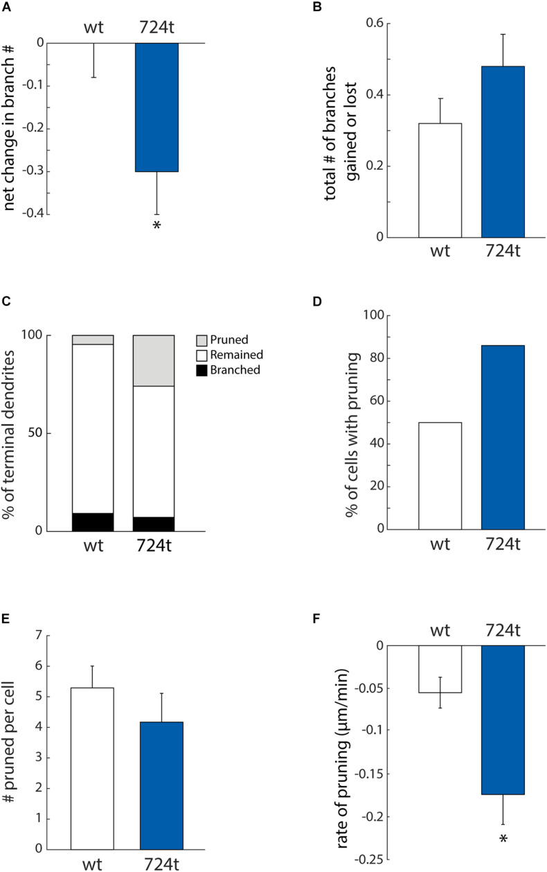
Mutations in GRIN2B, which encodes the GluN2B subunit of NMDA receptors, lead to autism spectrum disorders (ASD), but the pathophysiological mechanisms remain unclear. Recently, we showed that a GluN2B variant that is associated with severe ASD (GluN2B724t) impairs dendrite morphogenesis. To determine which aspects of dendrite growth are affected by GluN2B724t, we investigated the dynamics of dendrite growth and branching in rat neocortical neurons using time-lapse imaging. GluN2B724t expression shifted branch motility toward retraction and away from extension. GluN2B724t and wild-type neurons formed new branches at similar rates, but mutant neurons exhibited increased pruning of dendritic branches. The observed changes in dynamics resulted in nearly complete elimination of the net expansion of arbor size and complexity that is normally observed during this developmental period. These data demonstrate that ASD-associated mutant GluN2B interferes with dendrite morphogenesis by reducing rates of outgrowth while promoting retraction and subsequent pruning. Because mutant dendrites remain motile and capable of growth, it is possible that reducing pruning or promoting dendrite stabilization could overcome dendrite arbor defects associated with GRIN2B mutations.

Keywords: GRIN2B gene; GluN2B (NMDA receptor subunit NR2B); NMDA receptor; autism; dendrite development; live imaging; neurodevelopment.


Conflict of interest statement

The authors declare that the research was conducted in the absence of any commercial or financial relationships that could be construed as a potential conflict of interest.

Figures

FIGURE 1
FIGURE 1
Developing neurons expressing ASD-mutant GluN2B have smaller dendrites. (A) Schematic of GluN2BWT (left) and GluN2B724t (right). M1, M2, M3, and M4 represent membrane domains. To focus on the mutation site, full amino and carboxy termini are not depicted when indicated by line breaks. (B) Representative images of neurons (5–9 DIV) transfected with mutant (right) or wildtype (left) GFP-tagged GluN2B (not shown) and tdTomato (color coded with an intensity scale, as indicated by the color key). Arrows point to terminal ends of dendrites. Imaging times are displayed in hours, relative to the start of imaging. Scale bar, 25 μm.
FIGURE 2
FIGURE 2
Examples of terminal dendrite dynamics. Both wild-type and mutant neurons were observed extending, retracting, branching, and pruning. (A–D) Representative images of dendrite behavior. Top panels, low magnification view of individual neurons, visualized by imaging of tdTomato cytoplasmic fills. Areas within the red boxes are magnified in the time-lapse panels below each neuron. Dashed lines indicate the positions of each dendrite tip at the beginning of imaging. White arrows indicate the position of the dendrite tip in each frame. Growth cones were considered the tips of dendrites, rather than individual filopodia. Imaging times are displayed relative to the start of imaging (0:00) and span 4 h. Intensities are colored as in Figure 1. Scale bars, 10 μm. (A) wildtype dendrite extending and remaining terminal (white arrows). (B) Wildtype dendrite retracting and remaining terminal (white arrows). (C) GluN2B724t dendrite emerging, growing, and then retracting (white arrows). (D) GluN2B724t dendrite retracting and pruning (white arrows).
FIGURE 3
FIGURE 3
ASD-associated mutant GluN2B leads to a bias toward retraction of growing dendrites, while wild-type dendrites tend to elongate. (A,B) ASD-associated mutation in GluN2B disrupts dendrite growth kinetics. Quantification of the average change in length of terminal dendrites for neurons expressing GluN2BWT (white) or GluN2B724t (blue) from eight independent experiments. (A) Over 4 h, neurons expressing GluN2B724t had reduced net growth. Data represent means ± S.E. (*p = 0.018, n = 14 GluN2BWT neurons, and 14 GluN2B724t neurons). (B) ASD-associated mutation in GluN2B biases net growth of dendrites toward retraction and away from extension. Top and middle panels, histograms represent the number of dendrites that lengthen or shorten, separated into bins of 10 μm, with negative values representing a terminal dendrite becoming shorter over 4 h of live imaging. Bottom panel, cumulative probability distribution of changes in dendrite length. GluN2BWT (n = 113 dendrites), and GluN2B724t (n = 93 dendrites). Dashed orange lines, zero change in length. (C,D) ASD-mutant GluN2B reduces the net rate of dendrite outgrowth but not overall motility of terminal dendrites. Lengths of terminal dendrites were measured every 12 min for 1 h. Data represent means ± S.E. (n = 113 GluN2BWT dendrites, and 93 GluN2B724t dendrites). (C) ASD-associated mutant dendrites elongated at a reduced rate, compared to neurons expressing GluN2BWT (*p = 0.0008). (D) Overall motility of terminal dendrites was similar for neurons transfected with either wildtype or mutant GluN2B (p = 0.7147). Motility represents all movements, regardless of direction, and was quantified by averaging the absolute values of instantaneous growth rates. Rates of extension [(E), p = 0.5255] and retraction [(F), p = 0.3121] were similar in ASD and WT neurons. Time spent extending [(G), left, p = 0.0017] and retracting [(G), middle, p = 0.0283] were significantly different, however. Time spent not actively extending or retracting was similar for ASD and WT neurons [(G), right, p = 0.2138]. Each frame represents 12 min.
FIGURE 4
FIGURE 4
ASD-associated mutation in GluN2B increases dendrite pruning but does not affect formation of new branches. (A) ASD mutant neurons (blue bars) display a net loss of terminal dendrite branches, while wild-type neurons (white bars) have a stable number of terminal dendrites. Positive values represent gain of dendrite branches, while negative values represent loss of dendrite branches. Data represent the mean ± S.E. (*p = 0.0302, n = 14 GluN2BWT, and 14 GluN2B724t neurons). (B) To calculate the total change in the number of terminal dendrites per cell per hour, both gains and losses of dendrites are represented as positive values. The ASD-associated mutation did not have a significant impact on the total change in the number of terminal dendrites (p = 0.2553). (C) Outcomes for terminal dendrites. A higher percentage of ASD-mutant dendrites were pruned, while a higher percentage of wild-type dendrites remained stable as terminal dendrites. The percentages of dendrites that branched were similar for mutant and wild-type dendrites. (A–C) For each cell, data were collected every hour for 4 h. (D) An increased percentage of ASD-associated mutant neurons had at least one pruned terminal dendrite over a 4-h time period, as compared to wild-type neurons. (E) Neurons that had at least one pruned dendrite had a similar number of pruned dendrites. Data represent the mean ± S.E. (p = 0.114, n = 7 GluN2BWT, and 12 Glun2B724t neurons). (F) Pruned dendrites retracted at a significantly faster rate for neurons expressing GluN2B724t compared to neurons expressing GluN2BWT (p = 0.008, n = 37 GluN2BWT, and 50 GluN2B724t dendrites).

References

    1. Abrahams B. S., Arking D. E., Campbell D. B., Mefford H. C., Morrow E. M., Weiss L. A., et al. (2013). SFARI Gene 2.0: a community-driven knowledgebase for the autism spectrum disorders (ASDs). Mol. Autism 4:36. 10.1186/2040-2392-4-36 - DOI - PMC - PubMed
    1. Baumert R., Ji H., Paulucci-Holthauzen A., Wolfe A., Sagum C., Hodgson L., et al. (2020). Novel phospho-switch function of delta-catenin in dendrite development. J. Cell Biol. 219:e201909166. - PMC - PubMed
    1. Bell S., Maussion G., Jefri M., Peng H., Theroux J. F., Silveira H., et al. (2018). Disruption of GRIN2B impairs differentiation in human neurons. Stem Cell Reports 11 183–196. 10.1016/j.stemcr.2018.05.018 - DOI - PMC - PubMed
    1. Berry C. T., Sceniak M. P., Zhou L., Sabo S. L. (2012). Developmental up-regulation of vesicular glutamate transporter-1 promotes neocortical presynaptic terminal development. PLoS One 7:e50911. 10.1371/journal.pone.0050911 - DOI - PMC - PubMed
    1. Bourgeron T. (2009). A synaptic trek to autism. Curr. Opin. Neurobiol. 19 231–234. 10.1016/j.conb.2009.06.003 - DOI - PubMed