. 2021 Aug;13(4):451-458.
doi: 10.1177/1758573219884010. Epub 2019 Nov 13.

Current concepts in elbow fracture dislocation

Affiliations

Current concepts in elbow fracture dislocation

Adam C Watts et al. Shoulder Elbow. 2021 Aug.

Abstract

Background: Elbow fracture dislocations are complex injuries that can provide a challenge for experienced surgeons. Current classifications fail to provide a comprehensive system that encompasses all of the elements and patterns seen in elbow fracture dislocations.

Methods: The commonly used elbow fracture dislocation classifications are reviewed and the three-column concept of elbow fracture dislocation is described. This concept is applied to the currently recognised injury patterns and the literature on management algorithms.

Results: Current elbow fracture dislocation classification systems only describe one element of the injury, or only include one pattern of elbow fracture dislocation. A new comprehensive classification system based on the three-column concept of elbow fracture dislocation is presented with a suggested algorithm for managing each injury pattern.

Discussion: The three-column concept may improve understanding of injury patterns and treatment and leads to a comprehensive classification of elbow fracture dislocations with algorithms to guide treatment.

Keywords: Elbow fracture dislocation; elbow instability; management.


Conflict of interest statement

Declaration of Conflicting Interests: The author(s) declared no potential conflicts of interest with respect to the research, authorship, and/or publication of this article.

Figures
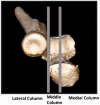
Figure 1.
Figure 1.
Three-dimensional computed tomography reconstruction demonstrating the three columns of the proximal forearm: lateral (radial head), middle (anterolateral coronoid facet), and medial (anteromedial coronoid facet).
Figure 2.
Figure 2.
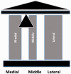
Representative illustration of the three-column model demonstrating the natural fulcrum in between the middle and medial columns.
Figure 3.
Figure 3.
Wrightington A fracture dislocation: loss of the medial column (anteromedial coronoid facet) with associated proximal avulsion of the lateral ligament complex and posterior band of medial collateral ligament results in varus instability. This pattern is commonly seen in posteromedial rotatory fracture dislocation.
Figure 4.
Figure 4.
(a) Wrightington C fracture dislocation: combined loss of the middle (anterolateral coronoid facet) and lateral (radial head) columns with disruption of the lateral ligament complex results in valgus instability. This pattern is synonymous with the terrible triad fracture dislocation and will result in posterolateral rotatory instability if not surgically stabilised. (b) Restoration of the radial head and lateral collateral ligament complex will reliably restore stability in a Wrightington C fracture dislocation without the need to fix the anterolateral coronoid fracture.
Figure 5.
Figure 5.
(a) and (b) Plain radiograph and 3D CT reconstruction demonstrating an isolated anteromedial coronoid facet fracture (Wrightington A). (c) Plain radiographs demonstrating fixation of Wrightington A fracture.
Figure 6.
Figure 6.
(a) Plain radiograph demonstrating an ulnar fracture extending proximal to and involving both coronoid facets with an intact radial head (Wrightington B). (b) Plain radiographs demonstrating fixation of Wrightington B fracture.
Figure 7.
Figure 7.
(a) Plain radiograph demonstrating an ulnar fracture extending proximal to and involving both coronoid facets with an associated radial head fracture (Wrightington B+). (b) Plain radiographs demonstrating fixation of Wrightington B+ fracture.
Figure 8.
Figure 8.
(a) and (b) Plain radiographs (pre and post fixation) demonstrating an ulnar fracture distal to and therefore not involving the coronoid with apex posterior angulation and an associated radial head fracture (Wrightington D+). What differentiates a type D from type B is an intact coronoid and hence a stable ulnohumeral articulation.
Figure 9.
Figure 9.
Management algorithm based on Wrightington Classification.

References

    1. O'Driscoll SW, Bell DF, Morrey BF. Posterolateral rotatory instability of the elbow. J Bone Joint Surg 1991; 73: 440–446. - PubMed
    1. O'Driscoll SW, Jupiter JB, King GJ, et al.The unstable elbow. Instr Course Lect 2001; 50: 89–102. - PubMed
    1. Hwang J-T, Shields MN, Berglund LJ, et al.The role of the posterior bundle of the medial collateral ligament in posteromedial rotatory instability of the elbow. Bone Joint J 2018; 100-B: 1060–1065. - PubMed
    1. Bado JL. The Monteggia lesion. Clin Orthop Relat Res 1967; 50: 71–86. - PubMed
    1. Ring D, Jupiter JB, Simpson NS. Monteggia fractures in adults. J Bone Joint Surg 1998; 80: 1733–1744. - PubMed

LinkOut - more resources