Review
. 2021 Jul 30:8:696438.
doi: 10.3389/fmolb.2021.696438. eCollection 2021.

RNA Polymerase III Subunit Mutations in Genetic Diseases

Affiliations
Review

RNA Polymerase III Subunit Mutations in Genetic Diseases

Elisabeth Lata et al. Front Mol Biosci. .

Abstract

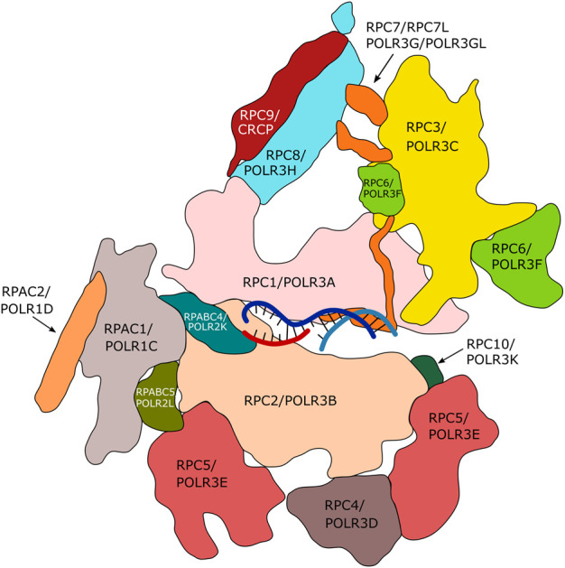
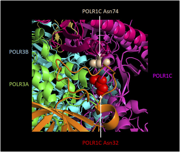
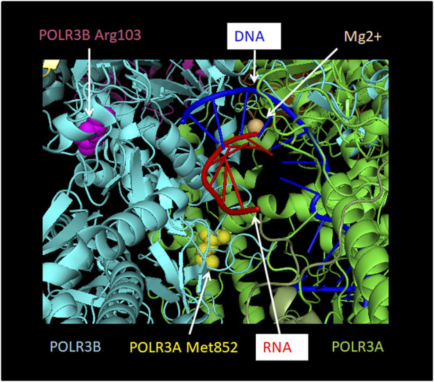
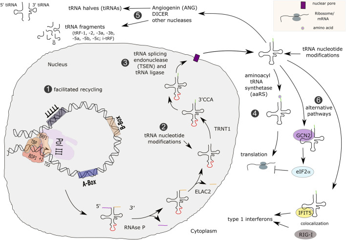
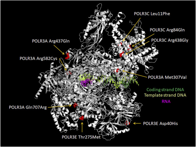
RNA polymerase (Pol) III transcribes small untranslated RNAs such as 5S ribosomal RNA, transfer RNAs, and U6 small nuclear RNA. Because of the functions of these RNAs, Pol III transcription is best known for its essential contribution to RNA maturation and translation. Surprisingly, it was discovered in the last decade that various inherited mutations in genes encoding nine distinct subunits of Pol III cause tissue-specific diseases rather than a general failure of all vital functions. Mutations in the POLR3A, POLR3C, POLR3E and POLR3F subunits are associated with susceptibility to varicella zoster virus-induced encephalitis and pneumonitis. In addition, an ever-increasing number of distinct mutations in the POLR3A, POLR3B, POLR1C and POLR3K subunits cause a spectrum of neurodegenerative diseases, which includes most notably hypomyelinating leukodystrophy. Furthermore, other rare diseases are also associated with mutations in genes encoding subunits of Pol III (POLR3H, POLR3GL) and the BRF1 component of the TFIIIB transcription initiation factor. Although the causal relationship between these mutations and disease development is widely accepted, the exact molecular mechanisms underlying disease pathogenesis remain enigmatic. Here, we review the current knowledge on the functional impact of specific mutations, possible Pol III-related disease-causing mechanisms, and animal models that may help to better understand the links between Pol III mutations and disease.

Keywords: Pol III subunits (POLR3A, POLR3B, POLR3C, POLR3E, POLR3F, POLR3GL, POLR3H, POLR3K, POLR1C); Pol III-related hypomyelinating leukodystrophy (POLR3-HLD); RNA polymerase III (Pol III); innate immunity; neurodegenerative disease.


Conflict of interest statement

The authors declare that the research was conducted in the absence of any commercial or financial relationships that could be construed as a potential conflict of interest.

Figures

FIGURE 1
FIGURE 1
Subunits of human RNA polymerase III. Fourteen of the seventeen subunits of human Pol III are appropriately assigned. Subunits POLR2E, POLR2F and POLR2H are not displayed. The Figure is inspired by the structure of human Pol III published by Girbig et al. (2021) (PDB7AE3). The RPC4/RPC5 hetero-dimeric complex is required by termination/reinitiation. The RPC3/RPC6/RPC7 hetero-trimeric subcomplex is required for transcription initiation.
FIGURE 2
FIGURE 2
Promoters directing human RNA polymerase III transcription. Pol III type 1 and type 2 genes contain gene-internal promoter elements. (A) Type 1 gene transcription of 5S ribosomal (r)RNA is directed by A- and C-boxes that are located relative to the transcription start site (TSS) as indicated. These promoter elements are bound by TFIIIA, permitting the recruitment of TFIIIC and subsequently of TFIIIB-β (composed of TBP, BDP1 and BRF1), which altogether recruit Pol III. (B) Type 2 genes (tRNA genes, VA1, VA2, SINEs) contain A- and B-boxes as promoter elements at varying positions relative to the TSS. They are bound by TFIIIC, subsequently allowing recruitment of TFIIIB-β and in turn of Pol III. (C) Type 3 gene regulatory elements are entirely located upstream (5′) of the TSS. They are composed of a TATA-box at -30, as well as a proximal sequence element (PSE) and a distal sequence element (DSE) at variable distances with respect to the TSS, depending on the gene. Transcriptional activators (STAF/ZNF143; SNAPc/PTF) bind to the DSE and PSE, respectively, and regulate transcriptional activity. The TATA-box is the only promoter element required for directing Pol III to the TSS (Teichmann et al., 1997). It is bound by TFIIIB-α (composed of TBP, BDP1 and BRF2), which in turn recruits Pol III. (D) Hybrid promoter-directed transcription is regulated by gene-internal elements of type 2 promoters (A- and B-boxes) and additionally by gene regulatory elements upstream of the TSS. All these elements vary in their distance to the TSS depending on the gene. In the presence of the PSE (tRNASec gene), TFIIIB-α is recruited (not shown in the Figure). In the case of all other enhancer-activator combinations with gene-internal A- and B-boxes (EBER 1 and 2; 7SL; vault; BC1 and BC200), TFIIIB-β is recruited, allowing the subsequent recruitment of RNA polymerase III. For all promoter types, the stretch of T’s represents the transcription termination site. Arrows in panels C. and D. symbolize protein-protein-interactions that contribute to activation of Pol III transcription from these promoters. Promoter types were reviewed in Dumay-Odelot et al. (2014).
FIGURE 3
FIGURE 3
Neuro-anatomical structures affected or for which myelination is preserved in POLR3-related disorders. (A) Schematic of a sagittal view of the human brain. Structures involved/preserved in POLR3-HLD are depicted in distinct colours and labeled with a number. On the right side, the names of anatomical structures corresponding to each number are shown in the same colour as the structure, followed by a description of how the structure is affected/preserved in POLR3-HLD. (B) Schematic of a coronal view of the human brain. Structures involved/preserved in the striatal variant of POLR3-related disorders (caudate and putamen) or in POLR3-HLD (other structures) are shown in distinct colours and labeled with a number. The legend on the right side follows the same description as in (A). White matter (in white on the brain schematic) is indicated by an arrow. Corticospinal tracts are displayed as brown dashed lines. The figure was adapted from images available on https://smart.servier.com.
FIGURE 4
FIGURE 4
Schematic representing possible mechanisms underlying POLR3-HLD. In wild-type conditions (healthy individuals), Pol III synthesizes small ncRNAs that play essential roles in housekeeping processes such as translation and co-translational targeting of nascent peptides, which are necessary for the production of myelin. In individuals with POLR3-HLD, it is hypothesized that mutations in Pol III subunits (POLR3A, POLR3B, POLR1C or POLR3K) result in reduced Pol III transcription and decreased levels of Pol III transcripts. The “tRNA-centric” hypothesis postulates that lower levels of tRNAs (either globally, for specific anticodons or for specific isodecoders) will impact translation and synthesis of proteins that are essential for myelination. Alternatively or in addition, reduced levels of other Pol III transcripts may contribute to POLR3-HLD pathogenesis through suboptimal performance of their respective functions that will particularly affect oligodendrocytes and/or neurons. An example is shown for 7SL RNA, where reduced levels of this ncRNA could impair translocation of secreted or transmembrane proteins to the ER, which could impact production of myelin. The schematic of the neuron was adapted from images available on https://smart.servier.com.
FIGURE 5
FIGURE 5
Localization of POLR1C Asn32Ile and Asn74Ser mutations that are associated with POLR3-HLD. Amino acids in POLR1C which cause an assembly defect in Pol III (Thiffault et al., 2015) upon mutation are shown in red (POLR1C Asn32) and in ochre (POLR1C Asn74). They are localized at the interface with Pol III subunits POLR3A (green) and POLR3B (turquoise). The remainder of POLR1C is colored in pink. The Figure was modified from PDB 7AE3 by employing Pymol.
FIGURE 6
FIGURE 6
Localization of POLR3A Met852Val and POLR3B Arg103His mutations relative to the active site. POLR3A is shown in green and POLR3B in turquoise. Pol III mutations described in the text that are found close to the active site and which may thus affect catalytic activity are depicted. Methionine 852 of POLR3A as part of the bridge helix is highlighted as a sphere in yellow. Arginine 103 of POLR3B is highlighted as a sphere in magenta. RNA is shown in red and DNA in blue. The Mg2+ ion of the active site is shown as a sphere in light orange. The Figure was modified from PDB 7AE3 by employing Pymol.
FIGURE 7
FIGURE 7
Transcription, maturation and functions of tRNAs. Type 2 promoter tRNA genes are transcribed by Pol III, possibly involving the facilitated recycling pathway, which allows transcription reinitiation in the absence of TFIIIC (1). Primary tRNAs contain 5′ leader and 3′ trailer sequences (colored lines) and a few human tRNAs contain introns (red line). tRNAs are processed by RNAse P, ELAC2 and TRNT1 (2), as well as spliced in the nucleus (3). Mature tRNAs are exported to the cytoplasm and loaded with amino acids for participating in translation (4). Other functions for tRNAs that were described involve enzymatic processing for yielding tRNA fragments (tRFs) or tRNA halves (tiRNAs) (5). In addition, they can enter alternative functional pathways (6).
FIGURE 8
FIGURE 8
Transcription of the 7SL RNA gene, assembly of the signal recognition particle and functions in the secretory pathway. 7SL RNA gene transcription is directed by gene-internal A- and B-boxes as well as by an ATF/CREB-response element and a distal sequence element (DSE) upstream of the TSS. Transcription is initiated by TFIIIC- and TFIIIB-β-dependent recruitment of Pol III (1). The SRP 9/14/19/68/72 pre-signal recognition particle (pre-SRP) is assembled in the nucleus (2), exported to the cytoplasm and completed with SRP54 (3). SRP recognizes hydrophobic signal peptides in nascent polypeptide chains of secretory pathway proteins (4), arrests translation, translocates arrested ribosomes to the endoplasmic reticulum (ER), where translation resumes and proteins are translocated either into the lumen or into the membrane of the ER (5). Proteins of the secretory pathway are either incorporated into membranes or exported from cells (6). ERGIC refers to the ER-Golgi-intermediate compartment.
FIGURE 9
FIGURE 9
Transcription of the BC200 RNA and its functions in mRNA transport and regulation of translation. BC200 RNA gene transcription depends on TFIIIC bound to gene-internal A- and B-boxes, which allows the subsequent recruitment of TFIIIB-β and Pol III. Sequences upstream of the TSS including a TATA-like box at -30 regulate transcription activity (1). BC200 RNA assembles with SRP 9/14 in the nucleus (2) and is exported to the cytoplasm where it was shown to interact with a variety of proteins. BC200 was shown to interact with FMRP, SYNCRIP and Pur-alpha, suggesting a role in mRNA transport similar to what was demonstrated for BC1 in complex with these proteins (3). In association with eukaryotic initiation factor 4A (eIF4A) and poly A binding protein (PABP), BC200 RNA prevents the assembly of the 48S ribosomal subunit (4). This repression of translation can be counteracted by hnRNP E1/E2 binding to BC200 RNA in vitro (5).
FIGURE 10
FIGURE 10
Cytoplasmic transcription by RNA polymerase III in innate immune defense. Upon infection (1), the varicella zoster virus (VZV) DNA is found in the nucleus and the cytoplasm (Mahalingam et al., 1999; Zerboni et al., 2014). Although not specifically reported for VZV, cytoplasmic liberation of DNA from capsids may involve proteasomal degradation (2), as was described for herpes simplex virus 1 (HSV-1; Horan et al., 2013). Cytoplasmic AT-rich VZV DNA is transcribed by Pol III (3), the 5′ tri-phosphorylated RNA folds into stem-loop structures, interacts with the retinoic acid-inducible gene I (RIG-I) (4), which, via mitochondrial antiviral signaling (MAVS) protein and NF-kB/IRF3 transcriptional activators (5) increases transcription of type I interferon genes (6).
FIGURE 11
FIGURE 11
Distribution of mutations in RNA polymerase III subunits associated with impaired innate immune defense. Spatial distribution of amino acid mutations that are associated with severe immune deficiency (POLR3E Asp40His) or with varicella zoster virus (VZV) encephalitis and/or pneumonitis (other displayed mutations). Mutated amino acids are depicted as spheres and highlighted in red. Individual mutations in subunits POLR3A, 3C, 3E are appropriately designated. The mutation POLR3F Arg50Trp cannot be displayed since the corresponding sequence was not resolved in the cryo-EM structure (PDB:7ae1). The Figure was created using Pymol.

References

    1. Abascal-Palacios G., Ramsay E. P., Beuron F., Morris E., Vannini A. (2018). Structural Basis of RNA Polymerase III Transcription Initiation. Nature 553, 301–306. 10.1038/nature25441 - DOI - PubMed
    1. Ablasser A., Bauernfeind F., Hartmann G., Latz E., Fitzgerald K. A., Hornung V. (2009). RIG-I-dependent Sensing of poly(dA:dT) through the Induction of an RNA Polymerase III-Transcribed RNA Intermediate. Nat. Immunol. 10, 1065–1072. 10.1038/ni.1779 - DOI - PMC - PubMed
    1. Adang L. A., Sherbini O., Ball L., Bloom M., Darbari A., Amartino H., et al. (2017). Revised Consensus Statement on the Preventive and Symptomatic Care of Patients with Leukodystrophies. Mol. Genet. Metab. 122, 18–32. 10.1016/j.ymgme.2017.08.006 - DOI - PMC - PubMed
    1. Akopian D., Shen K., Zhang X., Shan S.-o. (2013). Signal Recognition Particle: an Essential Protein-Targeting Machine. Annu. Rev. Biochem. 82, 693–721. 10.1146/annurev-biochem-072711-164732 - DOI - PMC - PubMed
    1. Al Yazidi G., Tran L. T., Guerrero K., Vanderver A., Schiffmann R., Wolf N. I., et al. (2019). Dystonia in RNA Polymerase III‐Related Leukodystrophy. Mov Disord. Clin. Pract. 6, 155–159. 10.1002/mdc3.12715 - DOI - PMC - PubMed

LinkOut - more resources