. 2021 Jul 20;11(14):e4100.
doi: 10.21769/BioProtoc.4100.

GeneWeld: Efficient Targeted Integration Directed by Short Homology in Zebrafish

Affiliations

GeneWeld: Efficient Targeted Integration Directed by Short Homology in Zebrafish

Jordan M Welker et al. Bio Protoc. .

Abstract

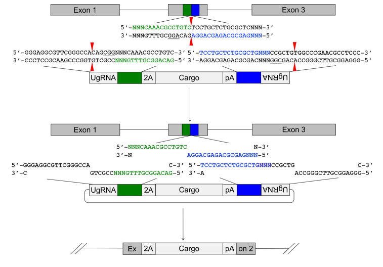
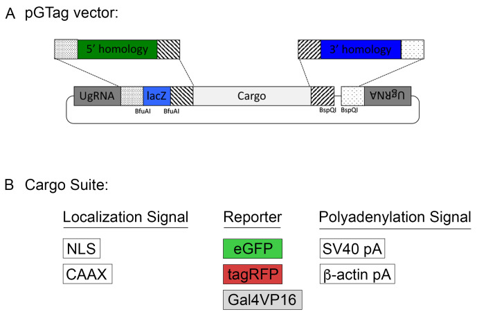
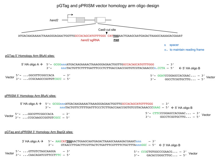
Efficient precision genome engineering requires high frequency and specificity of integration at the genomic target site. Multiple design strategies for zebrafish gene targeting have previously been reported with widely varying frequencies for germline recovery of integration alleles. The GeneWeld protocol and pGTag (plasmids for Gene Tagging) vector series provide a set of resources to streamline precision gene targeting in zebrafish. Our approach uses short homology of 24-48 bp to drive targeted integration of DNA reporter cassettes by homology-mediated end joining (HMEJ) at a CRISPR/Cas induced DNA double-strand break. The pGTag vectors contain reporters flanked by a universal CRISPR sgRNA sequence to liberate the targeting cassette in vivo and expose homology arms for homology-driven integration. Germline transmission rates for precision-targeted integration alleles range 22-100%. Our system provides a streamlined, straightforward, and cost-effective approach for high-efficiency gene targeting applications in zebrafish. Graphic abstract: GeneWeld method for CRISPR/Cas9 targeted integration.

Keywords: CRISPR/Cas9; Homology mediated-end joining; Knock-in; Targeted integration; Zebrafish.


Conflict of interest statement

Competing interestsJJE, MM, and KJC have a financial conflict of interest with Recombinetics, Inc.; JJE and SCE with Immusoft, Inc.; JJE, MM, WAW, KJC, and SCE with LifEngine and LifEngine Animal Technologies.

Figures

Figure 1.
Figure 1.. Targeted integration of pGTag vector cargo DNA into a 5’ coding exon.
Short homology arms complementary to the 5’ (green) and 3’ (blue) sequences of the genomic target site are cloned on the 5’ and 3’ sides of the vector cargo DNA. The short homology arm cargo cassette is flanked by two universal guide RNA UgRNA sites. CRISPR/Cas9 simultaneously targets double-strand breaks at the sgRNA genomic target site and at the UgRNA sites flanking the cargo on the plasmid donor. Exonuclease end resection liberates single-stranded DNA in the vector homology arms that is complementary to the resected strands on the 5’ and 3’ sides of the genomic double-strand break. The complementary sequences direct homology mediated end joining integration of the cargo DNA at the exon target site. PAM sequences are underlined, and small red arrows indicate Cas9 cut sites in the genome and vector.
Figure 2.
Figure 2.. Screenshot of zebrafish hand2 Transcript page on the ensembl.org genome browser
(https://useast.ensembl.org/Danio_rerio/Transcript/Summary?db=core;g=ENSDARG00000008305;r=1:38193147-38195012;t=ENSDART00000020409)
Figure 3.
Figure 3.. Exon sequences and Download page for zebrafish hand2 gene on the ensembl.org genome browser
Figure 4.
Figure 4.. Injection tray mold.
The injection tray mold (A) is set on top of melted 1.2% agarose (B). Solidified injection plate with troughs to hold embryos (C).
Figure 5.
Figure 5.. Microinjection needle calibration and zebrafish single-cell embryo microinjection.
(A) Backloaded injection needle with closed tip. (B) A small portion at the tip of the needle is removed using forceps to create an open end. (C) A single droplet of injection solution is expelled by pressing on the injection apparatus pedal. The tip of a 1 μl Drummond capillary tube that was used to capture 10 drops is shown. (D) Embryos lined up in an injection tray trough with labels indicating the chorion, yolk, and single-cell embryo. (E) Needle inserted through the chorion and into the embryo. The tip of the injection needle is positioned at the yolk interface (white arrow) between the single cell on top and the yolk below. The image in (E) was published in Almeida et al. (2021) .
Figure 6.
Figure 6.. hand2 exon 1 sgRNA validation.
A. Sequence of the hand2 reverse strand sgRNA site located in exon 1. B. PCR amplicons with primers flanking the sgRNA target site. Diffuse bands in injected embryos represent heteroduplex DNA caused by indel mutations at the target site.
Figure 7.
Figure 7.. Validation of sgRNA mutagenesis efficiency by ICE analysis.
PCR amplicons from hand2 exon 1 targeted embryos #3 (A) and #6 (B) were Sanger sequenced and the results analyzed with Synthego ICE Analysis, revealing 84% and 80% of sequences contained indel mutations. Plots on the right show the range of indel mutations recovered.
Figure 8.
Figure 8.. The pGTag vectors allow one step cloning of homology arms.
Figure 9.
Figure 9.. Screenshot of a targeted gene displayed in ApE, highlighting the target sequence (yellow), PAM (orange), coding sequence (purple), and the gene sequence of the upstream homology arm (highlighted white).
Figure 10.
Figure 10.. Screenshot of the gene sequence of the upstream homology arm (purple), the PAM (orange), and the remaining target sequence to the cut site (yellow).
ccc was added as a spacer with a non-homologous sequence.
Figure 11.
Figure 11.. Screenshot of the gene sequence of the upstream homology arm (purple), the PAM (orange), and the remaining target sequence to the cut site with the padding nucleotides (tg) to keep the integration in frame (yellow).
Figure 12.
Figure 12.. Screenshot of the gene sequence of the upstream homology arm (purple), the PAM (orange), and the remaining target sequence to the cut site with the padding nucleotides (tg) to keep the integration in frame (yellow) and the BfuAI sites added to each end.
Figure 13.
Figure 13.. Screenshot of the gene sequence of the upstream homology arm (purple), the PAM (orange), and the remaining target sequence to the cut site with the padding nucleotides (tg) to keep the integration in frame (yellow) and the BfuAI sites added to each end.
The sequence of Oligo A is in white.
Figure 14.
Figure 14.. Screenshot of the gene sequence of the upstream homology arm (purple), the PAM (orange), and the remaining target sequence to the cut site with the padding nucleotides (tg) to keep the integration in frame (yellow) and the BfuAI sites added to each end.
The sequence of Oligo B is highlighted. Use the reverse complement of the highlighted sequence.
Figure 15.
Figure 15.. Screenshot of a targeted gene, highlighting the target sequence (yellow), PAM (orange), coding sequence (purple), and the gene sequence of the downstream homology arm (in white).
Figure 16.
Figure 16.. Screenshot of the gene sequence in the downstream homology arm from the targeted gene. This comprises part of the target sequence (yellow) and additional 3’ coding sequence (purple).
aaa was added as padding nucleotides.
Figure 17.
Figure 17.. Screenshot of the gene sequence in the downstream homology arm from the targeted gene with part of the target sequence (yellow) and additional 3’ coding sequence (purple).
BspQI enzyme overhang sequences are added to each end.
Figure 18.
Figure 18.. Screenshot of the gene sequence in the downstream homology arm from the targeted gene with part of the target sequence (yellow) and additional 3’ coding sequence (purple).
The sequence for Oligo A is in white.
Figure 19.
Figure 19.. Screenshot of the gene sequence in the downstream homology arm from the targeted gene with part of the target sequence (yellow) and additional 3’ coding sequence (purple).
The sequence for Oligo B is highlighted in white. The reverse complement should be ordered.
Figure 20.
Figure 20.. Example of pGTag and pPRISM vector homology arm design showing complementary 5’ overhangs for cloning into the BfuAI and BspQI type II restriction enzyme sites.
Diagram of CRISPR/Cas9 target site in the hand2 gene. gRNA sequence in red and PAM sequence underlined and in bold. Annealed homology arm oligos A and B are shown with overhangs (green) complementary to the vector overhangs after enzyme digestion. n, spacer nucleotides; n., nucleotides included to maintain the reading frame of pGTag integration alleles.

References

    1. Aida T., Nakade S., Sakuma T., Izu Y., Oishi A., Mochida K., Ishikubo H., Usami T., Aizawa H., Yamamoto T. and Tanaka K.(2016). Gene cassette knock-in in mammalian cells and zygotes by enhanced MMEJ. BMC Genomics 17(1): 979. - PMC - PubMed
    1. Almeida M. P., Welker J. M., Siddiqui S., Luiken J., Ekker S. C., Clark K. J., Essner J. J. and McGrail M.(2021). Endogenous zebrafish proneural Cre drivers generated by CRISPR/Cas9 short homology directed targeted integration. Sci Rep 11(1): 1732. - PMC - PubMed
    1. Balciuniene J., Nagelberg D., Walsh K. T., Camerota D., Georlette D., Biemar F., Bellipanni G. and Balciunas D.(2013). Efficient disruption of Zebrafish genes using a Gal4-containing gene trap. BMC Genomics 14: 619. - PMC - PubMed
    1. Bedell V. M., Wang Y., Campbell J. M., Poshusta T. L., Starker C. G., Krug 2nd R. G., Tan W., Penheiter S. G., Ma A. C., Leung A. Y., Fahrenkrug S. C., Carlson D. F., Voytas D. F., Clark K. J., Essner J. J. and Ekker S. C.(2012). In vivo genome editing using a high-efficiency TALEN system . Nature 491(7422): 114-118. - PMC - PubMed
    1. Beumer K. J., Trautman J. K., Bozas A., Liu J. L., Rutter J., Gall J. G. and Carroll D.(2008). Efficient gene targeting in Drosophila by direct embryo injection with zinc-finger nucleases . Proc Natl Acad Sci U S A 105(50): 19821-19826. - PMC - PubMed

LinkOut - more resources