. 2018 Sep 5;8(17):e2990.
doi: 10.21769/BioProtoc.2990.

Artificial Inoculation of Epichloë festucae into Lolium perenne, and Visualisation of Endophytic and Epiphyllous Fungal Growth

Affiliations

Artificial Inoculation of Epichloë festucae into Lolium perenne, and Visualisation of Endophytic and Epiphyllous Fungal Growth

Yvonne Becker et al. Bio Protoc. .

Abstract

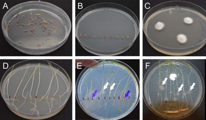
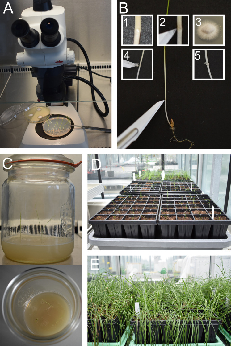
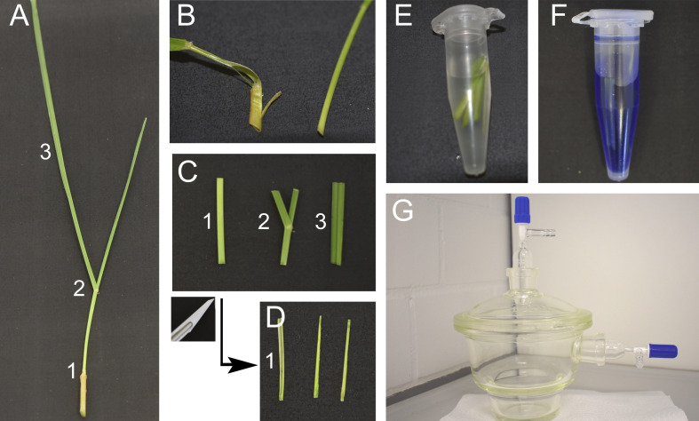
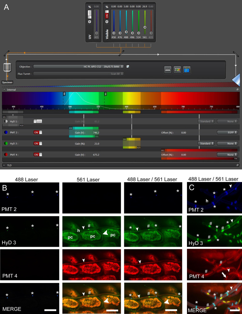
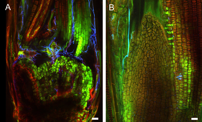
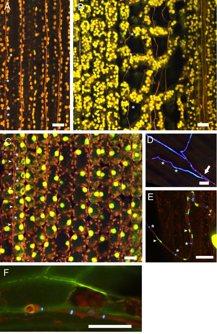
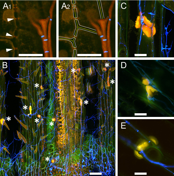
Natural hosts for the fungal endophyte Epichloë festucae include Festuca rubra (fine fescue) and Festuca trachyphylla (hard fescue). Some strains also form stable associations with Lolium perenne (perennial ryegrass). L. perenne is a suitable host to study fungal endophyte-grass interactions, such as endophytic fungal growth within the plant and epiphyllous growth on the plant surface. Here we provide a detailed protocol based on work by, for artificial inoculation of E. festucae into L. perenne, and newly developed staining and visualization techniques for observing endophytic and epiphyllous hyphae and the expressorium, an appressorium-like structure used by the fungus to exit the plant. The staining method uses a combination of glucan binding aniline blue diammonium salt (AB) and chitin binding wheat germ agglutinin-conjugated Alexa Fluor®488 -(WGA-AF488). This protocol will be a useful tool to study Epichloë-grass interactions, particularly the comparison of different Epichloë-grass associations, various endophyte-host developmental stages, as well as the analysis of mutant Epichloë strains.

Keywords: Confocal Laser Scanning Microscopy; Epichloë; Epiphyllous Growth; Expressorium; Fungal endophyte; Inoculation.


Conflict of interest statement

Competing interestsThe authors declare no conflict of interests.

Figures

Figure 1.
Figure 1.. Preparation of perennial ryegrass seeds and Epichloë mycelium.
A. Sterilized and dried seeds. B. Seeds placed on 3% water agar. C. E. festucae Fl1 mycelium on PDA agar grown for one week at 20 °C. D. Germinated ryegrass after one week on 3% water agar. E. Contaminated plate after inoculation. The white arrows indicate E. festucae Fl1 mycelium growing out from inoculation points, the blue arrows indicate contaminations growing out from the seeds. F. Non-contaminated plate one-week post inoculation. The white arrows indicate E. festucae Fl1 mycelium.
Video 1.
Video 1.. Mycelia blocks cut from the colony edge
Video 2.
Video 2.. Mycelia is placed on a scalpel blade and inserted directly after cutting a small slit in the apical meristem
Figure 2.
Figure 2.. Inoculation of perennial ryegrass seedlings and plant cultivation.
A. Set up a dissecting microscope under a laminar flow cabinet. B. Inoculate a small piece of E. festucae mycelium into a small incision made at the SAM, visible as a small knot. C and D. Two weeks after inoculation (one week in the dark, one week in light) plant seedlings in sterile jars or in root trainers in potting mix. E. Check for infection when several tillers have developed.
Video 3.
Video 3.. Preparing leaf samples for clearing and staining
Figure 3.
Figure 3.. Staining of endophytes in perennial ryegrass for CLSM.
A. Cut tillers close to the root. Tillers can be separated in three parts, the pseudostem (1), the area around the ligule containing the blade meristem (2) and the blade (3). B. Peel the outermost leaf sheath and discard. C. Cut a 2 cm sized piece from the base of the pseudostem (1), the area around the ligule (2) and from the blade (3). D. Cut the pseudostem section (1) in half longitudinally. E. Incubate the leaf samples in 95% ethanol overnight and treat with 10% KOH for 3 h, then wash them three times with PBS. F. Prepare the staining solution. G. Vacuum infiltrate the samples.
Video 4.
Video 4.. Vacuum infiltration for staining of endophytes in grass.
Seal the lid by turning the upper stopcock. Open the side stopcock connecting the chamber to the water jet pump. Turn the water on, vacuum is now applied to the chamber. Check after a few seconds that vacuum is established, you cannot slide the lid to the left or right. Wait 2 min. For ventilation of the chamber, close the side stopcock in order to avoid water being sucked into the chamber. Turn the water off. Open the upper stopcock on the lid slowly. You should hear a hissing sound when air flows into the chamber. Repeat the procedure two more times.
Figure 4.
Figure 4.. Confocal laser scanning microscopy (CLSM) of E. festucae in hard fescue.
A. Settings for CLSM (here using a Leica TCS SP8 microscope) of samples stained with WGA-AF488, aniline blue and propidium iodide. B and C. CLS micrographs depicting the different and merged fluorescence signals captured with detectors (PMT 2, PMT 4 and HyD 3) that collect light of defined wave lengths (as shown in A). The laser used to excite the samples is also indicated. (B) Endophytic hyphae (h) of E. festucae Fl1 in between plant cells (pc) of Festuca trachyphylla leaf blade. The emission fluorescence from WGA-AF488 excited by the 488 nm argon ion laser is captured with the PMT 2 and also the HyD 3 detector by crossover of fluorescence emission. WGA-AF488 stains chitin only present in septa (asterisk) of endophytic hyphae, plant cells are not stained. The emission fluorescence from AB, PI and plant (chlorophyll) autofluorescence excited by the 561 nm DPSS laser is captured with the HyD 3 detector. PI stains nuclei (hash) of endophytic hyphae. AB stains β-D-1,3-glucans in fungal (small arrowhead) and plant cell walls (big arrowhead). Autofluorescence of chlorophyll and fungal cytoplasm is captured with the HyD 3 and PMT 4 detector. Note that the green (HyD 3) and red (PMT 4) pseudocolors are combined to orange in the merged images. (C) Epiphyllous hyphae (h) of E. festucae Fl1 on the Festuca trachyphylla blade. WGA-AF488 stains chitin in septa (asterisk) and the cell wall (small arrowhead) of epiphyllous hyphae. The weak WGA-AF488 signal captured with PMT 2 indicates that the hyphae shown here just start unmasking and/or accumulating chitin at the cell wall. The HyD 3 detector captures the PI nuclei signal (hash), the WGA-AF488 signal of septa and cell wall (asterisk) and the AB stained fungal cell wall (small arrowhead). The PMT 4 detector captures the AB stained fungal cell wall (small arrowhead) and the fungal cytoplasm (arrow). Scale bars: B = 20 µm, C = 5 µm.
Figure 5.
Figure 5.. Early infection of perennial ryegrass by E. festucae Fl1.
Samples were taken two weeks after inoculation and stained with WGA-AF488 and AB. A. Image taken at the inoculation side. WGA-AF488 stains chitin in epiphyllous hyphae as well as septa of endophytic hyphae of E. festucae. B. Endophytic hyphae colonizing a leaf primordium. Cell wall glucan of endophytic hyphae stained with AB is shown in orange, septa and epiphyllous hyphae are shown in blue. Scale bars = 20 µm.
Figure 6.
Figure 6.. Endophytic and epiphyllous hyphae of E. festucae associated with perennial ryegrass.
Samples were taken two weeks after inoculation and stained with WGA-AF488 and AB (A, B, D, F) and WGA-AF488, AB and PI (C, E). A. Endophytic hyphae in the leaf sheath. The cell walls of endophytic hyphae are stained with AB and the chitin in septa is stained with WGA-AF488, shown in blue, and marked by asterisks. Autofluorescence of the plant chloroplasts is shown in orange. B. Endophytic hyphae in the leaf blade; note that the hyphae are not straight between the spongy mesophyll. Staining and pseudocolors are as in A. C. Endophytic hyphae in the blade, stained additionally with PI for visualization of nuclei. Fungal nuclei are in yellow and marked by arrows. Plant nuclei are shown in yellow and marked by hash symbols. D. Cell walls of the endophytic hyphae are not stained by WGA-AF488 and so are hyphae after emergence from the plant stained by AB, shown in red pseudocolor and marked by an arrow. Cell walls and septa of epiphyllous hyphae on the leaf sheath contain chitin stained with WGA-AF488, shown in blue, septa are marked by asterisks. E. Epiphyllous hyphae on the leaf blade, fungal nuclei are stained additionally with PI (arrows). Note that the hyphal cell walls are chitin-free shortly after emerging on the outer surface and therefore only septa bind WGA-AF488 and appear blue (asterisks), while the fungal cell wall is stained by AB and shown in red. F. Expressorium near blade meristem rupturing the cuticle, which shows green autofluorescence. Scale bars = 20 µm.
Figure 7.
Figure 7.. Callose production at cell walls of perennial ryegrass.
Samples were taken eight weeks after inoculation and stained with WGA-AF488 and AB. A. E. festucae-independent callose production at plasmodesmata; endophytic hyphae of E. festucae wild type Fl1 are shown in orange, septa in blue; A1. Arrowheads pointing to callose; A2. Artist’s view of A1, in which plant middle lamella and cell walls were added graphically. B. E. festucaenoxA mutant inducing formation of callose papilla (asterisk) where a hypha exits the plant; epiphyllous hyphae are shown in blue. C-E. Callose papillae in E. festucaenoxA mutant (C), in E. festucaenoxB mutant (D), in E. festucaenoxR mutant (E). L. perenne reacts with induction of callose production to all nox mutants in a similar way. Scale bar in A = 10 µm, in B = 100 µm, in C-E = 20 µm.

References

    1. Becker M., Becker Y., Green K. and Scott B.(2016). The endophytic symbiont Epichloë festucae establishes an epiphyllous net on the surface of Lolium perenne leaves by development of an expressorium, an appressorium-like leaf exit structure . New Phytol 211(1): 240-254. - PMC - PubMed
    1. Figueroa-Lopez A. M., Cordero-Ramirez J. D., Quiroz-Figueroa F. R. and Maldonado-Mendoza I. E.(2014). A high-throughput screening assay to identify bacterial antagonists against Fusarium verticillioides . J Basic Microbiol 54 Suppl 1: S125-133. - PubMed
    1. Florea S., Schardl C. L. and Hollin W.(2015). Detection and isolation of Epichloë species, fungal endophytes of grasses . Curr Protoc Microbiol 38: 19.A 11 11-19A 11 24. - PubMed
    1. Latch G. C. M. and Christensen M. J.(1985). Artificial infection of grasses with endophytes. Ann Appl Biol 107(1): 17-24.
    1. Scott B., Becker Y., Becker M. and Cartwright G.(2012). Morphogenesis, growth, and development of the grass symbiont Epichlöe festucae. In: Martín, J. P. and Pietro, A. D.(Eds.). Morphogenesis and Pathogenicity in Fungi. Springer, 243-264.

LinkOut - more resources