. 2021 Jun 24;2(6):989-995.
doi: 10.34067/KID.0000882021.

Ly6chi Infiltrating Macrophages Promote Cyst Progression in Injured Conditional Ift88 Mice

Affiliations

Ly6chi Infiltrating Macrophages Promote Cyst Progression in Injured Conditional Ift88 Mice

Ernald Jules G Aloria et al. Kidney360. .

Abstract

  1. Ly6chi infiltrating macrophage numbers are increased in injured, conditional Ift88 mice, compared with controls.

  2. Loss of Ly6chi infiltrating macrophages slows injury-accelerated cystic disease.

  3. Ly6chi infiltrating macrophages drive cystic disease in non-Pkd1–deficient cystic models.


Conflict of interest statement

M. Mrug reports having consultancy agreements with Chinook, Goldilocks Therapeutics, Natera, Otsuka Corporation, and Sanofi; receiving research funding from Chinook, Goldilocks Therapeutics, Otsuka Corporation, and Sanofi; receiving honoraria from Chinook, Natera, Otsuka Corporations, and Sanofi; and serving as a scientific advisor for—or member of—the PKD Foundation, on the Sanofi STAGED-PKD Steering Committee, and on the advisory board for Santa Barbara Nutrients. All remaining authors have nothing to disclose.

Figures

Figure 1.
Figure 1.
Conditional Ift88 mice have a delayed, but increased, Ly6chi infiltrating macrophage response after injury compared with cilia wild-type (WT) mice. (A) Gating strategy used to identify infiltrating (Inf) and resident (Res) macrophages (ϕ). Infiltrating macrophages were further gated into Ly6chi and Ly6cint/lo subtypes on the basis of differential expression of Ly6c. (B) Quantification of the number of Ly6chi infiltrating macrophages (as a percentage of total sorted cells) at different time points after injury is shown as the mean±SEM. Each dot represents an individual mouse; red dots represent males, blue dots represent females. N=7–12 mice per group. ***P<0.001, two-way ANOVA. FSC-A, forward scatter area; FSC-H, forward scatter height; GR1, GR1 antibody; IR, ischemia reperfusion.
Figure 2.
Figure 2.
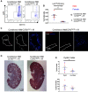
CCR2 deficiency significantly decreases cyst severity in adult-induced, injured, conditional Ift88 mice. (A) Representative flow cytometry plots of infiltrating macrophages isolated from injured, conditional Ift88 mice on the CCR2cont or CCR2rfp/rfp knockout background 56 days postinjury. (B) Quantification of the number of Ly6chi infiltrating macrophages (as a percentage of total sorted cells) from each group is shown as the mean±SEM. For quantification, each dot represents an individual mouse (two replicates, six mice per group). (C) Representative confocal images of a cystic region from injured, conditional Ift88 CCR2cont and injured, conditional Ift88 CCR2rfp/rfp mice. Sections were stained with F4/80 (white) and Hoescht (dark blue). N=3 animals per group. Images were captured with a 20× objective. (D) Kidneys from conditional Ift88 mice on the CCR2cont or CCR2rfp/rfp knockout background were harvested 56 days after renal injury, and the cystic index and number of renal cysts were quantified using hematoxylin and eosin–stained sections and ImageJ software. (E) Quantification of cystic index (cystic area/total kidney area) and cystic number for each group is shown as the mean±SEM. For quantification, each dot represents an individual mouse. Three replicates, eight to ten mice per group. Red dots represent males, blue dots represent females. Starred points indicate the mice corresponding to the hematoxylin and eosin–stained sections shown in (D). *P<0.05, **P<0.01, t test.

References

    1. Yoder BK, Hou X, Guay-Woodford LM: The polycystic kidney disease proteins, polycystin-1, polycystin-2, polaris, and cystin, are co-localized in renal cilia. J Am Soc Nephrol 13: 2508–2516, 2002. 10.1097/01.ASN.0000029587.47950.25 - DOI - PubMed
    1. Karihaloo A, Koraishy F, Huen SC, Lee Y, Merrick D, Caplan MJ, Somlo S, Cantley LG: Macrophages promote cyst growth in polycystic kidney disease. J Am Soc Nephrol 22: 1809–1814, 2011. 10.1681/ASN.2011010084 - DOI - PMC - PubMed
    1. Swenson-Fields KI, Vivian CJ, Salah SM, Peda JD, Davis BM, van Rooijen N, Wallace DP, Fields TA: Macrophages promote polycystic kidney disease progression. Kidney Int 83: 855–864, 2013. 10.1038/ki.2012.446 - DOI - PMC - PubMed
    1. Zimmerman KA, Bentley MR, Lever JM, Li Z, Crossman DK, Song CJ, Liu S, Crowley MR, George JF, Mrug M, Yoder BK: Single-cell RNA sequencing identifies candidate renal resident macrophage gene expression signatures across species. J Am Soc Nephrol 30: 767–781, 2019. 10.1681/ASN.2018090931 - DOI - PMC - PubMed
    1. Schulz C, Gomez Perdiguero E, Chorro L, Szabo-Rogers H, Cagnard N, Kierdorf K, Prinz M, Wu B, Jacobsen SE, Pollard JW, Frampton J, Liu KJ, Geissmann F: A lineage of myeloid cells independent of Myb and hematopoietic stem cells. Science 336: 86–90, 2012. 10.1126/science.1219179 - DOI - PubMed

Publication types