Review
. 2020 Jun 16;2(2):270-292.
doi: 10.1016/j.jaccao.2020.04.011. eCollection 2020 Jun.

Magnetic Resonance Imaging to Detect Cardiovascular Effects of Cancer Therapy: JACC CardioOncology State-of-the-Art Review

Affiliations
Review

Magnetic Resonance Imaging to Detect Cardiovascular Effects of Cancer Therapy: JACC CardioOncology State-of-the-Art Review

Iwan Harries et al. JACC CardioOncol. .

Abstract

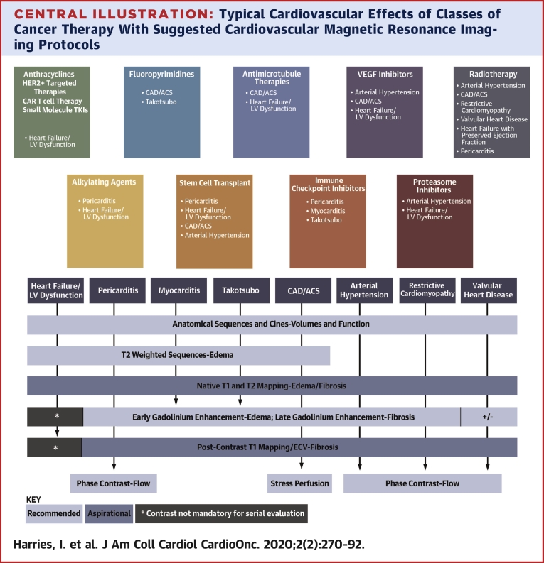
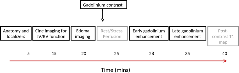
This paper aims to empower and inform cardio-oncologists by providing a practical guide to the clinical application of cardiac magnetic resonance (CMR) in the rapidly evolving field of cardio-oncology. Specifically, we describe how CMR can be used to assess the cardiovascular effects of cancer therapy. The CMR literature, relevant societal guidelines, indication-specific imaging protocols, and methods to overcome some of the challenges encountered in performing and accessing CMR are reviewed.

Keywords: CMR, cardiac magnetic resonance; ECV, extracellular volume fraction; EGE, early gadolinium enhancement; ICI, immune checkpoint inhibitors; LGE, late gadolinium enhancement; LVEF, left ventricular ejection fraction; MACE, major adverse cardiac event; SSFP, balanced steady state free precession; cardio-oncology; cardiotoxicity; cardiovascular magnetic resonance; chemotherapy; left ventricular dysfunction; tissue characterization.


Figures

None
Graphical abstract
Central Illustration
Typical Cardiovascular Effects of Classes of Cancer Therapy With Suggested Cardiovascular Magnetic Resonance Imaging Protocols The typical cardiovascular effects of various classes of cancer therapy, together with suggested cardiovascular magnetic resonance (CMR) imaging protocols and key metrics provided by each sequence. “Recommended” and “aspirational” CMR imaging sequences to characterize the respective pathologies are suggested. For specific data on the timing and incidence of toxicity, please see relevant literature (1,66,72,80,81,91,110, 111, 112, 113). ∗Contrast administration may not necessarily be required for serial evaluation of left ventricular function. ACS = acute coronary syndrome; CAD = coronary artery disease; ECV = extracellular volume fraction; HER2+ = human epidermal growth factor receptor; LV = left ventricular; TKI = tyrosine kinase inhibitor; VEGF = vascular endothelial growth factor.
Figure 1
Figure 1
Typical Composition of a CMR Imaging Protocol CMR protocols are composed of different imaging sequences and tailored to the clinical question posed. Example additional imaging sequences are represented in grey. Native (non-contrast) and contrast-enhanced sequences occur before and after gadolinium administration, respectively. CMR = cardiovascular magnetic resonance; LV = left ventricle; RV = right ventricle.
Figure 2
Figure 2
Example Anatomical CMR Sequences Sequential axial black blood (A to D) and coronal white blood (E to H) images through the thorax and upper abdomen. Abbreviation as in Figure 1.
Figure 3
Figure 3
Abnormalities Detected on Anatomical Sequences (A) T1-weighted black blood turbo spin echo showing diffuse pericardial thickening (arrows). (B) White blood T2-weighted axial sequence showing bilateral pleural effusions (arrows) due to constrictive pericarditis (same patient A). (C) Incidental right lung tumor (arrow). (D) Incidental right renal tumor (arrow).
Figure 4
Figure 4
Steady-State Free Precession “Cine” Images of the Left Ventricle Example long-axis views: (A) 4-chamber, (B) 2-chamber, and (C) 3-chamber. (D) A stack of short axis “slices” from base to apex.
Figure 5
Figure 5
Feature Tracking Strain Overlay on Steady-State Free Precession Images of the Left Ventricle Diastole (A to D) and systole (E to H). A and E show a 2-chamber view. B and F show a 4-chamber view. C and G show a mid-ventricular short axis view. D and H show a composite 3-dimensional reconstruction. Red line represents endocardial border; green represents epicardial border; and yellow represents tracked myocardial features.
Figure 6
Figure 6
Examples of Intracardiac Thrombus on CMR (A) Axial SSFP showing PICC-associated thrombus prolapsing through the tricuspid valve (arrow). (B) T1-weighted EGE image showing marked hypointensity of the same thrombus (arrow). (C) RV 2-chamber cine showing 2 separate thrombi (arrows) in the same patient. (D) T1-weighted RV 2-chamber EGE image of the same patient, with thrombus indicated by an arrow. EGE = early gadolinium enhancement; PICC = peripherally inserted central catheter; SSFP = steady-state free precession; other abbreviations as in Figure 1.
Figure 7
Figure 7
Patterns of Myocardial Scarring in Post-Contrast Images (Late Myocardial Enhancement) T1-weighted post-contrast short-axis mid ventricular images. (A) Ischemic LGE (arrows) due to transmural infarction in the right coronary artery territory. (B) Mid-myocardial LGE in dilated cardiomyopathy (arrow). (C) Subepicardial LGE (arrows). LGE = late gadolinium enhancement.
Figure 8
Figure 8
Native Short-Axis Mid-Ventricular T1 Map (A) Normal native myocardial measurements (1,010 ms) and (B) abnormal measurements (1,136 ms) in the mid-ventricular septum.
Figure 9
Figure 9
Overlapping Symptomatology of Cardiovascular Effects of Cancer Therapy Cardiovascular effects of cancer therapy can manifest with a variety of cardiovascular symptoms and occur in the context of cancer and cancer therapy, which can themselves cause similar symptoms, independent of cardiovascular disease, highlighting the diagnostic challenges faced by cardio-oncologists. ACS = acute coronary syndrome; CAD = coronary artery disease.
Figure 10
Figure 10
CMR and Angiographic Case Example of Ischemic Heart Disease (A) T1-weighted mid-ventricular short-axis stress perfusion image showing an extensive inducible perfusion defect in the inferior and inferoseptal walls (arrows). (B) T1-weighted short-axis image showing minimal scar according to small volume of LGE (arrows). (C) Coronary angiography of the distally occluded (∗) dominant right coronary artery. (D) Coronary angiography of the unobstructed left coronary artery. The left circumflex is recessive and the posterior descending and posterior left ventricular branches of the right coronary artery are missing. Abbreviations as in Figures 1 and 7.
Figure 11
Figure 11
CMR Case Example of Pericardial Constriction Real-time short-axis mid-ventricular SSFP cine imaging during deep breathing (A) during expiration, and (B) at onset of inspiration. Note the D-shaped LV due to septal flattening (arrow). (C) Axial phase-contrast imaging of the inferior vena cava, with a flow pattern (D) showing rapid and steep y descent with flow reversal in diastole (arrow) reminiscent of the jugular venous waveform encountered in pericardial constriction. Abbreviations as in Figures 1 and 6.

Similar articles

Cited by

References

    1. Zamorano J.L., Lancellotti P., Rodriguez Muñoz D. 2016 ESC Position Paper on cancer treatments and cardiovascular toxicity developed under the auspices of the ESC Committee for Practice Guidelines: The Task Force for cancer treatments and cardiovascular toxicity of the European Society of Cardiology (ESC) Eur Heart J. 2016;37:2768–2801. - PubMed
    1. Doherty J.U., Kort S., Mehran R. ACC/AATS/AHA/ASE/ASNC/HRS/SCAI/SCCT/SCMR/STS 2019 appropriate use criteria for multimodality imaging in the assessment of cardiac structure and function in nonvalvular heart disease: a report of the American College of Cardiology Appropriate Use Criteria Task Force, American Association for Thoracic Surgery, American Heart Association, American Society of Echocardiography, American Society of Nuclear Cardiology, Heart Rhythm Society, Society for Cardiovascular Angiography and Interventions, Society of Cardiovascular Computed Tomography, Society for Cardiovascular Magnetic Resonance, and the Society of Thoracic Surgeons. J Am Coll Cardiol. 2019;73:488–516. - PubMed
    1. Kramer C.M., Barkhausen J., Bucciarelli-Ducci C., Flamm S.D., Kim R.J., Nagel E. Standardized cardiovascular cardiac magnetic resonance (CMR) protocols: 2020 update. J Cardiovasc Magn Reson. 2020;22:17. - PMC - PubMed
    1. Schulz-Menger J., Bluemke D.A., Bremerich J. Standardized image interpretation and post-processing in cardiovascular magnetic resonance — 2020 update: Society for Cardiovascular Magnetic Resonance (SCMR): Board of Trustees Task Force on Standardized Post-Processing. J Cardiovasc Magn Reson. 2020;22:19. - PMC - PubMed
    1. Hundley W.G., Bluemke D., Bogaert J.G. Society for Cardiovascular Magnetic Resonance guidelines for reporting cardiovascular magnetic resonance examinations. J Cardiovasc Magn Reson. 2009;11:5. - PMC - PubMed