. 2021 Oct:368:104422.
doi: 10.1016/j.cellimm.2021.104422. Epub 2021 Aug 8.

Lactic acid suppresses MRGPRX2 mediated mast cell responses

Affiliations

Lactic acid suppresses MRGPRX2 mediated mast cell responses

Meesum Syed et al. Cell Immunol. 2021 Oct.

Abstract

MAS related G-protein coupled receptor X2 (MRGPRX2) is a G-protein coupled receptor (GPCR) expressed in human mast cells that has been implicated to play an important role in causing pseudo-allergic reactions as well as exacerbating inflammation during asthma and other allergic diseases. Lactic acid, a byproduct of glucose metabolism, is abundantly present in inflamed tissues and has been shown to regulate functions of several immune cells. Because the endogenous ligands for MRGPRX2 (substance P and LL-37) are elevated during pathologic conditions, such as cancer and asthma, and given that lactic acid levels are also enhanced in these patients, we explored the role of lactic acid in regulating mast cells response via MRGPRX2 and MrgprB2, the mouse orthologue of the human receptor. We found that lactic acid suppressed both the early (Ca2+ mobilization and degranulation) and late (chemokine/cytokine release) phases of mast cell activation; this data was confirmed in LAD2, human skin and mouse peritoneal mast cells. In LAD2 cells, the reduction in degranulation and chemokine/cytokine production mediated by lactic acid was dependent on pH. In agreement with our in vitro studies, lactic acid also reduced passive systemic anaphylaxis to compound 48/80 (a known MRGPRX2/MrgprB2 ligand) and skin inflammation in a mouse model of rosacea that is dependent on MrgprB2 expression on skin mast cells. Our data thus suggest that lactic acid may serve to inhibit mast cell-mediated inflammation during asthma and reduce immune response during cancer by affecting mast cell activation through MRGPRX2.

Keywords: Lactic acid; MAS-related G-protein coupled receptor-X2 (MRGPRX2); Mast cells; MrgprB2; Pseudo-allergic reactions.


Conflict of interest statement

CONFLICT OF INTEREST

The authors have no conflict of interest to declare.

Figures
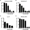
Figure 1.
Figure 1.. Intracellular Ca2+ mobilization induced by MRGPRX2 ligands is reduced by lactic acid in HEK 293T cells expressing MRGPRX2.
HEK-293T cells stably expressing the MRGPRX2 receptor were treated with vehicle (SIR buffer) or lactic acid (indicated concentrations) for 30 min. (A-D) Ca2+ mobilization assays were performed following incubation with (A) compound 48/80 (300 ng/ml), (B) substance P (100 nM), (C) LL-37 (0.5 μM) or (D) (R)-Zinc 3573 (0.5 μM). Data are plotted as the change in fluorescence intensity [minimum (Min) subtracted from maximum (Max) value] measurements. Results shown are mean ± S.E. of 3 independent experiments. Statistical significance was determined by unpaired Student’s t-test and values from the lactic acid-treated group was compared with the vehicle group. * p <0.05, ** p <0.01 and *** p <0.001.
Figure 2.
Figure 2.. Lactic acid inhibits MRGPRX2-induced Ca2+ mobilization and degranulation in LAD2 mast cells.
(A-D) Intracellular Ca2+ mobilization and (E-H) degranulation in LAD2 human mast cells was determined following pre-incubation with vehicle (SIR buffer) or varying concentrations of lactic acid for 30 min. Cells were stimulated with the MRGPRX2 agonist (A) compound 48/80 (300 ng/ml), (B) substance P (300 nM), (C) LL-37 (3 μM) or (D) (R)-Zinc 3573 (2 μM) and changes in fluorescence intensities were recorded for 90 sec. Data are plotted as the change in fluorescence intensity values following ligand additions. Vehicle- or lactic acid-treated cells were exposed to (E) compound 48/80 (300 ng/ml), (F) substance P (300 nM), (G) LL-37 (3 μM) or (H) (R)-Zinc 3573 (2 μM) and degranulation was quantified by β-hexosaminidase release. Values are plotted as percentages of total cell lysate β-hexosaminidase content. Data shown are mean ± S.E. of 3 independent experiments. Statistical significance was determined by unpaired Student’s t-test with valúes compared between the lactic acid- and vehicle-treated groups. * p <0.05, ** p <0.01 and *** p <0.001.
Figure 3.
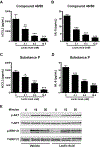
Figure 3.. Lactic acid suppresses cytokine/chemokine production but does not affect ERK1/2 or AKT activation in LAD2 cells.
(A-F) LAD2 cells (0.15 × 10⁶ cells/well) were exposed to vehicle (SIR buffer) or different concentrations of lactic acid for 30 min, and washed and exposed to, (A, B) compound 48/80 (300 ng/ml) or (C, D) substance P (300 nM) for 6 h. Supernatants were collected and analyzed for (A, C) CCL2 and (B, D) IL-8 by ELISA. Data shown are mean ± S.E. of 3 independent experiments performed in triplicates. Statistical analysis was done using unpaired Student’s t-test by comparing the vehicle- and lactic acid-treated groups. * p <0.05, ** p <0.01 and *** p <0.001. (E) LAD2 cells were treated with vehicle (SIR buffer) or lactic acid (8.3 mM) and then exposed compound 48/80 (300 ng/ml) for different time intervals. Western blotting was performed to detect phosphorylated ERK1/2 (p-ERK1/2) or AKT (p-AKT) proteins. The blots probed with ERK1/2 (T-ERK1/2) and AKT (T-AKT) antibodies for loading controls. Images of representative blots from 3 independent experiments are shown.
Figure 4.
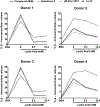
Figure 4.. Lactic acid attenuates MRGPRX2-mediated degranulation in human skin mast cells.
Human skin-derived mast cells from 4 different donors were pre-treated with vehicle (SIR buffer) or lactic acid (8.3 mM and 10.4 mM) and exposed to compound 48/80 (1000 ng/ml), substance P (1 pM), LL-37 (5 μM) or (R)-Zinc 3573 (10 μM). Line graphs show degranulation of mast cells from different donors as estimated by β-hexosaminidase release in supernatants.
Figure 5.
Figure 5.. The acidic proton moeity in lactic acid contributes to its effects on degranulation of LAD2 cells to MRGPRX2 agonists.
LAD2 cells were exposed to vehicle (SIR buffer, control) or varying concentrations of lactic acid or sodium lactate for 30 mins. The cells were washed and stimulated with the MRGPRX2 agonist (A) compound 48/80 (300 ng/ml), (B) substance P (300 nM). Degranulation was quantified by β-hexosaminidase release. Values are plotted as percentages of total cell lysate β-hexosaminidase content. Data shown are mean ± S.E. of 3 independent experiments. Statistical significance was determined by unpaired Student’s t-test with values. *** p <0.001 indicates values compared between the lactic acid- and vehicle-treated groups. ††† p <0.001 indicates values compared between the sodium lactate- and vehicle-treated groups.
Figure 6.
Figure 6.. Sodium lactate has no effect on chemokine/cytokine production in LAD2 cells.
LAD2 cells were exposed to vehicle (SIR buffer, indicated by “0”) or different concentrations of lactic acid or sodium lactate, washed and exposed to, (A, B) compound 48/80 (300 ng/ml) or (C, D) substance P (300 nM) for 6 h. Supernatants were collected and analyzed for (A, C) CCL2 and (B, D) IL-8 by ELISA. Data shown are mean ± S.E. of 3 independent experiments performed in triplicates. Statistical analysis was done using unpaired Student’s t-test by comparing the control vehicle- and lactic acid-treated groups. *** p <0.001 indicates values compared between the lactic acid- and vehicle-treated groups. p <0.05 and ††† p <0.001 indicates values compared between the sodium lactate- and vehicle-treated groups.
Figure 7.
Figure 7.. Lactic acid reduces systemic anaphylaxis to compound 48/80 in vivo.
(A) Vehicle-(PBS) or lactic acid- (125 mg/kg) treated C57BL/6 mice were exposed to compound 48/80, (C48/80, 3.75 mg/kg) and rectal temperature was measured at different time points. Line graph shows change in rectal temperature for the different groups of mice. Data shown are mean ± S.E. from 3 experiments (a total of n=10 mice/group). Statistical significance was determined by Student’s t-test. (B-D) Mouse peritoneal cells were exposed to vehicle (SIR buffer) or lactic acid (8.3 mM) and stimulated with the indicated concentrations of (B) compound 48/80, (C) substance P or (D) LL-37. Graphs shows degranulation of mast cells as estimated by β-hexosaminidase release in supernatants. Data shown are mean ± S.E. from 3 experiments (a total of n=3–5 mice/group). Statistical significance was determined by unpaired Student’s t-test. *** p <0.001.
Figure 8.
Figure 8.. Lactic acid prevents the development of LL-37-induced rosacea in mice.
Vehicle-(PBS, LL-37/Vehicle) or lactic acid (LL-37/Lactic acid) treated Balb/c mice were injected with LL-37 into the dorsal skin twice daily for 2 consecutive days. Mice that only received PBS on the dorsal skin were used as control. (A) H&E stained skin sections of mice from different cohorts are shown. Scale bar = 100 μm. (B) Graph represents epidermal thickness of the H&E stained skin sections as measured by Image J. (C, D) mRNA expression of selected gene targets from the excised skin was analyzed by real-time PCR. Values are plotted as fold change (2−ΔΔCt) normalized to GAPDH levels. (E) The paraffin embedded skin sections from different cohorts of mice were stained with toludine blue to detect mast cells. Representiative pictures of the skin sections are shown. Bold closed arrowheads indicate intact mast cells whereas open arrowheads represent degranulated mast cells. The inset figure is an enlarged image of the mast cell(s) shown in the pictures. Scale bar = 100 μm. (F) Graph shows the percentage of degranulated mast cells in the skin tissue of different cohorts of mice. Data are mean ± S.E. from n=3–9 mice/group. Statistical significance was determined by unpaired Student’s t-test comparing the vehicle- vs lactic acid-treated groups. * p <0.05 and ** p<0.01.

References

    1. Blank U The mechanisms of exocytosis in mast cells. Adv Exp Med Biol 2011; 716: 107–22. - PubMed
    1. Mukai K, Tsai M, Saito H, Galli SJ. Mast cells as sources of cytokines, chemokines, and growth factors. Immunol Rev 2018; 282(1): 121–50. - PMC - PubMed
    1. Galli SJ, Tsai M. IgE and mast cells in allergic disease. Nat Med 2012; 18(5): 693–704. - PMC - PubMed
    1. Elieh Ali Komi D, Shafaghat F, Kovanen PT, Meri S. Mast cells and complement system: Ancient interactions between components of innate immunity. Allergy 2020; 75(11): 2818–28. - PubMed
    1. Subramanian H, Gupta K, Ali H. Roles of Mas-related G protein-coupled receptor X2 on mast cell-mediated host defense, pseudoallergic drug reactions, and chronic inflammatory diseases. JAllergy Clin Immunol 2016; 138(3): 700–10. - PMC - PubMed

Publication types