. 2021 Aug 16;19(1):243.
doi: 10.1186/s12957-021-02349-5.

Iliosacral Bone Tumor Resection Using Cannulated Screw-Guided Gigli Saw - A Novel Technique

Affiliations

Iliosacral Bone Tumor Resection Using Cannulated Screw-Guided Gigli Saw - A Novel Technique

Tao Ji et al. World J Surg Oncol. .

Abstract

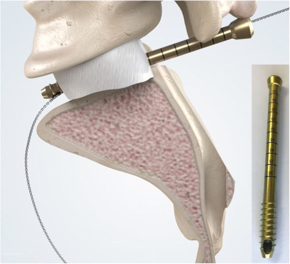
Background: Adequate margins are technically difficult to achieve for malignant tumors involving the sacroiliac joint due to limited accessibility and viewing window. In order to address the technical difficulties faced in iliosacral tumor resection, we proposed a technique for precise osteotomy, which involved the use of canulated screws and Gigli saw (CSGS) that facilitated directional control, anteroposterior linkage of resection points and adequate surgical margins. The purpose of the current study was to evaluate whether CSGS technique facilitated sagittal osteotomy at sacral side, and were adequate surgical margins achieved? Also functional and oncological outcomes was determined along with the noteworthy complications.

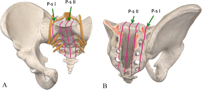
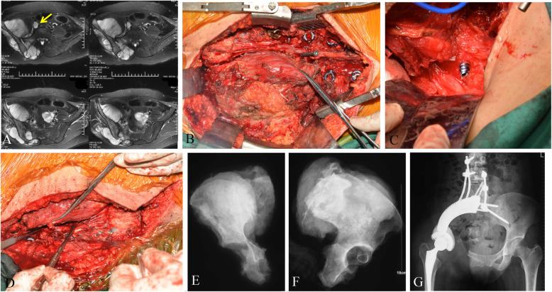
Methods: From April 2018 to November 2019, we retrospectively reviewed 15 patients who underwent resections for primary tumors of pelvis or sacrum necessitating iliosacral joint removal using the proposed CSGS technique. Chondrosarcoma was the most common diagnosis. The osteotomy site within sacrum was at ipsilateral ventral sacral foramina in 8 cases, midline of sacrum in 5 cases, and contralateral ventral sacral foramina and sacral ala with 1 case each. The average intraoperative blood loss was 3640 mL (range, 1200 and 6000 mL) with a mean operation duration of 7.4 hours (range, 5 to 12 hours). The mean follow-up was 23.0 months (range, 18 and 39 months) for alive patients.

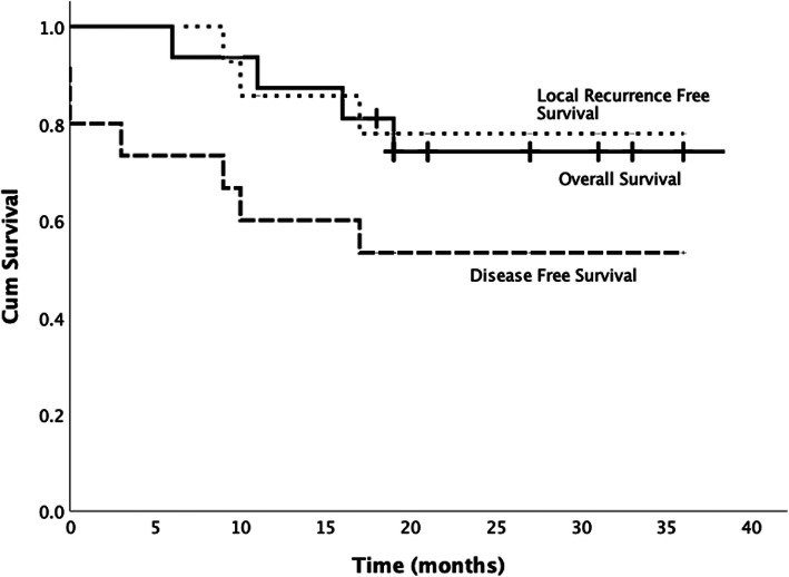
Results: Surgical margins were wide in 12 patients (80%), wide-contaminated in 1 patient (6.7%), and marginal in 2 patients (13.3%). R0 resection was achieved in 12 (80%) patients and R1 resection in 3 patients. There were three local recurrences (20%) occurred at a mean time of 11 months postoperatively. No local recurrence was observed at sacral osteotomy. The overall one-year and three-year survival rate was 86.7% and 72.7% respectively.Complications occurred in three patients.

Conclusions: The current study demonstrated that CSGS technique for tumor resection within the sacrum and pelvis was feasible and can achieve ideal resection accuracies. The use of CSGS was associated with high likelihood of negative margin resections in the current series. Intraoperative use of CSGS appeared to be technically straightforward and allowed achievement of planned surgical margins. It is worthwhile to consider the use of CSGS technique in resection of pelvic tumors with sacral invasion and iliosacral tumors, however further follow-up at mid to long-term is warranted to observe local recurrence rate.

Keywords: iliosacral tumor; limb salvage; osteotomy; surgical margin.


Conflict of interest statement

The authors declare that they have no competing interests.

Figures

Fig. 1
Fig. 1
A and B. The typical osteotomy sites at sacral side during iliosacral tumor resection according to the reported classification system [31]. Both anterior view (A) and posterior view (B) were shown
Fig. 2
Fig. 2
A-K. An illustrative case (patient no. 2) of CSGS technique. Preoperative MRI after neo-adjuvant chemotherapy showed tumor extent (A). A canulated screw was placed at middle point of L5/S1 level without penetration of the anterior cortex under fluoroscopic guidance (B). Penetration of the cannulated screw through the anterior cortex was confirmed by finger palpation by a pedicle sound (C). D and E showed a Gigli saw was introduced through the canulated screw and the planned osteotomy site (dash line on figure E). Then the iliac osteotomy was carried out through posterior approach (F and G). The tumor was removed after resection of ipsilateral half of L5/S1 disk (H). The specimen was showed (I and J). Postoperative X-ray showed osteotomy sites and reconstruction
Fig. 3
Fig. 3
A-D. The follow-up at one and half year of patient no. 2. The patient can walk independently with mild gait abnormal. Pelvic x-ray were shown. The transverse CT scan (B, C and D) of different levels showed bony cut at both sacrum and ilium
Fig. 4
Fig. 4
A-G. The number 10 patient was diagnosed of chondrosarcoma in right pelvis invading sacrum. Preoperative MRI (A) showed tumor thrombus in iliac vein (arrow). Following sacral laminectomy, affected nerve roots (S1-3) were identified and ligated during posterior approach. Then the canulated screw as placed (B). Intraoperatvie photo of anterior approach (C) showed the canulated screw advanced through the anterior sacrum under direct observation. Then the Gigli saw was placed and sagittal osteotomy at sacrum was performed (D). X-rays (E and F) of the specimen were shown. A custom-made pelvic endoprosthesis combined with SRS were used to reconstruct the defect (G)
Fig. 5
Fig. 5
A modified canulated screw was developed with purpose of direction-control of the Gigli saw. This can be helpful for posterior approach only procedures
Fig. 6
Fig. 6
The Kaplan-Meier survival curves shows overall survival, local-recurrence-free survival and disease-free survival

References

    1. Zhang Y, Guo W, Tang X, Yang R, Ji T, Yang Y, Wang Y, Wei R. En bloc resection of pelvic sarcomas with sacral invasion: a classification of surgical approaches and outcomes. Bone Joint J. 2018;100-B(6):798–805. doi: 10.1302/0301-620X.100B6.BJJ-2017-1212.R1. - DOI - PubMed
    1. Court C, Bosca L, Le Cesne A, Nordin JY, Missenard G. Surgical excision of bone sarcomas involving the sacroiliac joint. Clin Orthop Relat Res. 2006;451:189–194. doi: 10.1097/01.blo.0000229279.58947.91. - DOI - PubMed
    1. Ozaki T, Rodl R, Gosheger G, Hoffmann C, Poremba C, Winkelmann W, Lindner N. Sacral infiltration in pelvic sarcomas: joint infiltration analysis II. Clin Orthop Relat Res. 2003;407:152–158. doi: 10.1097/00003086-200302000-00023. - DOI - PubMed
    1. Ozaki T, Lindner N, Hillmann A, Link T, Winkelmann W. Transarticular invasion of iliopelvic sarcomas into the sacrum. Radiological analysis of 47 cases. Acta Orthop Scand. 1997;68(4):381–383. doi: 10.3109/17453679708996181. - DOI - PubMed
    1. Starantzis KA, Sakellariou VI, Rose PS, Yaszemski MJ, Papagelopoulos PJ. A new type of reconstruction of the hemipelvis after type 3 amputative sacrectomy using pedicled fibula: technical note. J Neurosurg Spine. 2014;21(2):195–202. doi: 10.3171/2014.1.SPINE13245. - DOI - PubMed