. 2021 Aug 16;16(1):150.
doi: 10.1186/s13014-021-01865-8.

Investigation of the clinical inter-observer bias in prostate fiducial marker image registration between CT and MR images

Affiliations

Investigation of the clinical inter-observer bias in prostate fiducial marker image registration between CT and MR images

Emilia Persson et al. Radiat Oncol. .

Abstract

Background and purpose: Inter-modality image registration between computed tomography (CT) and magnetic resonance (MR) images is associated with systematic uncertainties and the magnitude of these uncertainties is not well documented. The purpose of this study was to investigate the potential uncertainty of gold fiducial marker (GFM) registration for localized prostate cancer and to estimate the inter-observer bias in a clinical setting.

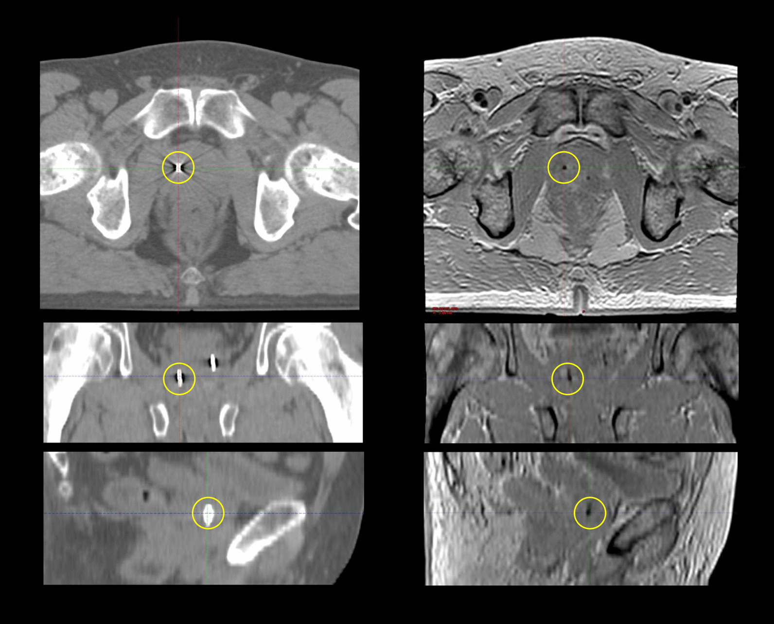
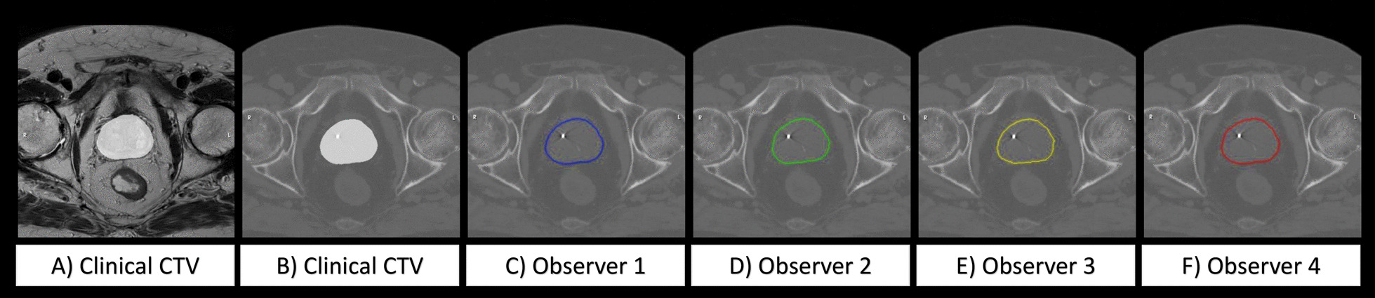
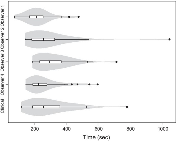
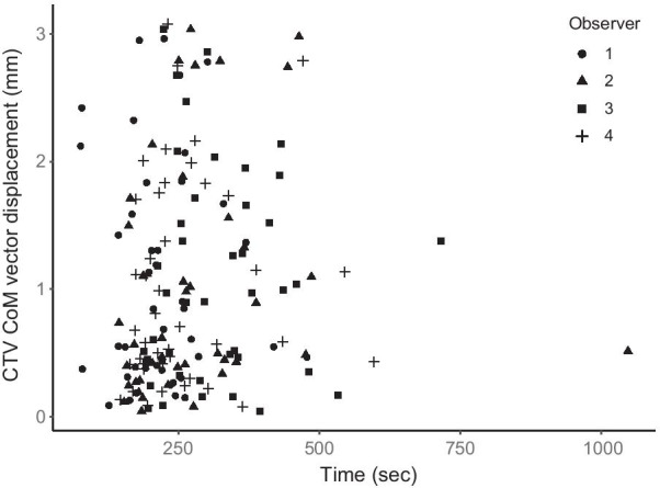
Methods: Four experienced observers registered CT and MR images for 42 prostate cancer patients. Manual GFM identification was followed by a landmark-based registration. The absolute difference between observers in GFM identification and the displacement of the clinical target volume (CTV) was investigated. The CTV center of mass (CoM) vector displacements, DICE-index and Hausdorff distances for the observer registrations were compared against a clinical baseline registration. The time allocated for the manual registrations was compared.

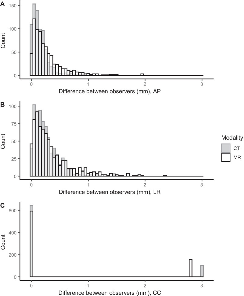
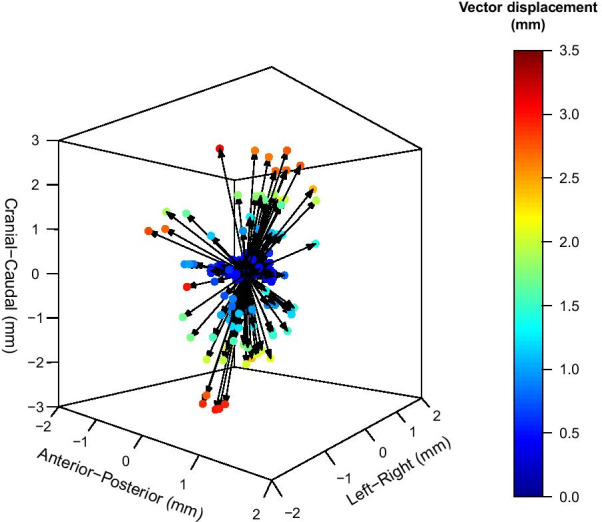
Results: Absolute difference in GFM identification between observers ranged from 0.0 to 3.0 mm. The maximum CTV CoM displacement from the clinical baseline was 3.1 mm. Displacements larger than or equal to 1 mm, 2 mm and 3 mm were 46%, 18% and 4%, respectively. No statistically significant difference was detected between observers in terms of CTV displacement. Median DICE-index and Hausdorff distance for the CTV, with their respective ranges were 0.94 [0.70-1.00] and 2.5 mm [0.7-8.7].

Conclusions: Registration of CT and MR images using GFMs for localized prostate cancer patients was subject to inter-observer bias on an individual patient level. A CTV displacement as large as 3 mm occurred for individual patients. These results show that GFM registration in a clinical setting is associated with uncertainties, which motivates the removal of inter-modality registrations in the radiotherapy workflow and a transition to an MRI-only workflow for localized prostate cancer.

Keywords: Computed tomography; Fiducial marker; Image registration; Inter-observer; Magnetic resonance imaging; Prostate cancer; Registration uncertainty.


Conflict of interest statement

The authors declare that they have no competing interests.

Figures

Fig. 1
Fig. 1
CT images (left) and multi echo gradient echo (MEGRE) MR images (right) used for gold fiducial marker (GFM) registration. The GFM, highlighted in yellow circles, generate a streak artefact in the CT images and a signal void in the MR images. Images are shown in transversal (top row), coronal (middle row, calculated projection) and sagittal (bottom row, calculated projection) planes
Fig. 2
Fig. 2
An example of a patient where the clinical target volume (CTV) has been transferred from the MR geometry (A) to the CT geometry (BF). All images are shown in the corresponding transversal slice through the center of the prostate volume. The clinical CTV is illustrated as a white structure in the center on the MR image (A) and on the CT image (B). The corresponding CTV is shown for observer 1, 2, 3 and 4 in blue, green, yellow and red contours (CF)
Fig. 3
Fig. 3
Absolute difference for each identified gold fiducial marker (GFM) center of mass (CoM) between observers, displayed in anterior-posterior (A), left-right (B) and cranio-caudal (C) directions. On the x-axis, the absolute difference between observers in mm is shown for identification in CT images (dark gray) and MR images (white with black outline). On the y-axis the number of observations in each bin are shown. Bin width was 0.05 mm for all graphs. Bins with an overlap of the CT and MR are colored in light gray
Fig. 4
Fig. 4
Clinical target volume (CTV) center of mass (CoM) vector displacement from the clinical baseline. The CTV CoM vector displacements for 167 observer registrations are displayed as colored dots ranging from dark blue (0 mm vector displacement) to dark red (3.5 mm vector displacement)
Fig. 5
Fig. 5
Time required for the image registrations. The times for the observer registrations are shown in order 1 to 4 from the top and at the bottom row the clinical baseline registration time is shown. The figure includes a boxplot overlaid on a violin plot. The medians are defined as solid black lines in the boxes and outliers as solid black dots. The width of the violin plot indicates the frequency
Fig. 6
Fig. 6
The relation between registration time and the clinical target volume (CTV) center of mass (CoM) vector displacement for observer 1 (circles), observer 2 (triangles), observer 3 (squares) and observer 4 (plus signs). Time is displayed in seconds on the x-axis and the CTV CoM vector displacement in mm on the y-axis. All observers were represented in the range of displacements and times. Observer 1 was clearly in the lower range of registration times, also seen in Fig. 5, but was not exclusively part of the lower nor higher range of CTV displacements

References

    1. Salembier C, Villeirs G, De Bari B, Hoskin P, Pieters BR, Van Vulpen M, et al. ESTRO ACROP consensus guideline on CT- and MRI-based target volume delineation for primary radiation therapy of localized prostate cancer. Radiother Oncol. 2018;127(1):49–61. doi: 10.1016/j.radonc.2018.01.014. - DOI - PubMed
    1. Korsager AS, Carl J, Riis Ostergaard L. Comparison of manual and automatic MR-CT registration for radiotherapy of prostate cancer. J Appl Clin Med Phys. 2016;17(3):294–303. doi: 10.1120/jacmp.v17i3.6088. - DOI - PMC - PubMed
    1. McLaughlin PW, Narayana V, Kessler M, McShan D, Troyer S, Marsh L, et al. The use of mutual information in registration of CT and MRI datasets post permanent implant. Brachytherapy. 2004;3(2):61–70. doi: 10.1016/j.brachy.2004.06.001. - DOI - PubMed
    1. Roberson PL, McLaughlin PW, Narayana V, Troyer S, Hixson GV, Kessler ML. Use and uncertainties of mutual information for computed tomography/ magnetic resonance (CT/MR) registration post permanent implant of the prostate. Med Phys. 2005;32(2):473–82. doi: 10.1118/1.1851920. - DOI - PubMed
    1. Vidakovic S, Jans HS, Alexander A, Sloboda RS. Post-implant computed tomography-magnetic resonance prostate image registration using feature line parallelization and normalized mutual information. J Appl Clin Med Phys. 2006;8(1):21–32. doi: 10.1120/jacmp.v8i1.2351. - DOI - PMC - PubMed

MeSH terms