. 2021 Oct 1:340:68-78.
doi: 10.1016/j.ijcard.2021.08.021. Epub 2021 Aug 14.

Exercise-induced late preconditioning in mice is triggered by eNOS-dependent generation of nitric oxide and activation of PKCε and is mediated by increased iNOS activity

Affiliations

Exercise-induced late preconditioning in mice is triggered by eNOS-dependent generation of nitric oxide and activation of PKCε and is mediated by increased iNOS activity

Yiru Guo et al. Int J Cardiol. .

Abstract

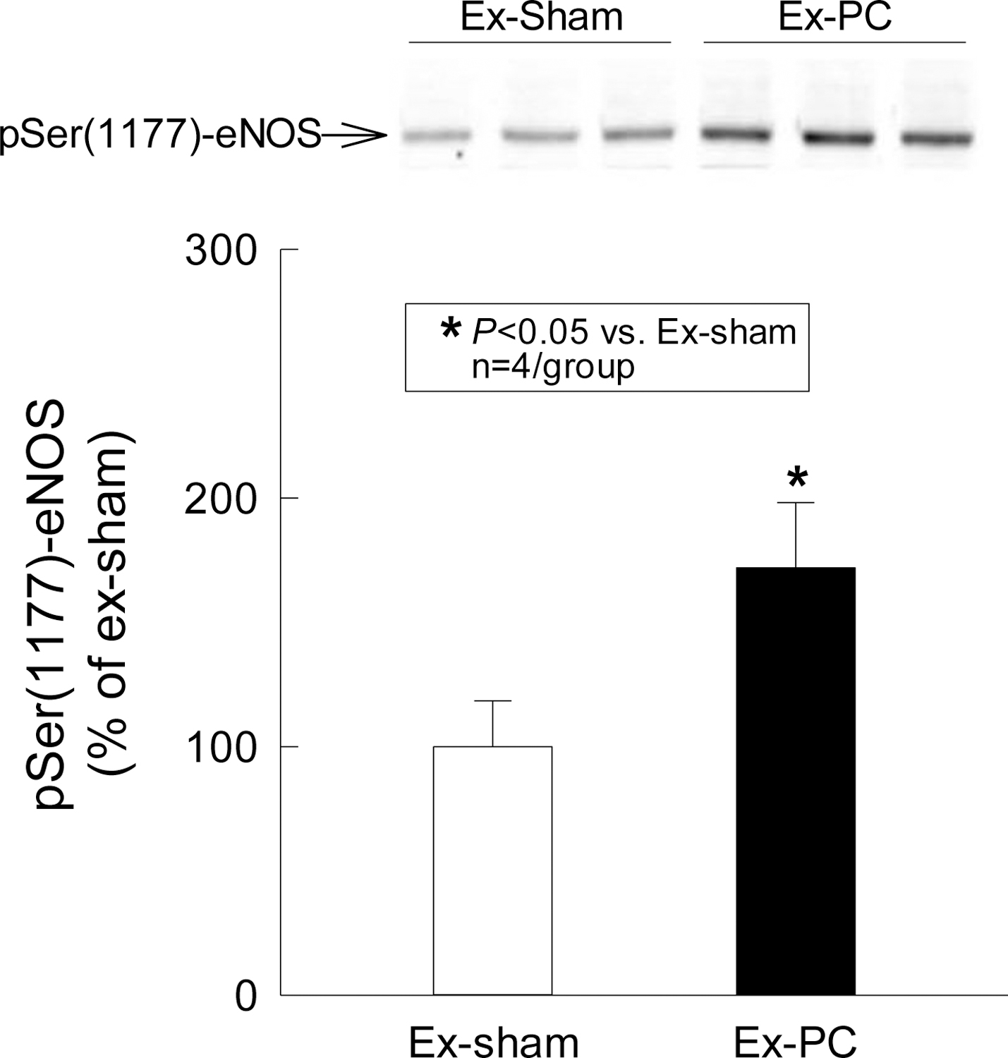
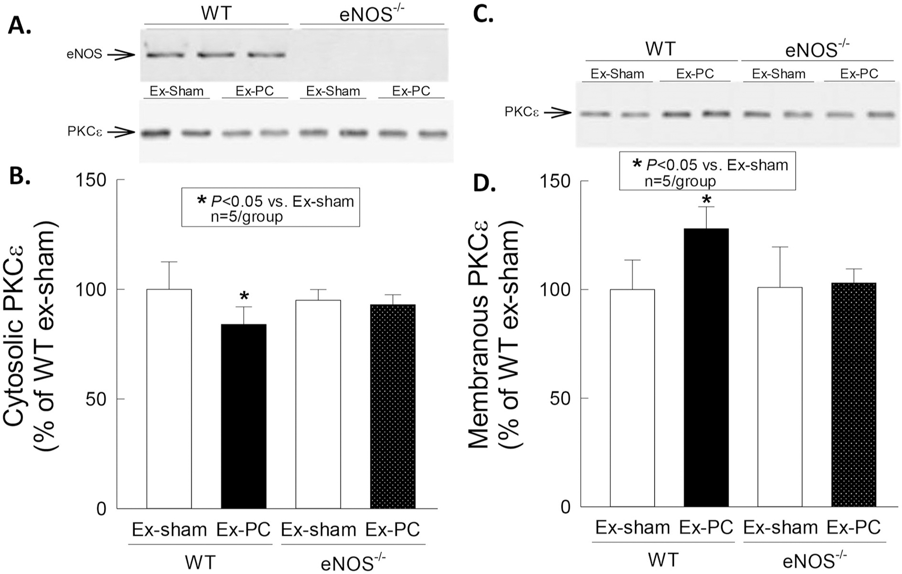
The purpose of this study was to assess whether short-term, mild exercise induces protection against myocardial infarction and, if so, what role the eNOS-PKCε-iNOS axis plays. Mice were subjected to 2 bouts/day of treadmill exercise (60 min at 15 m/min) for 2 consecutive days. At 24 h after the last bout of exercise, mice were subjected to a 30-min coronary artery occlusion and 24 h of reperfusion. In the exercise group (group III, wild-type mice), infarct size (25.5 ± 8.8% of risk region) was significantly (P < 0.05) reduced compared with the control groups (sham exercise, group II [63.4 ± 7.8%] and acute myocardial infarction, group I [58.6 ± 7.0%]). This effect was abolished by pretreatment with the NOS inhibitor L-NA (group VI, 56.1 ± 16.2%) and the PKC inhibitor chelerythrine (group VIII, 57.9 ± 12.5%). Moreover, the late PC effect of exercise was completely abrogated in eNOS-/- mice (group XIII, 61.0 ± 11.2%). The myocardial phosphorylated eNOS at Ser-1177 was significantly increased at 30 min after treadmill training (exercise group) compared with sham-exercised hearts. PKCε translocation was significantly increased at 30 min after exercise in WT mice but not in eNOS-/- mice. At 24 h after exercise, iNOS protein was upregulated compared with sham-exercised hearts. The protection of late PC was abrogated in iNOS-/- mice (group XVI, 56.4 ± 12.9%) and in wildtype mice given the selective iNOS inhibitor 1400 W prior to ischemia (group X 62.0 ± 8.8% of risk region). We conclude that 1) even short, mild exercise induces a delayed PC effect that affords powerful protection against infarction; 2) this cardioprotective effect is dependent on activation of eNOS, eNOS-derived NO generation, and subsequent PKCε activation during PC; 3) the translocation of PKCε is dependent on eNOS; 4) the protection 24 h later is dependent on iNOS activity. Thus, eNOS is the trigger and iNOS the mediator of PC induced by mild exercise.

Keywords: Exercise; Ischemia/reperfusion injury; NOS, nitric oxide synthase; Preconditioning; Protein kinase C.


Conflict of interest statement

Declaration of Competing Interest

The authors declare no competing interests.

Figures

Fig. 1.
Fig. 1.
A. Experimental protocol for phase I. Six groups of WT or eNOS−/− mice underwent either the sham exercise or the exercise protocol. Myocardial samples were harvested either 30 min (groups I-IV, tissue assay for eNOS activity and PKCε) or 24 h (groups V and VI, tissue assay for eNOS and iNOS expression and NOx) after the last bout of sham exercise or exercise. B. Experimental protocol for phase II. Seventeen groups of mice underwent five different protocols. Mice in groups I (wild-type [WT] naïve, n = 15), XI (eNOS−/−, naïve, n = 10) and XIV (iNOS−/−, naïve, n = 10) underwent a 30-min coronary occlusion followed by 24 h of reperfusion (Protocol I, Naïve). Mice in groups II (WT, sham exercise, n = 9), XII (eNOS−/−, sham exercise, n = 8) and XV (iNOS−/−, sham exercise, n = 12) underwent the same protocol as Protocol II (Sham Ex) except that on days 5 and 6 they ran at 1 m/min (instead of 15 m/min) for 75 min, twice each day. In groups III (WT, exercise PC, n = 17), XIII (eNOS−/−, exercise PC, n = 8), and XVI (iNOS−/−, exercise PC, n = 8) animals were subjected to Protocol III (Ex-PC), which consisted of three stages. In stage 1 (acclimation stage), all mice were acclimated to the treadmill for 4 consecutive days (days 14) by running at 15 m/min (0% grade) for 10 min and 1 m/min for 2 h/day (with electric shock on [0.1–0.2 mA]). An electronic counter counted the shock numbers. In stage 2 (exercise stage), on days 5 and 6, the mice ran for 10 min at 7 m/min (warm-up), then for 60 min at 15 m/min, and then for 5 min at 7 m/min (cooldown), twice a day (9:00 a.m. and 2:00 p.m.) without electrical shock (the distance covered by one bout of exercise was about 1000 m). In stage 3 (coronary occlusion stage), 24 h after exercise or sham exercise (day 7), mice underwent a 30-min occlusion followed by 24 h of reperfusion. Mice in groups V (WT, sham exercise + L-NA, n = 11) and VIII (WT, sham exercise + CHE, n = 8) received L-NA or chelerythrine, respectively, 30 min before each bout of sham exercise on days 5 and 6 (Protocol IV, Sham Ex + Drug). Mice in groups VI (WT, exercise + L-NA, n = 7), IX (WT, exercise + CHE, n = 9), IV (WT, exercise + NS, n = 9), and VII (WT, exercise + DMSO, n = 9) received L-NA, chelerythrine, or the respective vehicle 30 min before each bout of exercise on days 5 and 6 (Protocol V, Ex-PC + Drug). Moreover, mice in groups X (WT, exercise +1400 W) received 1400 W 30 min before coronary occlusion on day 7 and then subjected to a 30-min occlusion and followed by 24 h reperfusion.
Fig. 2.
Fig. 2.
Serine phosphorylation of eNOS by exercise PC. Cardiac samples were obtained 30 min after the 4th session of sham exercise or exercise and the membranous fraction was prepared for immunoblotting with a specific anti-pSer(1177)-eNOS antibody. Exercise induced a 70% increase in pSer(1177)-eNOS. Data are means ± SD.
Fig. 3.
Fig. 3.
Membranous translocation of PKCε by exercise PC. Cardiac samples were obtained 30 min after the 4th session of sham exercise or exercise, and both membranous and cytosolic fractions were prepared for immunoblotting with a specific anti-PKCε antibody. Exercise induced a decrease in the cytosolic content of PKCε and a corresponding increase in the membranous content of this isoform in WT but not in eNOS−/− mice. Data are means ± SD.
Fig. 4.
Fig. 4.
Upregulation of eNOS by exercise PC. Cardiac samples were obtained 24 h after the 4th session of sham exercise or exercise and the membranous fraction was subjected to immunoblotting with a specific anti-eNOS antibody. Exercise induced a 40% increase in eNOS protein expression. Data are means ± SD.
Fig. 5.
Fig. 5.
Upregulation of iNOS in exercise-induced PC. Cardiac samples were collected 24 h after the 4th session of sham exercise or exercise. The cytosolic fraction of protein was subjected to immunoblotting with a specific anti-iNOS antibody. Note the elevation of iNOS protein expression in exercise-induced PC. Data are means ± SD.
Fig. 6.
Fig. 6.
A, Representative example of a heart from group II (sham exercise group) subjected to a 30-min coronary occlusion and 24 h of reperfusion 24 h after a sham exercise protocol. The infarcted region was delineated by perfusing the aortic root with TTC; the region at risk was delineated by perfusing the aortic root with Phthalo blue after tying the previously occluded artery. As a result of this procedure, the nonischemic portion of the left ventricle was stained dark blue, the viable tissue within the region at risk was stained bright red, and the infarcted tissue was light yellow. Note the large, confluent areas of infarction spanning most of the thickness of the LV wall, with thin rims of viable subendocardial tissue. The scale at the bottom is in mm. B, Representative example of a heart from group III (exercise group), which was subjected to 2 sessions/day of treadmill exercise (60 min at 15 m/min) for 2 consecutive days and, 24 h later, to a 30-min occlusion and 24 h of reperfusion. Postmortem perfusion was performed as described in the legend to Fig. 1B. In contrast to the confluent infarction shown in Fig. 6A, in this heart the 30-min occlusion resulted in patchy areas of infarction, a pattern that was characteristic of both groups VI and VII, indicating that the sequence of four bouts of exercise-induced a powerful late PC effect against infarction. The scale at the bottom is in mm.
Fig. 7.
Fig. 7.
Myocardial infarct size in groups I (WT, control), II (WT, sham exercise), III (WT, exercise PC), VI (WT, exercise + NS), V (WT, sham exercise + L-NA), VI (WT, exercise + L-NA), VII (WT, exercise + DMSO), VIII (WT, sham exercise + CHE), IX (WT, exercise + CHE), X (WT, exercise + 1400 W), XI (eNOS/, control), XII (eNOS/, sham exercise), XIII (eNOS/, exercise PC), XIV (iNOS/, control), XV (iNOS/, sham exercise), XVI (iNOS/, exercise PC). The experimental protocols are specified in the legend to Fig. 1B. Open circles represent individual mice, whereas solid circles represent means ± SD.

Comment in

References

    1. Bolli R, The early and late phases of preconditioning against myocardial stunning and the essential role of oxyradicals in the late phase: an overview, Basic Res. Cardiol 91 (1996) 57–63, 10.1007/BF00788866. - DOI - PubMed
    1. Yellon DM, Downey JM, Preconditioning the myocardium: from cellular physiology to clinical cardiology, Physiol. Rev 83 (2003) 1113–1151, 10.1152/physrev.00009.2003. - DOI - PubMed
    1. Bolli R, The late phase of preconditioning, Circ. Res 87 (2000) 972–983, 10.1161/01.res.87.11.972. - DOI - PubMed
    1. Dawn B, Bolli R, Role of nitric oxide in myocardial preconditioning, Ann. N. Y. Acad. Sci 962 (2002) 18–41, 10.1111/j.1749-6632.2002.tb04053.x. - DOI - PubMed
    1. Guo Y, Stein AB, Wu WJ, Zhu X, Tan W, Li Q, et al., Late preconditioning induced by NO donors, adenosine A1 receptor agonists, and delta1-opioid receptor agonists is mediated by iNOS, Am. J. Physiol. Heart Circ. Physiol 289 (2005) H2251–H2257, 10.1152/ajpheart.00341.2005. - DOI - PMC - PubMed