Review
. 2021 Dec 1;111(5):e54-e62.
doi: 10.1016/j.ijrobp.2021.08.003. Epub 2021 Aug 14.

Preclinical Risk Evaluation of Normal Tissue Injury With Novel Radiosensitizers

Affiliations
Review

Preclinical Risk Evaluation of Normal Tissue Injury With Novel Radiosensitizers

Sonja Dragojevic et al. Int J Radiat Oncol Biol Phys. .

Abstract

Genotoxic damage induced by radiation triggers a highly coordinated DNA damage response, and molecular inhibitors of key nodes within this complex response network can profoundly enhance the antitumor efficacy of radiation. This is especially true for drugs targeting the catalytic subunit of DNA-dependent protein kinase, which is a core component of the nonhomologous end-joining DNA repair pathway, and ataxia telangiectasia mutated, which coordinates cell cycle arrest, apoptosis, and DNA repair functionalities after radiation exposure. Unlike the more modest in vitro radiosensitizing effects seen with classic sensitizing agents such as cisplatin, 5-fluorouracil, or taxanes, DNA-dependent protein kinase or ataxia telangiectasia mutated inhibitors provide much more robust sensitizing effects in vitro, as might be anticipated from targeting these key DNA repair modulators. However, patients with homozygous inactivating mutations of ataxia telangiectasia mutated or mice with homozygous defects in DNA-dependent protein kinase (severe combined immunodeficiency) have profoundly enhanced acute normal tissue radiation reactions. Therefore, there is significant potential that the combination of small molecule inhibitors of these kinases with radiation could cause similar dose-limiting acute normal tissue toxicities. Similarly, although less understood, inhibition of these DNA repair response pathways could markedly increase the risk of late radiation toxicities. Because these potent radiosensitizers could be highly useful to improve local control of otherwise radiation-resistant tumors, understanding the potential for elevated risks of radiation injury is essential for optimizing therapeutic ratio and developing safe and informative clinical trials. In this review, we will discuss 2 straightforward models to assess the potential for enhanced mucosal toxicity in the oral cavity and small intestine established in our laboratories. We also will discuss similar strategies for evaluating potential drug-radiation interactions with regard to increased risks of debilitating late effects.


Figures

Fig. 1.
Fig. 1.
Schematic of the mouse models of normal tissue injury after the effects of potent radiosensitizing agents inhibiting the DNA damage response (DDR) pathways.
Fig. 2.
Fig. 2.
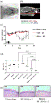
(a) Animals were set-up for radiation using fluoro (x-ray) imaging of the head and targeting oral cavity. (b) Radiation dosimetry map in sagittal plane through head with dose levels of 105% (red), 100% (green), and 90% (blue) shown. (c) Weight of animals treated with TMZ (25 mg/kg) + RT (2.85Gy) × 3 days or RT (2.85 Gy) + M3814 (105 mg/kg, 10 minutes prior and 7 hours after RT) × 3 days. Black arrows show the days when mice received treatment (n = 5 for each group) (d) Maximum weight loss relative to starting weight for individual animals is shown after the indicated 3-fraction treatment regimens. (e) Hematoxylin and eosin staining of tongue mucosa 8 days after treatment with sham, RT (2.85 Gy × 3), or RT + M3814 as in Figure 2d.
Fig. 3.
Fig. 3.
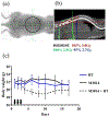
(a) A radiograph of animal set-up for radiation of small intestine (black circle showing 1.5 cm collimator target area). (b) Sagittal view of dose distribution for radiation of the abdomen. Dose distribution curves for 105% (red), 100% (green), and 90% (blue) doses are shown. (c) Weight of animals treated (black arrows treatment day) with RT (2.85 Gy × 3) alone, M3814 (90 mg/kg, 10 minutes prior, and 7 hours after RT), and M3814 + RT × 3 (n = 3 for each treatment group).
Fig. 4.
Fig. 4.
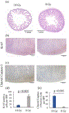
The effect of ionizing radiation on intestinal tissue. (a) Jejunal cross-sections obtained on day 4 after total body ionizing radiation exposure of 0 Gy versus 10 Gy were stained with hematoxylin and eosin and for (b) Ki67 and (c) cleaved caspase-3. Positively stained cells per high power field (HPF) are shown for (d) Ki-67 and (e) cleaved caspase-3.
Fig. 5.
Fig. 5.
(a) Effect of ionizing radiation on intestinal permeability and inflammation. Mice were exposed to 0 or 10 Gy of total body radiation. On day 4 after irradiation, the effect of radiation was evaluated on (b) intestinal permeability by measuring serum levels 4 hours after oral gavage with FITC-dextran and by (c) measuring plasma C-reactive protein levels.
Fig. 6.
Fig. 6.
Sciatic nerve injury model. (a) X ray imaging was used for targeting the sciatic nerve with 1 cm circular collimator (black circle showing the targeted area) in regard to knee and hip joints (black arrows showing knee and hip joints). (b) Radiation dosimetry map for sciatic nerve showing dose distribution curves for 300% (red), 200% (green), and 100% (blue) of prescribed radiation dose.
Fig. 7.
Fig. 7.
Brainstem radiation injury model. Dosimetry maps for radiation of brainstem in (a) axial, (b) sagittal, and (c) coronal planes. The blue arc on the coronal image (far right image) represents a partial arc (230°) radiation therapy beam with the isocenter located in the pons. The animals were planned with a 2.5 mm circular collimator and radiation dose of 6.0 Gy.

References

    1. Bezjak A, Paulus R, Gaspar LE, et al. Safety and efficacy of a five-fraction stereotactic body radiotherapy schedule for centrally located non-small-cell lung cancer: NRG oncology/RTOG 0813 trial. J Clin Oncol 2019;37:1316–1325. - PMC - PubMed
    1. Timmerman R, McGarry R, Yiannoutsos C, et al. Excessive toxicity when treating central tumors in a phase II study of stereotactic body radiation therapy for medically inoperable early-stage lung cancer. J Clin Oncol 2006;24:4833–4839. - PubMed
    1. Shiloh Y, Ziv Y. The ATM protein kinase: Regulating the cellular response to genotoxic stress, and more. Nat Rev Mol Cell Biol 2013;14:197–210. - PubMed
    1. Reid DA, Keegan S, Leo-Macias A, et al. Organization and dynamics of the nonhomologous end-joining machinery during DNA double-strand break repair. Proc Natl Acad Sci U S A 2015;112:E2575–E2584. - PMC - PubMed
    1. Zenke FT, Zimmermann A, Sirrenberg C, et al. Pharmacological inhibitor of DNA-pk, m3814, potentiates radiotherapy and regresses human tumors in mouse models. Mol Cancer Ther 2020;19:1091–1101. - PubMed

Publication types

Substances