. 2021 Dec 1;29(12):3512-3524.
doi: 10.1016/j.ymthe.2021.08.020. Epub 2021 Aug 14.

Cell type-selective targeted delivery of a recombinant lysosomal enzyme for enzyme therapies

Affiliations

Cell type-selective targeted delivery of a recombinant lysosomal enzyme for enzyme therapies

Andrew D Baik et al. Mol Ther. .

Abstract

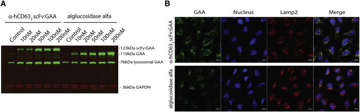
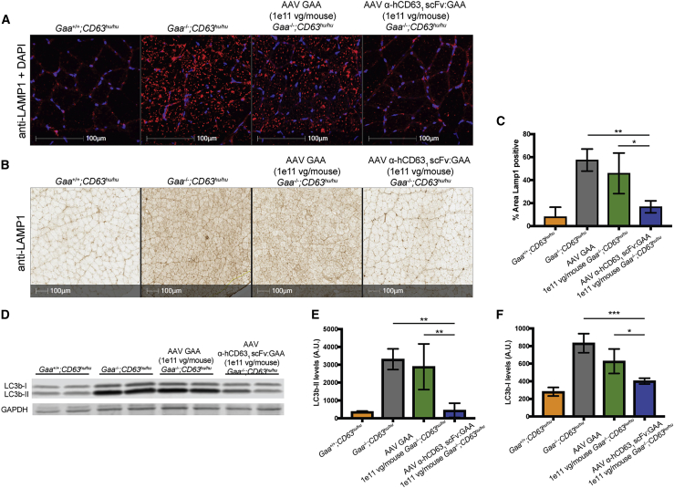
Lysosomal diseases are a class of genetic disorders predominantly caused by loss of lysosomal hydrolases, leading to lysosomal and cellular dysfunction. Enzyme replacement therapy (ERT), where recombinant enzyme is given intravenously, internalized by cells, and trafficked to the lysosome, has been applied to treat several lysosomal diseases. However, current ERT regimens do not correct disease phenotypes in all affected organs because the biodistribution of enzyme uptake does not match that of the affected cells that require the enzyme. We present here targeted ERT, an approach that utilizes antibody-enzyme fusion proteins to target the enzyme to specific cell types. The antibody moiety recognizes transmembrane proteins involved in lysosomal trafficking and that are also preferentially expressed in those cells most affected in disease. Using Pompe disease (PD) as an example, we show that targeted ERT is superior to ERT in treating the skeletal muscle phenotypes of PD mice both as a protein replacement therapeutic and as a gene therapy.

Keywords: enzyme therapy; genetic therapy; glycogen storage disease II; hydrolases; lysosomes; protein transport.


Conflict of interest statement

Declaration of interests All authors were employees of Regeneron Pharmaceuticals, Inc., while engaged in the study and may hold stock and/or stock options in the company. A.D.B. and K.D.C. have patent applications for internalizing enzymes and uses thereof.

Figures

None
Graphical abstract
Figure 1
Figure 1
Antibody:GAA proteins are endocytosed by an antibody-mediated, effector-dependent and CI-MPR-independent mechanism and are processed after internalization to the mature, lysosomal GAA form (A) α-hCD63 IgG:GAA is internalized via CD63. GAA activity was detected in HEK293 cell lysate after overnight incubation of cells with α-hCD63:GAA. Eliminating the interaction between the antibody and CD63 by using an IgG4 isotype control fused to either GAA or CD63-deficient HEK293 cells reduced the GAA activity in the lysate. n = 3. (B) Saturation of CI-MPR by addition of free M6P blocks uptake of GAA but does not block uptake of α-hCD63:GAA in human skeletal myoblasts. n = 3. (C) In C2C12 mouse myoblasts, α-mCD63 IgG:GAA and α-ITGA7:GAA can mediate internalization of GAA independently of M6P. n = 3. (D) α-hCD63 IgG:GAA is processed into the mature, lysosomal 76-kDa GAA form and persists for at least 12 days in Pompe disease patient fibroblasts (probed with anti-GAA, upper). The antibody portion, which is cleaved off, is rapidly degraded (probed with anti-hIgG, lower). Note that an unrelated background band is also detected by the anti-GAA antibody, as seen in lane “Day −1.”
Figure 2
Figure 2
An α-hCD63 scFv:GAA fusion protein delivers GAA to lysosomes (A) α-hCD632 scFv:GAA and alglucosidase alfa are both processed to lysosomal forms after internalization. HEK293 were incubated with either α-hCD632 scFv:GAA or alglucosidase alfa overnight, and cell lysates were probed for GAA uptake and processing with an anti-GAA antibody (green) and loading control anti-GAPDH (red). A dose-dependent accumulation of the lysosomal GAA form is observed in either GAA molecule. (B) α-hCD632 scFv:GAA colocalizes with lysosomes after internalization. HEK293 cells were incubated with either α-hCD632 scFv:GAA or alglucosidase alfa, cells were fixed 4 h after internalization, and immunofluorescence was performed for GAA (green), nuclei, and Lamp2. Scale bars, 10 μm.
Figure 3
Figure 3
α-hCD63 scFv:GAA outperforms alglucosidase alfa in glycogen removal on a molar basis (A) α-hCD632 scFv:GAA removed more glycogen at equivalent molar doses to alglucosidase alfa in skeletal muscle. 2- to 3-month old PD mice were dosed weekly with either α-hCD632 scFv:GAA or alglucosidase alfa, and muscle glycogen was assayed 1 week after the last dose. n = 4–8 per muscle (n = 8 mice in each treatment group). Error bars represent means ± SD. ∗p < 0.01 between groups. (B) Quadriceps show lower LAMP1 staining after treatment with α-hCD632 scFv:GAA. (C) LAMP1-stained quadriceps were quantified for areal staining. Error bars represent means ± SD. ∗p < 0.01 between groups.
Figure 4
Figure 4
Treatment of PD mice with AAV α-hCD631 scFv:GAA restores glycogen to wild-type levels in key muscles (A) Serum levels of 1e10 vg/mouse and 1e11 vg/mouse of each AAV were quantified by western blot and are higher in the AAV α-hCD631 scFv:GAA groups at the same dose. (B) PAS-H stain for glycogen of 1e11 vg-treated quadricep muscle sections 3 months after AAV infection show uniform removal of glycogen in the α-hCD631 scFv:GAA group. (C) α-hCD631 scFv:GAA-treated mice have lower glycogen levels in skeletal muscle than GAA-treated mice at equivalent doses. Normalization of muscle glycogen to wild-type levels is seen at the 1e11 vg/mouse dose of α-hCD631 scFv:GAA. Glycogen in muscle tissue lysates was assayed 3 months after AAV administration. Error bars represent means ± SD. ∗∗p < 0.01 between 1e10 vg/mouse groups. ∗p < 0.05 between 1e11 vg/mouse groups. n = 2 for wild type, n = 4 for treatment groups.
Figure 5
Figure 5
Treatment of PD mice with AAV α-hCD631 scFv:GAA reduces lysosomal area and autophagy (A and B) Confocal images (A) and wide-field images (B) of anti-LAMP1 staining to detect lysosomes in section of quadriceps in mice 3 months after administration of AAV GAA or α-hCD631 scFv:GAA show that lysosome staining is decreased with scFv:GAA treatment. Scale bars, 100 μm. (C) Quantification of the LAMP-1-positive area in wide-field images. n = 2 for wild type, n = 4 for treatment groups. Error bars represent means ± SD. (D) Autophagy is decreased with α-hCD631 scFv:GAA treatment. Representative western blotting for LC3b to monitor autophagy in quadriceps lysate from two mice per group. (E and F) Quantification of LC3b-II (E) and LC3b-I (F) levels in quadriceps lysates. n = 2 for wild type, n = 4 for treatment groups. Error bars represent means ± SD. ∗p < 0.05, ∗∗p < 0.01, ∗∗∗p < 0.001.
Figure 6
Figure 6
Treatment of PD mice with AAV α-hCD631 scFv:GAA restores muscle function in PD mice (A) Rotarod test performance of mice treated with either AAV GAA or AAV α-hCD631 scFv:GAA shows recovery of scFv:GAA-treated mice within 2 months of AAV administration. (B) Forelimb grip strength measurements show continued strength improvement within 1 month of treatment in α-hCD63SC:GAA-treated mice. PD mice were treated with 1e11 vg AAV α-hCD631 scFv:GAA or 1e11 vg AAV GAA or were left untreated, and these groups were compared to wild-type mice. (C) Ex vivo peak tetanic force of the tibialis anterior muscle 6 months after AAV treatment shows a recovery in muscle strength in the AAV α-hCD631 scFv:GAA-treated group to comparable to wild-type levels. (A and B) Error bars represent medians ± SD, n = 7 for wild type, n = 11 for treatment groups. (C) Error bars represent means ± SD, n = 4 for untreated and wild type, n = 5 for treatment group. ∗p < 0.05, ∗∗p < 0.01, ∗∗∗p < 0.001.

References

    1. Hirschhorn R., Reuser A. In: Scriver C.R., Bly W.S., Childs B., Beaudet A.L., Valle D., Kinzler K.W., Vogelstein B., editors. McGraw-Hill; New York: 2000. Glycogen Storage Disease Type II: Acid Alpha-glucosidase (acid maltase) Deficiency; pp. 3389–3420. (The Metabolic and Molecular Bases of Inherited Disease).
    1. Prater S.N., Banugaria S.G., DeArmey S.M., Botha E.G., Stege E.M., Case L.E., Jones H.N., Phornphutkul C., Wang R.Y., Young S.P., Kishnani P.S. The emerging phenotype of long-term survivors with infantile Pompe disease. Genet. Med. 2012;14:800–810. - PMC - PubMed
    1. Chien Y.-H., Lee N.-C., Chen C.-A., Tsai F.-J., Tsai W.-H., Shieh J.-Y., Huang H.-J., Hsu W.-C., Tsai T.-H., Hwu W.-L. Long-Term Prognosis of Patients with Infantile-Onset Pompe Disease Diagnosed by Newborn Screening and Treated since Birth. J. Pediat. 2015;166:985–991. e1-2. - PubMed
    1. DeRuisseau L.R., Fuller D.D., Qiu K., DeRuisseau K.C., Donnelly W.H., Jr., Mah C., Reier P.J., Byrne B.J. Neural deficits contribute to respiratory insufficiency in Pompe disease. Proc. Natl. Acad. Sci. USA. 2009;106:9419–9424. - PMC - PubMed
    1. Koeberl D.D., Luo X., Sun B., McVie-Wylie A., Dai J., Li S., Banugaria S.G., Chen Y.T., Bali D.S. Enhanced efficacy of enzyme replacement therapy in Pompe disease through mannose-6-phosphate receptor expression in skeletal muscle. Mol. Genet. Metab. 2011;103:107–112. - PMC - PubMed

Publication types

MeSH terms

LinkOut - more resources