. 2021 Sep 29;41(39):8249-8261.
doi: 10.1523/JNEUROSCI.1787-20.2021. Epub 2021 Aug 16.

Dorsal Root Ganglia Macrophages Maintain Osteoarthritis Pain

Affiliations

Dorsal Root Ganglia Macrophages Maintain Osteoarthritis Pain

Ramin Raoof et al. J Neurosci. .

Erratum in

Abstract

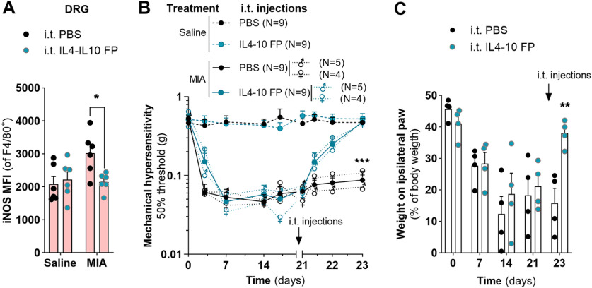
Pain is the major debilitating symptom of osteoarthritis (OA), which is difficult to treat. In OA patients joint tissue damage only poorly associates with pain, indicating other mechanisms contribute to OA pain. Immune cells regulate the sensory system, but little is known about the involvement of immune cells in OA pain. Here, we report that macrophages accumulate in the dorsal root ganglia (DRG) distant from the site of injury in two rodent models of OA. DRG macrophages acquired an M1-like phenotype, and depletion of DRG macrophages resolved OA pain in male and female mice. Sensory neurons innervating the damaged knee joint shape DRG macrophages into an M1-like phenotype. Persisting OA pain, accumulation of DRG macrophages, and programming of DRG macrophages into an M1-like phenotype were independent of Nav1.8 nociceptors. Inhibition of M1-like macrophages in the DRG by intrathecal injection of an IL4-IL10 fusion protein or M2-like macrophages resolved persistent OA pain. In conclusion, these findings reveal a crucial role for macrophages in maintaining OA pain independent of the joint damage and suggest a new direction to treat OA pain.SIGNIFICANCE STATEMENT In OA patients pain poorly correlates with joint tissue changes indicating mechanisms other than only tissue damage that cause pain in OA. We identified that DRG containing the somata of sensory neurons innervating the damaged knee are infiltrated with macrophages that are shaped into an M1-like phenotype by sensory neurons. We show that these DRG macrophages actively maintain OA pain remotely and independent of joint damage. The phenotype of these macrophages is crucial for a pain-promoting role. Targeting the phenotype of DRG macrophages with either M2-like macrophages or a cytokine fusion protein that skews macrophages into an M2-like phenotype resolves OA pain. Our work reveals a mechanism that contributes to the maintenance of OA pain distant from the affected knee joint and suggests that dorsal root ganglia macrophages are a target to treat osteoarthritis chronic pain.

Keywords: chronic pain; macrophage; osteoarthritis; sensory neuron.


Figures

Figure 1.
Figure 1.
Macrophages infiltrate the DRG in MIA-induced OA pain. A, Quantification of histopathological changes in the knee joints over 4 weeks after intra-articular MIA injection in mice. Knee joints were dissected for histology at indicated time points and scored according to the Histologic-Histochemical Grading System. One-way ANOVA with Dunnett's post hoc. B, Exemplar image of mice knee joints at week 3 after intra-articular injection of MIA or saline. The knee joints were stained with safranin-O. The femur is on the left side, and the tibia is at the right. Left, Image shows the normal cartilage structure (red staining)of the joint after saline injection. Right, Image shows MIA-induced knee joint lesion that was observed in the joint of the MIA-injected mice. Scale bar, 200 µm. C, Course of mechanical hyperalgesia after injection of MIA in the ipsilateral or saline in the contralateral knee. Mechanical hyperalgesia was followed over time using Von Frey. Two-way ANOVA with Sidak's post hoc. D, Course of weight distribution determined after intra-articular injection of MIA or saline in the contralateral knee using dynamic weight-bearing apparatus. Two-way repeated-measures ANOVA with Sidak's post hoc. E, Gating strategy to identify the immune cells (as displayed in F) in the lumbar DRG (L3–L5) after MIA injection. A time gate unified the acquisition analysis, followed by a rough separation of events based on CD45 expression but excluding the counting beads. Cells were further gated by FCS/SSC (to identify the size of each cell), single cells, and CD45 and Ly6G expression before analysis of CD11b and F4/80. CD3 and CD19 cells were identified from the CD11b-negative cells. F, Lumbar ipsilateral DRG (L3–L5), from male and female mice, containing the sensory neurons innervating the knees that received MIA were isolated at each time point. The absolute number of CD45+ cells, classified to subsets, is divided by the total DRGs analyzed per mouse. The control group consists of mice DRG that received saline and isolated at the different time points (1, 2, 3, and 4 weeks). Two-way ANOVA with Dunnett's post hoc. G, Light-sheet rendering of lumbar spinal cord with the spinal nerves and DRG attached of mice injected at one knee intra-articular with MIA (ipsilateral), and the other knee with saline (contralateral). Neurons are stained with neurofilament M (green) and macrophages with F4/80 (red). White arrows indicate the ipsilateral DRG, and yellow arrows indicate the contralateral DRG containing neurons innervating the saline-injected knee (Movie 1). Scale bar, 500 µm. **p < 0.01; ***p < 0.001.
Figure 2.
Figure 2.
Groove surgery induced OA-like pain and macrophage accumulation in the DRG. A, Quantification of histopathological changes in the obese rat knee joints 12 weeks after groove or sham surgery. The cartilage degeneration is scored according to the Histologic-Histochemical Grading System. Unpaired two-tailed t tests. B, Representative picture of histopathological changes in the rat knee joints 12 weeks after groove or sham surgery. The knee joints were stained with safranin-O. The arrow indicates the cartilage degeneration induced by groove surgery. Scale bar, 50 µm. C, Mechanical hyperalgesia at the hindpaw after groove (ipsilateral) or sham (contralateral) operation in rat knee joints before and 12 weeks after surgery. Two-way ANOVA with Sidak's post hoc. D, Paw weight distribution of the affected ipsilateral leg was evaluated before and 12 weeks after groove surgery in rat knee joints using dynamic weight bearing. Unpaired two-tailed t tests. E, Quantification of macrophages in infiltrated lumbar (L3–L5) DRG containing the sensory neurons innervating the knees at 12 weeks after groove (ipsilateral) or sham operation contralateral by assessment of CD68+ and Iba1+ immunostaining. Paired two-tailed t tests. Exemplar immunofluorescent microscopy images of Iba1+ macrophages (green) in the lumbar (L3–L5) DRG in rats at 12 weeks after groove or sham operations. Neurons were visualized with neurotrace Nissl stain (red). *p < 0.05; **p < 0.01; ***p < 0.001.
Figure 3.
Figure 3.
DRG infiltrating macrophages maintain osteoarthritis pain. A, B, Flow cytometry analysis of single-cell suspension of DRG and blood isolated from MMdtr mice treated with DT, intraperitoneal or intrathecal, to validate monocyte and macrophage depletion at week 4 after MIA injection. A, Number of F4/80+ macrophages, nonmyeloid cells (CD45+CD11b), and other myeloid cells (CD11b+F4/80) in the DRG of MMdtr mice after intraperitoneal and intrathecal injections of DT. Two-way ANOVA with Dunnett's post hoc. B, Flow cytometry analysis of CD45+CD11b+CD115+ monocytes in blood after intraperitoneal and intrathecal injections of DT in MMdtr mice. One-way ANOVA with Dunnett's post hoc. C, Course of mechanical hyperalgesia in WT versus MMdtr mice, in female and male mice, after intra-articular injection of MIA in the ipsilateral knee and saline in the contralateral knee. Monocytes/macrophages were systemically depleted by daily intraperitoneal injections of DT starting at day 21. Two-way ANOVA with Tukey's post hoc comparing MMdtr to WT in each time point. Statistical analyses using a repeated-measures ANOVA indicated that the course of mechanical hyperalgesia was not significantly different between males and females. D, Weight distribution of the ipsilateral hindpaws at indicated time points in WT versus MMdtr mice after intra-articular injection of MIA in the left hindpaw and saline in the right hindpaw. Mice were treated with daily intraperitoneal injection of DT starting at day 21 to deplete monocytes/macrophages. Two-way ANOVA with Sidak's post hoc. E, Course of mechanical hyperalgesia in WT versus MMdtr mice, in female and male mice, after intra-articular injection of MIA in the ipsilateral knee and saline in the contralateral knee. Mice received daily intrathecal injections of DT starting at day 21 to deplete macrophages in the lumbar DRG. Two-way ANOVA with Tukey's post hoc comparing MMdtr to WT in each time point. Statistical analyses using a repeated-measures ANOVA indicated that the course of mechanical hyperalgesia was not significantly different between males and females. F, Weight distribution of the ipsilateral hindpaws at indicated time points in WT versus MMdtr mice after intra-articular injection of MIA in the ipsilateral knee and saline in the contralateral knee. Mice received daily intrathecal injection of DT starting at day 21 to deplete macrophages in the lumbar DRG. Two-way ANOVA with Sidak's post hoc. i.p., Intraperitoneal; i.t., intrathecal. **p < 0.01; ***p < 0.001.
Figure 4.
Figure 4.
DRG infiltrating macrophages acquire an M1-like phenotype independent of nociceptors. A, B, Median fluorescence intensity (MFI) of (A) iNOS and (B) CD206 of the F4/80+ macrophage population in the DRG of mice at the indicated time points after intra-articular injection of MIA. One-way ANOVA with Dunnett's post hoc comparing MFI in indicated time point. C, D, MFI of (A) iNOS and (B) CD206 of bone-marrow-derived macrophages in vitro cocultured for 24 h with isolated sensory neurons that innervated the MIA-injected joint (ipsilateral) or healthy joint (contralateral) 1 week after MIA injection. One-way ANOVA with Dunnett's post hoc. E, Representative immunofluorescent microscopy images of peripherin (green; small-diameter neurons) and NF200 (red; large-diameter neurons) in littermate controls (above) and Nav1.8-DTA mice (below). F, Course of mechanical hyperalgesia after intra-articular injection of MIA in wild-type (week 0/1, n = 7; week 3, n = 6). versus Nav1.8-DTA mice (week 0/1, n = 7; week 3, n = 3). Two-way ANOVA with Sidak's post hoc. G, Weight distribution of WT versus Nav1.8-DTA mice before and at 1 and 3 weeks after intra-articular injection of MIA using static weight-bearing apparatus. Two-way ANOVA with Sidak's post hoc. H, Absolute number of CD45+ leukocytes classified to subset per lumbar DRG of WT compared with Nav1.8-DTA mice (purple background) at indicated time points after intra-articular MIA injection. Two-way ANOVA with Dunnett's post hoc, comparing DRG infiltrated cells in WT versus Nav1.8 mice, in each time point. I, J, MFI of (H) iNOS and (I) CD206 of F4/80+ macrophage population accumulated in the DRG of WT versus Nav1.8-DTA before and at 1 and 3 weeks after intra-articular injection of MIA. Two-way ANOVA with Sidak's post hoc. *p < 0.05; **p < 0.01; ***p < 0.001.
Figure 5.
Figure 5.
The M1 phenotype of DRG macrophages regulates the pain-promoting function. A, Median fluorescence intensity (MFI) of iNOS in macrophages that were differentiated in vitro into an M1-like phenotype or M2-like phenotype, and M1 macrophages cocultured for 24 h with mCherry2 expressing M2-like macrophages. Expression of iNOS was determined after gating out the mCherry2-positive macrophages. One-way ANOVA with Tukey's post hoc. B, Course of mechanical hyperalgesia in naive mice (males and females) injected intrathecally with 30,000 M0 or M1-like macrophages. Two-way ANOVA with Sidak's post hoc comparing mice that received M0 to M1 injected in each time point. C, Exemplar pictures of F4/80 staining (green) and neuronal staining (red) of untreated mice and mice (6 and 24 h) after intrathecal injection with M0 or M1 macrophages. Representative images shown for each mouse. D, MFI of CD206 in macrophages that were differentiated in vitro into an M1-like phenotype or M2-like phenotype, and M1 macrophages cocultured for 24 h with mCherry2 expressing M2-like macrophages. Expression of CD206 was determined after gating out the mCherry2-positive macrophages. One-way ANOVA with Tukey's post hoc. E, iNOS Median fluorescence intensity (MFI) of F4/80+ macrophage population in the DRG of mice 24 h after intrathecal injection of M2-like macrophages. All animals received intra-articular injection of MIA or saline at day 0 and a single intrathecal injection of macrophages at day 21. DRGs were isolated at day 22. Two-way ANOVA with Sidak's post hoc. F, Course of mechanical hyperalgesia after intra-articular injection of MIA in the left hindpaw and saline in the right hindpaw. Mice were injected intrathecally with M0 macrophages or M2-like macrophages at day 21. Arrow indicates the moment of intrathecal injection of macrophages. Two-way ANOVA with Sidak's post hoc comparing M0 to M2 injection in saline- and MIA-treated mice in each time point. Each group consisted of two males and three females. G, Weight distribution of ipsilateral hindpaws during the course of MIA-induced OA in mice treated with intrathecal injection of M0 macrophages, M1-like macrophages, or M2-like macrophages at day 21. Arrow indicates the moment of intrathecal injection of macrophages. Two-way ANOVA with Sidak's post hoc. i.t., Intrathecal. *p < 0.05; **p < 0.01; ***p < 0.001.
Figure 6.
Figure 6.
Inhibiting inflammatory DRG macrophages with a fusion protein of IL4-10 resolves OA pain. A, iNOS Median fluorescence intensity (MFI) of F4/80+ macrophages in the DRG of mice 48 h after intrathecal injection of IL4-10 FP. All animals received an intra-articular injection of MIA or saline at day 0, intrathecal injections were done at day 21, and DRGs were isolated at day 23. Two-way ANOVA with Sidak's post hoc. B, Course of mechanical hyperalgesia after intra-articular injection of MIA in the left hindpaw and saline in the right hindpaw. Mice were intrathecally injected with IL4-10 FP or PBS. Two-way ANOVA with Sidak's post hoc comparing PBS to IL4-10 FP in saline- and MIA-treated mice in each time point. Statistical analyses using a repeated-measures ANOVA indicated that the course of mechanical hyperalgesia was not significantly different between males and females. C, Weight distribution of hindpaws during the course of MIA-induced OA in mice treated with intrathecal injection of IL4-10 FP or PBS at day 21. Two-way ANOVA with Sidak's post hoc. i.t., Intrathecal. *p < 0.05; **p < 0.01; ***p < 0.001.

References

    1. Abrahamsen B, Zhao J, Asante CO, Cendan CM, Marsh S, Martinez-Barbera JP, Nassar MA, Dickenson AH, Wood JN (2008) The cell and molecular basis of mechanical, cold, and inflammatory pain. Science 321:702–705. 10.1126/science.1156916 - DOI - PubMed
    1. Adães S, Almeida L, Potes CS, Ferreira AR, Castro-Lopes JM, Ferreira-Gomes J, Neto FL (2017) Glial activation in the collagenase model of nociception associated with osteoarthritis. Mol Pain 13:1744806916688219. 10.1177/1744806916688219 - DOI - PMC - PubMed
    1. Arango Duque G, Descoteaux A (2014) Macrophage cytokines: involvement in immunity and infectious diseases. Front Immunol 5:491. 10.3389/fimmu.2014.00491 - DOI - PMC - PubMed
    1. Arendt-Nielsen L, Simonsen O, Laursen MB, Roos EM, Rathleff MS, Rasmussen S, Skou ST (2018) Pain and sensitization after total knee replacement or nonsurgical treatment in patients with knee osteoarthritis: identifying potential predictors of outcome at 12 months. Eur J Pain 22:1088–1102. 10.1002/ejp.1193 - DOI - PubMed
    1. Atri C, Guerfali FZ, Laouini D (2018) Role of human macrophage polarization in inflammation during infectious diseases. Int J Mol Sci 19:1801. - PMC - PubMed

Publication types