Review
. 2022 May 27;14(5):a039859.
doi: 10.1101/cshperspect.a039859.

Fourteen Stations of Auxin

Affiliations
Review

Fourteen Stations of Auxin

Jiří Friml. Cold Spring Harb Perspect Biol. .

Abstract

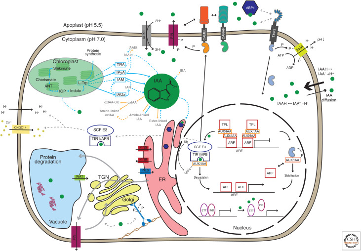
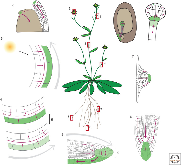
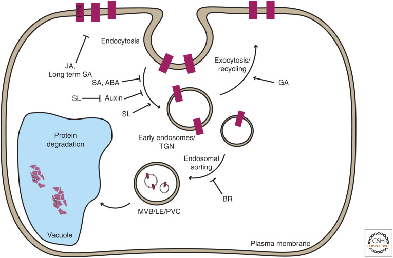
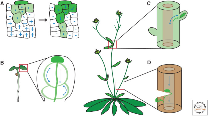
Auxin has always been at the forefront of research in plant physiology and development. Since the earliest contemplations by Julius von Sachs and Charles Darwin, more than a century-long struggle has been waged to understand its function. This largely reflects the failures, successes, and inevitable progress in the entire field of plant signaling and development. Here I present 14 stations on our long and sometimes mystical journey to understand auxin. These highlights were selected to give a flavor of the field and to show the scope and limits of our current knowledge. A special focus is put on features that make auxin unique among phytohormones, such as its dynamic, directional transport network, which integrates external and internal signals, including self-organizing feedback. Accented are persistent mysteries and controversies. The unexpected discoveries related to rapid auxin responses and growth regulation recently disturbed our contentment regarding understanding of the auxin signaling mechanism. These new revelations, along with advances in technology, usher us into a new, exciting era in auxin research.


Figures

Figure 1.
Figure 1.
Auxin biosynthesis, homeostasis, and signaling. The main precursor for IAA (indole-3-acetic acid) biosynthesis, L-tryptophan (L-Trp), is synthesized in chloroplasts. The dominant IAA biosynthesis pathway in Arabidopsis is via indole-3-pyuvic acid (IPyA). Free IAA is either oxidized or conjugated to, for example, amino acids and sugars (for review, see Zhang and Peer 2017 and Casanova-Sáez et al. 2021). IAA can either enter cells through the plasma membrane (PM)-localized H+/auxin symporter AUX1/LAX or diffuse through the PM in its protonated form (IAAH). Inside the cell, IAA is predominantly in an anionic form, necessitating export, which is mediated mainly by PIN proteins at the PM (for review, see Hammes et al. 2021). PIN proteins undergo constant endocytic cycling and are degraded in the vacuole (for review, see Adamowski and Friml 2015). Auxin influences these processes by an unclear mechanism involving myosin phosphorylation (Han et al. 2021). So-called short PINs and PILS mediate auxin in- and efflux in the endoplasmic reticulum (ER) (for review, see Abdollahi Sisi and Růžička 2020). WAT1 is an auxin transporter located in the tonoplast (Ranocha et al. 2013). In the nucleus, auxin binds to the canonical TIR1/AFB-Aux/IAA receptor complex and activates transcription via ubiquitination and degradation of Aux/IAA transcriptional repressors, which releases repressive ARF-Aux/IAA-TPL (TOPLESS) complexes (for review, see Morffy and Strader 2021). This pathway also regulates transcription of CAMEL. The CAMEL/CANAR complex at the PM interacts with and phosphorylates PINs (Hajný et al. 2020). Furthermore, auxin in the nucleus also releases the repressive complex between ETTIN/ARF3 (ETT) and other transcription factors such as INDEHISCENT (IND), causing transcriptional reprogramming required for various developmental processes. Transmembrane kinase (TMK) proteins are components of cell surface auxin signaling. Auxin can trigger the cleavage of the carboxy-terminal kinase domain of TMK1, which then translocates to the nucleus, where it regulates transcription by binding to the noncanonical AUX/IAAs (for review, see McLaughlin et al. 2021). TMK1 also mediates the auxin effect on AHA phosphorylation leading to apoplast acidification (Li et al. 2021b; Lin et al. 2021). It is unclear how auxin triggers the TMK pathway. The possible function of ABP1 as an auxin receptor in the apoplast and its role in the ER remains controversial (for review, see Napier 2021). TIR1/AFB auxin signaling, by an unknown mechanism, also mediates apoplast alkalinization and PM depolarization (Li et al. 2021b; Serre et al. 2021) and Ca2+ influx (Dindas et al. 2018; for review, see Dubey et al. 2021). Green-filled circles depict auxin molecules. Grey dashed arrows indicate hypothetical regulations. Blue arrows indicate IAA biosynthesis and orange arrows indicate metabolic pathways. Figure based on data in Skalický et al. (2018) and Gallei et al. (2020).
Figure 2.
Figure 2.
PIN-dependent auxin transport network generating local auxin maxima and gradients. (1) Maternally produced auxin is transported to the embryo proper, where it accumulates and regulates development (Robert et al. 2018). At early globular stage, auxin production starts at the embryo apex, auxin transport routes reverse, and a new auxin maximum is generated for the root pole specification (Robert et al. 2013; Wabnik et al. 2013). Auxin production is indicated by green stars. (2) Converging auxin fluxes in the epidermis of the shoot apical meristem generate auxin maxima for organ initiation. From the developing primordia, auxin is canalized toward preexisting vasculature (Benková et al. 2003). (3) Light diverges auxin fluxes toward the shaded side of the shoot (Ding et al. 2011), where auxin accumulates (Friml et al. 2002b) and promotes bending. It remains unclear whether this is the primary, causal mechanism. (4) Gravity stimulation diverges auxin fluxes toward the lower side of the shoot (Rakusová et al. 2011), where auxin accumulates and promotes bending. At a later stage, auxin accumulation leads to PIN relocation to the inner cell sides, thus equalizing the auxin distribution and terminating bending (Rakusová et al. 2016). (5) Gravity stimulation diverges auxin fluxes in root columella toward the lower root side, where auxin accumulates (Luschnig et al. 1998; Friml et al. 2002b). Auxin promotes its own flow along the lower root side (Baster et al. 2013) delivering auxin to the elongation zone where it inhibits growth (Fendrych et al. 2018), leading to downward bending of the root. (6) Multiple, redundant PIN fluxes converge to generate an auxin gradient in the central root meristem, which maintains cell divisions and fate specification (Friml et al. 2002a). (7) Auxin flow through the lateral root primordium generates an auxin maximum at the tip, which is partly dissipated by the flow through the epidermis (Benková et al. 2003). Gray arrows indicate direction of growth after gravity or light stimulation. Green shading shows auxin accumulation. Darker green indicates higher auxin levels than lighter green. g denotes direction of gravity.
Figure 3.
Figure 3.
Hormonal regulation of PIN trafficking. PIN auxin exporters undergo constant, subcellular endocytic recycling, which assists in maintaining polar localization (Kleine-Vehn et al. 2011) and integrates various signals. PINs can as well be directed from the plasma membrane (PM) to the vacuole through the trans-Golgi network (TGN)/endosomal network for lytic degradation. Environmental and developmental signals, including hormones, can modulate various steps of PIN trafficking, thus changing PIN incidence at the PM. This eventually fine-tunes directionality and capacity of auxin transport leading to adaptation of plant growth and development. Figure based on data reviewed in Semeradova et al. (2020).
Figure 4.
Figure 4.
Canalization processes for vasculature formation and regeneration. (A) Auxin source (dark green color) polarizes originally homogenous cells to create a directional transport (blue arrows) away from the source. The self-organizing property of auxin transport allows to canalize auxin from an initially broad domain into a narrow channel with high auxin-transporting capacity. (B) Auxin maximum in a cotyledon tip drives auxin canalization in a conserved pattern, demarcating the position of future vasculature. Transport-independent patterning mechanisms are likely also involved (Verna et al. 2019; for review, see Lavania et al. 2021). (C) The shoot apex is a well-known source of auxin, which keeps lateral buds inhibited. Once the apex is removed, the closest lateral bud is released from the inhibition and becomes a new dominant auxin source. Auxin canalization from this bud guides vasculature formation, connecting the lateral bud to the preexisting stem vasculature (Balla et al. 2011). (D) Wounding of stem vasculature results in a local auxin accumulation above the wound. Subsequently, auxin is canalized around the wound to initiate reconnection of the preexisting vasculature (Sauer et al. 2006; Mazur et al. 2016). Local external auxin application to the side of the stem also triggers auxin channel formation and guides formation of vasculature (Mazur et al. 2020b). Other cases of auxin canalization are during organogenesis at the shoot apical meristem and during embryogenesis (see Fig. 2).

References

    1. Abas L, Kolb M, Stadlmann J, Janacek DP, Lukic K, Schwechheimer C, Sazanov LA, Mach L, Friml J, Hammes UZ. 2021. Naphthylphthalamic acid associates with and inhibits PIN auxin transporters. Proc Natl Acad Sci 118: e2020857118. 10.1073/pnas.2020857118 - DOI - PMC - PubMed
    1. Abdollahi Sisi N, Růžička K. 2020. ER-localized PIN carriers: regulators of intracellular auxin homeostasis. Plants (Basel) 9: 1527. 10.3390/plants9111527 - DOI - PMC - PubMed
    1. Abel S, Theologis A. 2010. Odyssey of auxin. Cold Spring Harb Perspect Biol 2: a004572. 10.1101/cshperspect.a004572 - DOI - PMC - PubMed
    1. Adamowski M, Friml J. 2015. PIN-dependent auxin transport: action, regulation, and evolution. Plant Cell 27: 20–32. 10.1105/tpc.114.134874 - DOI - PMC - PubMed
    1. Antoniadi I, Novák O, Gelová Z, Johnson A, Plíhal O, Simerský R, Mik V, Vain T, Mateo-Bonmatí E, Karady M, et al. 2020. Cell-surface receptors enable perception of extracellular cytokinins. Nat Commun 11: 4284. 10.1038/s41467-020-17700-9 - DOI - PMC - PubMed

LinkOut - more resources