. 2021 Oct;26(10):5516-5531.
doi: 10.1038/s41380-021-01248-1. Epub 2021 Aug 16.

The acute phase protein lactoferrin is a key feature of Alzheimer's disease and predictor of Aβ burden through induction of APP amyloidogenic processing

Affiliations

The acute phase protein lactoferrin is a key feature of Alzheimer's disease and predictor of Aβ burden through induction of APP amyloidogenic processing

Andrew Tsatsanis et al. Mol Psychiatry. 2021 Oct.

Abstract

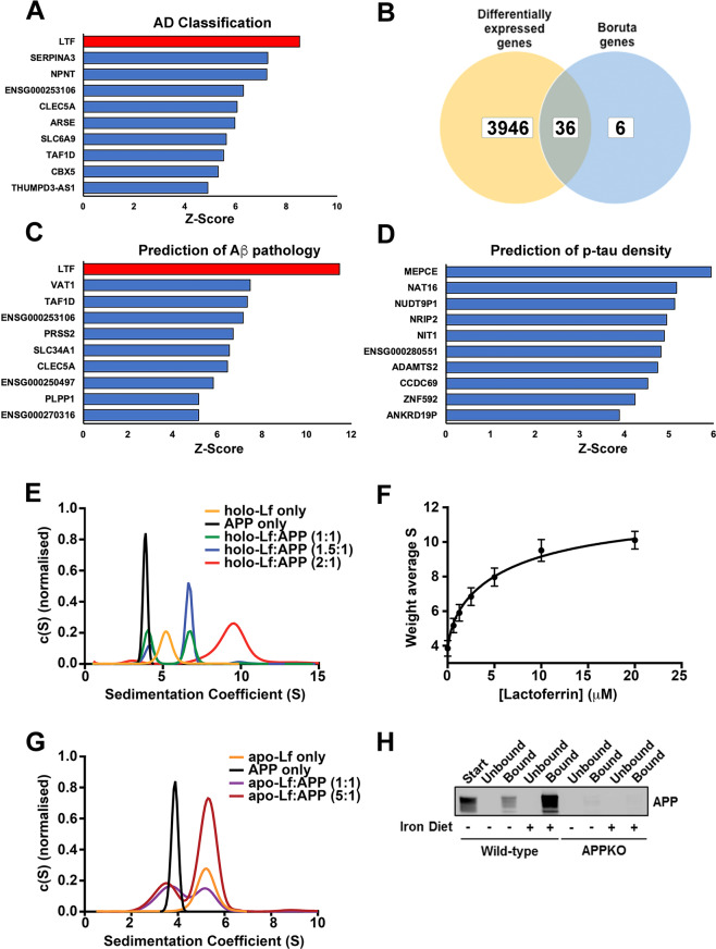
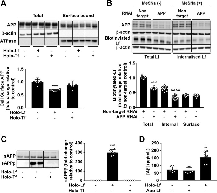
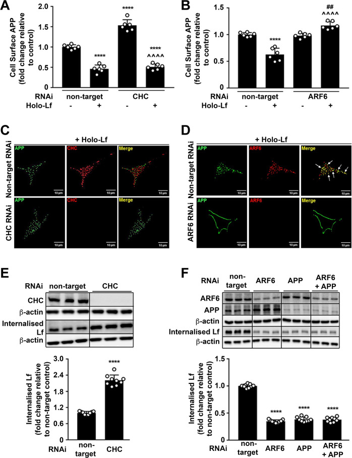
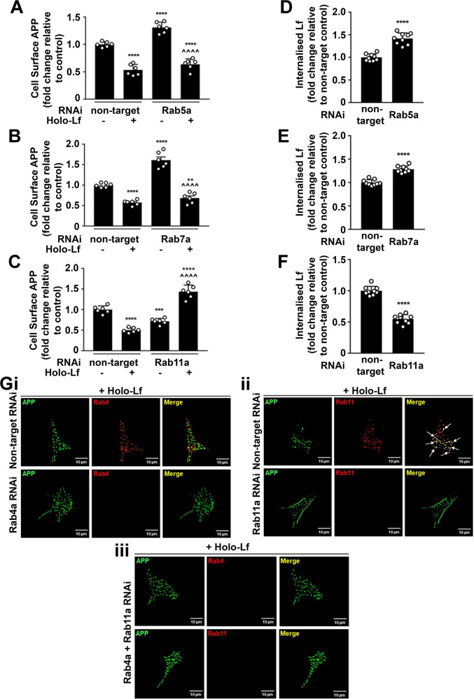
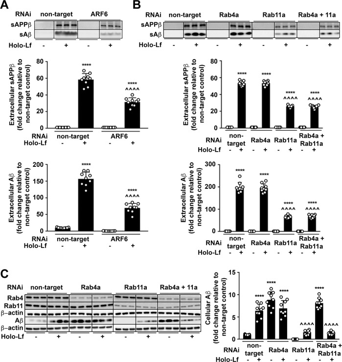
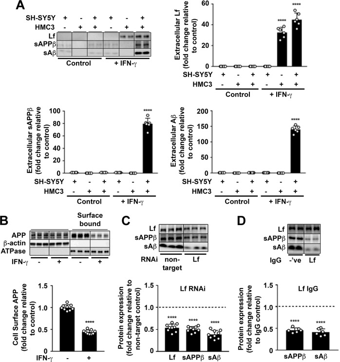
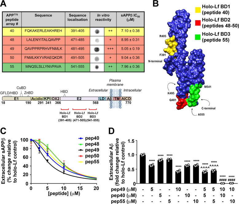
Amyloidogenic processing of the amyloid precursor protein (APP) forms the amyloid-β peptide (Aβ) component of pathognomonic extracellular plaques of AD. Additional early cortical changes in AD include neuroinflammation and elevated iron levels. Activation of the innate immune system in the brain is a neuroprotective response to infection; however, persistent neuroinflammation is linked to AD neuropathology by uncertain mechanisms. Non-parametric machine learning analysis on transcriptomic data from a large neuropathologically characterised patient cohort revealed the acute phase protein lactoferrin (Lf) as the key predictor of amyloid pathology. In vitro studies showed that an interaction between APP and the iron-bound form of Lf secreted from activated microglia diverted neuronal APP endocytosis from the canonical clathrin-dependent pathway to one requiring ADP ribosylation factor 6 trafficking. By rerouting APP recycling to the Rab11-positive compartment for amyloidogenic processing, Lf dramatically increased neuronal Aβ production. Lf emerges as a novel pharmacological target for AD that not only modulates APP processing but provides a link between Aβ production, neuroinflammation and iron dysregulation.


Conflict of interest statement

The authors have no known financial and personal relationships that may inappropriately influence their work. BG is a director of Pacific Analytics PTY LTD and SMRTR PTY LTD, Australia; a founding member of the International Cerebral Palsy Genetics Consortium and a member of the Australian Genomics Health Alliance. AIB is a shareholder in Prana Biotechnology Ltd, Cogstate Ltd, Brighton Biotech LLC, Grunbiotics Pty Ltd, Eucalyptus Pty Ltd and Mesoblast Ltd. He is a paid consultant for, and has a profit share interest in, Collaborative Medicinal Development Pty Ltd.

Figures

Fig. 1
Fig. 1. A biophysical interaction between APP and Lf supports machine learning analysis of a lead hit in classifying disease and predicting Aβ pathology.
A, B The top 10 genes identified by z-score using the feature selection algorithm, Boruta for classifying the ROSMAP cohort into neuropathologically positive AD cases and controls (A). Of these genes a small fraction were not identified by differential expression analysis using EdgeR (B). C Top 10 genes ranked by z-score predicting total amount of amyloid pathology. D Top 10 genes ranked by z-score predicting p-tau (AT8) immunoreactivity across all seven cortical regions and the hippocampus. E Sedimentation coefficient distributions of holo-Lf alone (5 μM; orange) and APP (2.5 μM) in the absence (black) and presence of Lf (green: 2.5 μM; blue: 3.75 μM; red: 5 μM). The c(S) distributions indicate two different complexes that form in a concentration dependent manner. See Supplementary Fig. S2 for example data sets showing data quality and the fit of the c(s) model to the data. F The weight average sedimentation coefficient obtained by integrating the c(S) distribution (as shown in E) calculated as a function of Lf concentration (Eq 1 in Supplementary data) with the assumption that APP contains two binding sites for Lf with two different dissociation constants. G Sedimentation velocity analysis of apo-Lf alone (2.5 μM; orange) and APP (2.5 μM) in the absence (black) and presence of apo-Lf (purple: 2.5 μM; maroon: 12.5 μM). H Confirmation of an interaction of APP with Lf in vivo using anti-APP (1:1000; 22C11) for detection and anti-Lf (1:200) for immunoprecipitation of brain homogenates from APP–/– mice and littermate controls either on a normal or high-iron diet. Specificity of interaction of APP to Lf was confirmed using anti-β-actin as the capture antibody (data not shown). Data are mean ± SEM of three experiments performed in triplicate.
Fig. 2
Fig. 2. Iron-bound Lf decreases cell-surface APP levels and promotes the amyloidogenic processing of APP.
A Biotinylation of surface proteins on primary murine neurons cultured in holo-Lf or holo-Tf (500 nM; 2 h) and followed by streptavidin immunoprecipitation shows a decrease in biotinylated APP with only holo-Lf when normalised against Na+/K+ ATPase surface protein content. B Biotinylated holo-Lf (200 nM; 1 h at 37 °C) was added to SH-SY5Y cells transfected with control non-target or APP RNAi (20 nM for 48 h) before being subjected to the ligand internalisation assay. APP depletion was confirmed by western blot (22C11). Total biotinylated holo-Lf (MeSNa (–)) and internalised holo-Lf (MeSNa (+)) (detected by Streptavidin-HRP) were quantified whilst surface-bound biotinylated Lf was determined by subtracting the internal from total fraction. C Primary murine neurons treated with holo-Lf or holo-Tf (500 nM; 2 h) were evaluated for sAPPβ release into the media. D Aβ production was also measured by ELISA on the media after treatment with apo- or holo-Lf (500 nM; 2 h). Data are mean ± SEM of three separate experiments performed in duplicate (A, B, D) or triplicate (C). Quantified data depict fold change compared to non-treated control cells, **p < 0.01 and ****p < 0.0001 or in (B) the non-targeted internal fraction, ^^^^p < 0.0001, as analysed statistically by two-tailed t-tests.
Fig. 3
Fig. 3. Holo-Lf-mediated APP internalisation is clathrin independent and ARF6 dependent.
A, B Cell-surface APP (ab15272) response to holo-Lf (500 nM; 2 h) as monitored by FACS in non-permeabilised SH-SY5Ys after control non-targeted of CHC knockdown by RNAi (40 nM; 72 h) (A) or ARF6 RNAi (20 nM; 48 h) (B). C, D Deconvoluted images of double immunofluorescence confocal microscopy of wt-APP695 SH-SY5Ys treated with holo-Lf. After RNAi depletion of CHC or ARF6 as in (A, B), surface APP was labelled with an APP antibody (22C11) (green) at 4 °C before replacing media with holo-Lf (1 μM; 1 h) at 37 °C. An APP secondary detection antibody was then added with total CHC (ab21679) (red) (C) or ARF6 (ab131261) (red) (D) co-labelling. Co-localisation of APP with CHC (C) and ARF6 (D) is represented as yellow in the merged image (white arrows). E, F Biotinylated holo-Lf (0.5 mg/ml; 1 h at 37 °C) added to SH-SY5Ys transfected with control non-target and CHC (40 nM; 72 h) (E) or ARF6 ± APP RNAi (20 nM; 48 h) (F) before being subjected to the ligand internalisation assay. Residual surface biotin was stripped with MeSNa so that only internalised biotinylated Lf could be detected in the total cell lysate when analysed by western blot with streptavidin-HRP. Data are mean ± SEM of three experiments performed at least in duplicate. Statistical analysis in A, B was by two-way ANOVA or two-tailed t-tests for E, F, ****p < 0.0001 depicts fold change compared with levels derived from non-targeting control, ^^^^p < 0.0001 compared to CHC RNAi (A) or holo-Lf-treated (B) non-target control and ##p < 0.0001 compared to ARF6 RNAi (B). C, D Images are a representative from multiple cells within experiments carried out in duplicate. Scale bar = 10 µm.
Fig. 4
Fig. 4. Holo-Lf promotes APP trafficking through the Rab11-positive recycling endosome.
AC Flow cytometric quantification of cell-surface APP levels (ab15272) on the cell surface of non-permeabilised SH-SY5Ys with and without holo-Lf (500 nM; 2 h) after treatment with RNAi (20 nM; 48 h) for Rab5a (A), Rab7a (B) Rab11a (C) and a non-targeted control. DF Within the same experimental parameters as AC, quantification of the effect of Rab5a (D), Rab7a (E) and Rab11a (F) knockdown on internalisation of biotinylated holo-Lf (0.5 mg/ml; 1 h at 37 °C) was measured by the ligand internalisation assay. Residual surface biotin was stripped with MeSNa so that only internalised biotinylated Lf could be detected in the total cell lysate when analysed by western blot (shown in Supplementary Fig. S6). G Representative deconvoluted images from double immunofluorescence confocal microscopy of wt-APP695 SH-SY5Ys reverse transfected with RNAi for control non-target, Rab4a (i) or Rab11a (ii). In double knockdown, cells were reverse transfected with Rab4a and then forward transfected with Rab11a (iii) RNAi (20 nM; 48 h). After surface labelling with anti-APP (22C11) (green) at 4 °C, cells were treated with holo-Lf (1 μM; 1 h at 37 °C) and then permeabilised to label with the antibody to APP (green) and anti-Rab4 (ab13252; red) (i, iii) or anti-Rab11 (ab3612; red) (ii, iii). AF Data are mean ± SEM of three experiments performed at least in duplicate. Statistical analysis by two-way ANOVA (AC) or two-tailed t-tests (DF), **p < 0.01, ***p < 0.001 and ****p < 0.0001 depict fold change compared to the untreated non-targeting control and ^^^^p < 0.0001 compared to Rab RNAi without holo-Lf. G Images are a representative from multiple cells within experiments carried out in duplicate. Scale bar = 10 µm.
Fig. 5
Fig. 5. Holo-Lf-mediated amyloidogenic processing of APP requires ARF6 and the Rab11-positive recycling endosome.
A Amyloidogenic processing of APP induced by holo-Lf (500 nM; 2 h) in wt-APP695 SH-SY5Ys pre-treated with control non-target or ARF6 RNAi (20 nM; 48 h) was measured by sAPPβ and Aβ levels in the media. B, C wt-APP695 SH-SY5Ys were reverse transfected with control non-target or Rab4a RNAi and/or forward transfected with Rab11a RNAi (20 nM; 48 h) before addition of holo-Lf (500 nM; 2 h). Extracellular sAPPβ and Aβ (B) as well as intracellular Aβ (C) protein levels were quantified. Data are mean ± SEM of three experiments performed in triplicate and depicted as fold change compared with levels derived from control non-target cells. Statistical analysis was by two-way ANOVA compared to untreated non-targeting control, ****p < 0.0001 or holo-Lf-treated non-targeting control, ^^^^p < 0.0001.
Fig. 6
Fig. 6. Secreted holo-Lf from activated microglia reduces neuronal surface-presented APP and increases APP amyloidogenic processing.
A In monocultures and a transwell co-culture with HMC3 microglia cultured in the upper inserts and wt-APP695 SH-SY5Ys in the lower wells, human recombinant IFN-γ (10 ng/ml; 24 h) was used to activate microglia, as confirmed by the MHC class II marker (Supplementary Fig. S9A). IFN-γ-induced changes in media levels of secreted Lf, sAPPβ and Aβ were quantified from western blotting. B From transwell co-culture as in (A), surface APP from wt-APP695 SH-SY5Y co-cultures were measured by surface biotinylation after microglial activation. C HMC3 cells transfected with control non-target or Lf RNAi (20 nM; 48 h) were added to transwell co-cultures with wt-APP695 SH-SY5Y and activated with IFN-γ (10 ng/ml; 24 h) to determine expression levels of sAPPβ and Aβ secretion from wt-APP695 SH-SY5Y. D In a neutralising antibody inhibition assay, a polyclonal for Lf or an isotypic control IgG (20 μg/ml) was added to the media of wt-APP695 SH-SY5Y in the transwell co-culture before microglial activation by IFN-γ (10 ng/ml; 24 h). Inhibition of Lf binding to APP by the antibody was determined by measuring secreted Lf and the APP amyloidogenic protein fragments (sAPPβ and Aβ) in neuronal media. Data are mean ± SEM of three experiments performed in duplicate as a minimum and normalised against a control protein. Statistical analysis was by two-tailed t-tests compared to corresponding cell line without IFN-γ (A, B), non-target control (C) or isotype IgG treatment (D), ****p < 0.0001.
Fig. 7
Fig. 7. Identification of the holo-Lf binding sites on APP required for holo-Lf-induced amyloidogenic processing of APP.
A APP peptide reactivity to holo-Lf (1 μg/ml; 2 h) was determined by visual observation (+++ strong, ++ moderate, + weak) and specificity of binding to holo-Lf by peptides 40, 48, 49, 50 and 55 was confirmed using apo-Lf and detection antibody treatment alone (see Supplementary Fig. S10A). B Model of holo-Lf binding sites (coloured as shown in (A)) overlaid on the APP-E2 structure [97]. C Potency of the holo-Lf binding peptides of APP was assessed by dose dependently pre-incubating the peptide with holo-Lf (500 nM; 2 h) in vitro before adding to wt-APP695 SH-SY5Y and evaluating sAPPβ secretion in the media after a further 2 h. wt-APP695 SH-SY5Y exposure of APP peptide alone at each respective concentration showed no change in sAPPβ levels (data not shown). IC50 for each peptide is shown in (A). D Covering the main holo-Lf binding sites, APP peptide 40, 49 and 55 were used to evaluate combinatory inhibition of holo-Lf-induced Aβ production in wt-APP695 SH-SY5Y. As in (C), peptide 49 with 40 or 55 (5 and 10 µM) was pre-incubated with holo-Lf (500 nM; 2 h) before addition to neuronal media for a further 2 h. Data are mean ± SEM of two experiments performed in duplicate with statistical analysis by two-way ANOVA comparing holo-Lf treated control, ****p < 0.0001, 5 µM peptide 49 alone, ^^^^p < 0.0001 or 10 µM peptide 49 alone, ####p < 0.0001.

References

    1. Cras P, Kawai M, Lowery D, Gonzalez-DeWhitt P, Greenberg B, Perry G. Senile plaque neurites in Alzheimer disease accumulate amyloid precursor protein. Proc Natl Acad Sci USA. 1991;88:7552–6. - PMC - PubMed
    1. Milward EA, Papadopoulos R, Fuller SJ, Moir RD, Small D, Beyreuther K, et al. The amyloid protein precursor of Alzheimer’s disease is a mediator of the effects of nerve growth factor on neurite outgrowth. Neuron. 1992;9:129–37. - PubMed
    1. Ninomiya H, Roch JM, Sundsmo MP, Otero DA, Saitoh T. Amino acid sequence RERMS represents the active domain of amyloid beta/A4 protein precursor that promotes fibroblast growth. J Cell Biol. 1993;121:879–86. - PMC - PubMed
    1. Duce JA, Tsatsanis A, Cater MA, James SA, Robb E, Wikhe K, et al. Iron-export ferroxidase activity of beta-amyloid precursor protein is inhibited by zinc in Alzheimer’s disease. Cell. 2010;142:857–67. - PMC - PubMed
    1. Wong BX, Tsatsanis A, Lim LQ, Adlard PA, Bush AI, Duce JA. beta-Amyloid precursor protein does not possess ferroxidase activity but does stabilize the cell surface ferrous iron exporter ferroportin. PloS One. 2014;9:e114174. - PMC - PubMed

Publication types

MeSH terms