. 2021 Oct;16(10):4611-4632.
doi: 10.1038/s41596-021-00588-0. Epub 2021 Aug 16.

Generic acquisition protocol for quantitative MRI of the spinal cord

Julien Cohen-Adad  1   2   3 Eva Alonso-Ortiz  4 Mihael Abramovic  5 Carina Arneitz  5 Nicole Atcheson  6 Laura Barlow  7 Robert L Barry  8   9   10 Markus Barth  11 Marco Battiston  12 Christian Büchel  13 Matthew Budde  14 Virginie Callot  15   16 Anna J E Combes  17 Benjamin De Leener  18   19 Maxime Descoteaux  20   21 Paulo Loureiro de Sousa  22 Marek Dostál  23 Julien Doyon  24 Adam Dvorak  25 Falk Eippert  26 Karla R Epperson  27 Kevin S Epperson  27 Patrick Freund  28 Jürgen Finsterbusch  13 Alexandru Foias  4 Michela Fratini  29   30 Issei Fukunaga  31 Claudia A M Gandini Wheeler-Kingshott  12   32   33 Giancarlo Germani  33 Guillaume Gilbert  34 Federico Giove  30   35 Charley Gros  4   6 Francesco Grussu  12   36 Akifumi Hagiwara  31 Pierre-Gilles Henry  37 Tomáš Horák  38 Masaaki Hori  39 James Joers  37 Kouhei Kamiya  40 Haleh Karbasforoushan  41   42 Miloš Keřkovský  23 Ali Khatibi  24   43 Joo-Won Kim  44 Nawal Kinany  45   46 Hagen Kitzler  47 Shannon Kolind  7   25   48 Yazhuo Kong  49   50   51 Petr Kudlička  38 Paul Kuntke  47 Nyoman D Kurniawan  6 Slawomir Kusmia  52   53   54 René Labounek  55   56 Maria Marcella Laganà  57 Cornelia Laule  58 Christine S Law  59 Christophe Lenglet  37 Tobias Leutritz  60 Yaou Liu  61   62 Sara Llufriu  63 Sean Mackey  59 Eloy Martinez-Heras  63 Loan Mattera  64 Igor Nestrasil  37   55 Kristin P O'Grady  17   65 Nico Papinutto  66 Daniel Papp  4   51 Deborah Pareto  67 Todd B Parrish  41 Anna Pichiecchio  32   33 Ferran Prados  12   53   68 Àlex Rovira  67 Marc J Ruitenberg  69 Rebecca S Samson  12 Giovanni Savini  33 Maryam Seif  28   60 Alan C Seifert  44 Alex K Smith  51 Seth A Smith  17   65 Zachary A Smith  70 Elisabeth Solana  63 Yuichi Suzuki  40 George Tackley  52 Alexandra Tinnermann  13 Jan Valošek  71 Dimitri Van De Ville  45   46 Marios C Yiannakas  12 Kenneth A Weber 2nd  59 Nikolaus Weiskopf  60   72 Richard G Wise  52   73 Patrik O Wyss  5 Junqian Xu  44
Affiliations

Generic acquisition protocol for quantitative MRI of the spinal cord

Julien Cohen-Adad et al. Nat Protoc. 2021 Oct.

Abstract

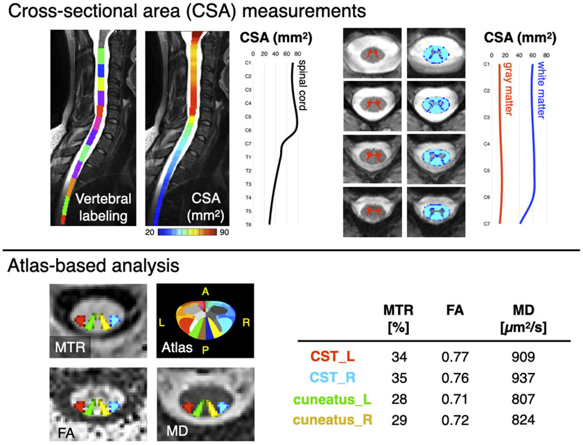
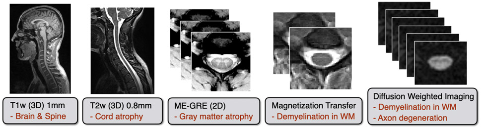
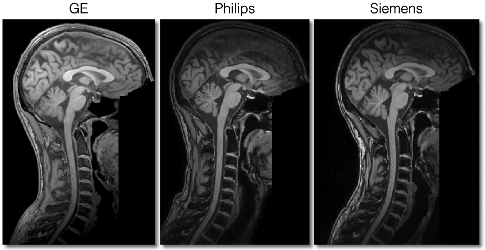
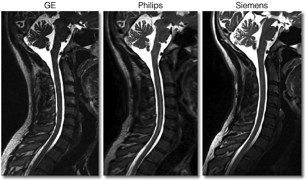
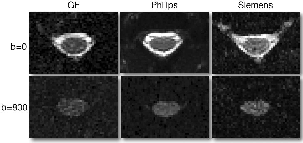
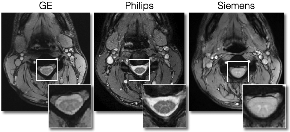
Quantitative spinal cord (SC) magnetic resonance imaging (MRI) presents many challenges, including a lack of standardized imaging protocols. Here we present a prospectively harmonized quantitative MRI protocol, which we refer to as the spine generic protocol, for users of 3T MRI systems from the three main manufacturers: GE, Philips and Siemens. The protocol provides guidance for assessing SC macrostructural and microstructural integrity: T1-weighted and T2-weighted imaging for SC cross-sectional area computation, multi-echo gradient echo for gray matter cross-sectional area, and magnetization transfer and diffusion weighted imaging for assessing white matter microstructure. In a companion paper from the same authors, the spine generic protocol was used to acquire data across 42 centers in 260 healthy subjects. The key details of the spine generic protocol are also available in an open-access document that can be found at https://github.com/spine-generic/protocols . The protocol will serve as a starting point for researchers and clinicians implementing new SC imaging initiatives so that, in the future, inclusion of the SC in neuroimaging protocols will be more common. The protocol could be implemented by any trained MR technician or by a researcher/clinician familiar with MRI acquisition.


Figures

Figure 1.
Figure 1.. Illustration of the MRI metrics that could be extracted from the spine generic protocol.
The top panel “Cross-sectional area (CSA) measurements” shows morphometric measures of the spinal cord and its gray and white matter. The bottom panel “Atlas-based analysis” on the left shows axial views of qMRI maps: Magnetization Transfer Ratio (MTR), Fractional Anisotropy (FA) and Mean Diffusivity (MD), with an overlay of 4 spinal tracts of general interest: the descending corticospinal tract (CST) and the ascending cuneatus, left (L) and right (R). The “Atlas” image corresponds to the white matter atlas , which includes 30 white matter tracts that could be used for computing metrics within specific tracts of interest. This atlas also includes 6 parcellations of the gray matter. The table shows average values of each metric in the corresponding tract.
Figure 2.
Figure 2.. List of sequences included in the spine generic protocol (in black) with possible applications (in red).
The total acquisition time is 20-30min, depending on the manufacturer/model.
Figure 3.
Figure 3.. Patient positioning.
Suggested subject positioning: Use a cushion to minimize cervical lordosis (bottom panel)
Figure 4.
Figure 4.
Positioning of FOV for T1w scans.
Figure 5.
Figure 5.
Positioning of FOV for T2w scans.
Figure 6.
Figure 6.
Positioning of FOV, shim box and saturation bands for the DWI scan.
Figure 7.
Figure 7.
Example of pulse oximeter trace on a Siemens scanner for triggered acquisition (small triangles).
Figure 8.
Figure 8.. Positioning of the FOV, shim box and saturation bands for the GRE-ME scan.
Siemens and GE users: the saturation band is already automatically positioned. Philips users: the saturation bands are “invisible” on this sequence, but they are nevertheless applied.
Figure 9.
Figure 9.
Sagittal views of good quality T1w scans for each manufacturer.
Figure 10.
Figure 10.
Sagittal views of good quality T2w scans for each manufacturer.
Figure 11.
Figure 11.
Axial views of good quality data for DWI scans at b=0 s/mm2 (top row) and b=800 s/mm2 (bottom row). The DW image corresponds to a diffusion gradient vector fairly orthogonal to the cord axis, hence the visible spinal cord. Notice the different noise patterns across the manufacturers, which is due to the different types of filters applied across manufacturers; these filters were present in an older version of the protocol, but have been removed in the latest version of the protocol in order to minimize differences across manufacturers.
Figure 12.
Figure 12.
Axial views of good quality data for MT0, MT1 and T1w scans. Notice the slight motion artifact on the Philips MT0 scan. Also notice the strong signal intensity at the periphery of the tissue on the Siemens scans, which is due to the inactivation of the intensity bias filter. This filter is not relevant when computing qMRI metrics such as MTR or MTsat.
Figure 13.
Figure 13.
Axial views of good quality ME-GRE scans for each manufacturer.

References

    1. Cercignani M, Dowell NG & Tofts PS Quantitative MRI of the Brain: Principles of Physical Measurement, Second edition. (CRC Press, 2018).
    1. Cohen-Adad J & Wheeler-Kingshott C Quantitative MRI of the Spinal Cord. (2014).
    1. Wheeler-Kingshott CA et al. The current state-of-the-art of spinal cord imaging: applications. Neuroimage 84, 1082–1093 (2014). - PMC - PubMed
    1. Stroman PW et al. The current state-of-the-art of spinal cord imaging: methods. Neuroimage 84, 1070–1081 (2014). - PMC - PubMed
    1. Cohen-Adad J & Wald LL Array Coils, in Quantitative MRI of the Spinal Cord 59–67 (2014).