. 2021 Winter;20(1):384-397.
doi: 10.22037/ijpr.2020.113272.14199.

AML-derived Extracellular Vesicles Confer De Novo Chemoresistance to Leukemic Myeloblast Cells by Promoting Drug Export Genes Expression and ROS Inhibition

Affiliations

AML-derived Extracellular Vesicles Confer De Novo Chemoresistance to Leukemic Myeloblast Cells by Promoting Drug Export Genes Expression and ROS Inhibition

Mohieddin Barzegar et al. Iran J Pharm Res. 2021 Winter.

Abstract

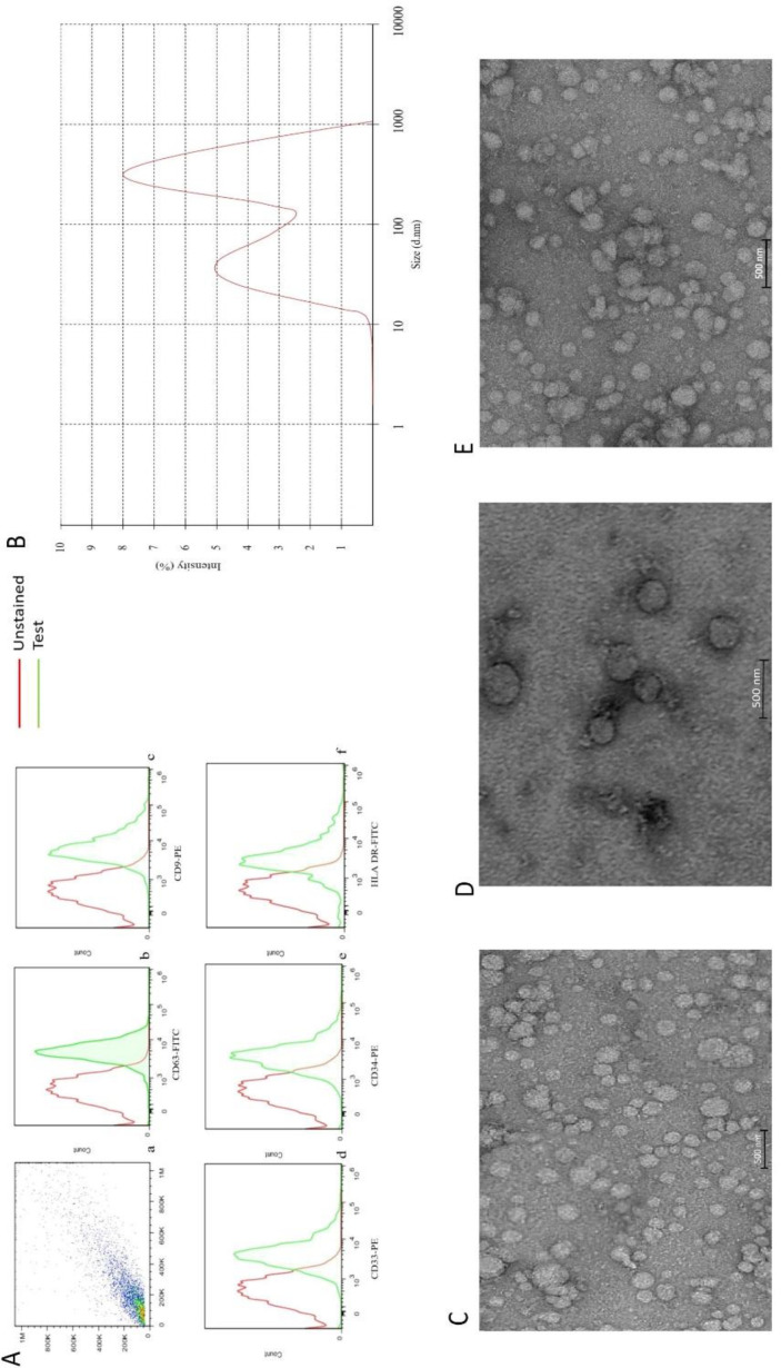
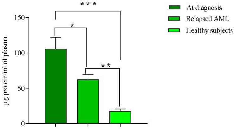
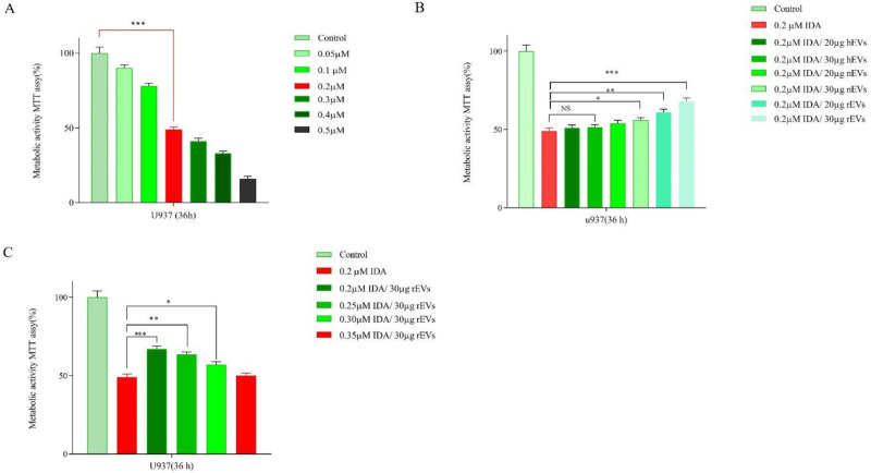
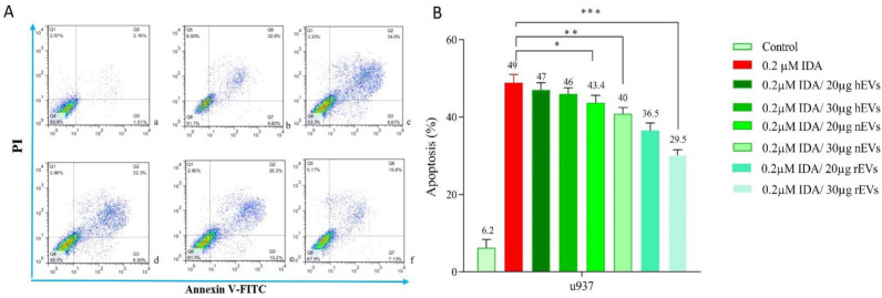
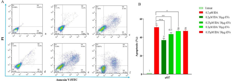
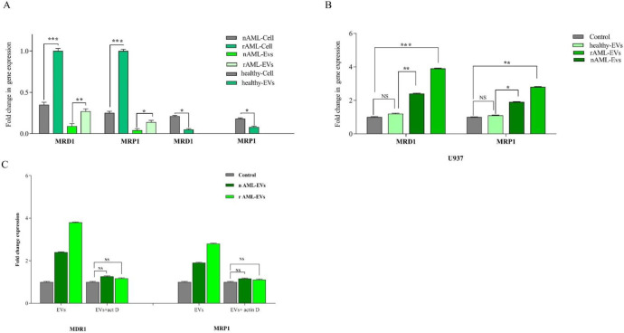
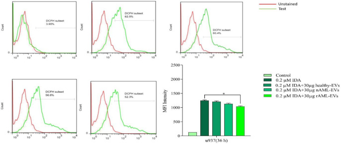
In spite of successful initial remission, chemo-resistance and relapse are still concerning points in acute myeloid leukemia (AML) treatment strategies. Multidrug resistance (MDR) appears to be the major contributor of chemo-resistance, arising in some sub-clones of cancers and could be developed in others. The aim of this study was to investigate the role of extracellular vesicles (EVs) derived from AML patients on the transmission of chemo-resistance phenotype. Ultracentrifugation was employed to isolate EVs from healthy controls, new cases, and relapsed AML patients. The EVs size, morphology, and immunophenotype were determined by dynamic light scattering, TEM, and flow cytometry respectively. Bradford assay was performed to measure the protein content of EVs. MTT assay and flow cytometry analysis were also used to determine the viability index, induction of apoptosis, and ROS generation in U937 cells. The expression level of two efflux pumps was assessed using qRT-PCR analysis. Findings of TEM, DLS, and flow cytometry confirmed that EVs had a desirable shape, size, and surface markers. EVs derived from both new cases and relapsed AML patients significantly reduced idarubicin-induced apoptosis in the U937 cells. The analysis of drug efflux pumps gens revealed that EVs over-express MRD1 and MRP1 in the target cells. These findings suggested a novel role of EVs in mediating the acquired chemo-resistance in AML patients by inducing the expression of the drug efflux pumps; however, further investigations will be required to elucidate other underlying mechanisms of resistance that are mediated by EVs.

Keywords: Acute myeloid leukemia; De novo; Extracellular vesicles; Multidrug resistance; Relapse.


Figures

Figure 1
Figure 1
(A) a flow cytometry gate AML- EVs. Flow-cytometric analyses showed that the AML-EVs express CD63, CD9, CD33 CD34, and HLA DR (b, c, d, e, and f). (B) Measurement of the size range of EVs by DLS with the average size of 340 nm. (C, D, and E) TEM analyses of EVs, three groups of newly diagnosed patients, healthy subjects and relapsed patients, respectively
Figure 2
Figure 2
Mean protein content of EVs measured by Bradford method. Concentration of protein in new cases and relapsed groups were significantly higher than healthy subjects. Of note, the concentration of protein in new cases was also higher than relapsed cases. Data are mean ± SE of three independent experiments. Statistical significance was defined at *P < 0.05, **P < 0.01 and ***P < 0.001 compared to corresponding control
Figure 3
Figure 3
(A) Effects of IDA (0.05-0.5 μM) on the viability of U937 cells. The growth suppressive effect of IDA on U937 cells was assessed using MTT assay. IC50 pharmaceutical dose for U937 cells was 0.2 µM. (B) Data indicated that the anti-proliferative effects of IDA was attenuated in the presence of different AML-derived EVs. (C) When U937 cells were treated with relapsed AML-EVs (30 µg) in combination with IDA, there was a more significant elevation in the IC50 value of IDA. Data are mean ± SE of three independent experiments. Statistical significance was defined at *P < 0.05, **P < 0.01 and ***P < 0.001 compared to corresponding control
Figure 4
Figure 4
Effects of IDA on the induction of apoptosis in U937 cells. The cells were treated with 0.2 μM IDA and 20 and 30 µg of healthy, new case AML and relapsed AML-EVs respectively. Data are mean ± SE of three independent experiments. Statistical significance were defined at *P < 0.05, **P < 0.01 and ***P < 0.001compared to corresponding control
Figure 5
Figure 5
The result of annexin V/PI staining in EVs - or IDA+ 30 µg relapsed AML-EVs‐treated U937 during co-culture. Cells were treated for 36 h with either EVs (30 µg) or IDA+ EVs (30 µg). The relapsed AML-EVs protected the cells from IDA-induced apoptosis and enhanced the IDA IC50 in U937 cells (P < 0.001).
Figure 6
Figure 6
(A) MDR-1 and MRP1 expression in healthy, new case AML and relapsed AML-cells and correspond EVs. We first analyzed the normalized expression of MRD1 and MRP1 in cellular and vesicular compartment of all subjects. The resulting data showed that the expression of these genes were lower in EVs as compared with AML parent cells. In new cases and relapsed patients, the fold changes of both genes in EVs were increased. (B) The expression of MRD1 and MRP1in U937 cell treated with new cases and relapsed EVs were significantly increased. Data are mean ± SE of three independent experiments. (C) U937 cells was treated with actinomycin D (1 mg/mL) prior to EV incubation. After 24 h of EV integration, we did not observe any significant change in MDR-1 and MRP1 expression, indicating that the increase of these genes was not due to RNA transfer. Statistical significance were defined at *P < 0.05, **P < 0.01 and ***P < 0.001compared to corresponding control
Figure 7
Figure 7
U937 cells were treated with or without 30 µg of healthy, new case AML and relapsed AML-EVs, and assayed for ROS production by using the fluorescent dye DCFDA. Representative histograms showed ROS generation in each experimental condition. Graph represented the mean values ± SE of the MFI of DCF. Statistical significance were defined at *P < 0.05

References

    1. Roug AS, Ommen HB. Clinical use of measurable residual disease in acute myeloid leukemia. Curr. Treat. Options Oncol. . 2019;20:1–12. - PubMed
    1. Saito Y, Kitamura H, Hijikata A, Tomizawa-Murasawa M, Tanaka S, Takagi S, Uchida N, Suzuki N, Sone A, Najima Y. Identification of therapeutic targets for quiescent, chemotherapy-resistant human leukemia stem cells. Sci. Transl. Med. . 2010;2:17ra9–ra9. - PMC - PubMed
    1. Rowe JM, Kim HT, Cassileth PA, Lazarus HM, Litzow MR, Wiernik PH, Tallman MS. Adult patients with acute myeloid leukemia who achieve complete remission after 1 or 2 cycles of induction have a similar prognosis: a report on 1980 patients registered to 6 studies conducted by the Eastern Cooperative Oncology Group. Cancer . 2010;116:5012–21. - PMC - PubMed
    1. Behrmann L, Wellbrock J, Fiedler W. Acute myeloid leukemia and the bone marrow niche—take a closer look. Front. Oncol. . 2018;8:444. - PMC - PubMed
    1. Landau DA, Carter SL, Getz G, Wu CJ. Clonal evolution in hematological malignancies and therapeutic implications. Leukemia . 2014;28:34–43. - PMC - PubMed

LinkOut - more resources