

Mediastinal bronchial artery aneurysm presenting as an incidental mediastinal mass: A rare finding
- PMID: 34401009
- PMCID: PMC8349918
- DOI: 10.1016/j.radcr.2021.06.076
Mediastinal bronchial artery aneurysm presenting as an incidental mediastinal mass: A rare finding
Abstract
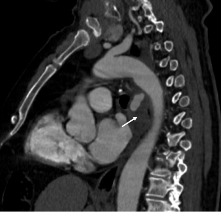
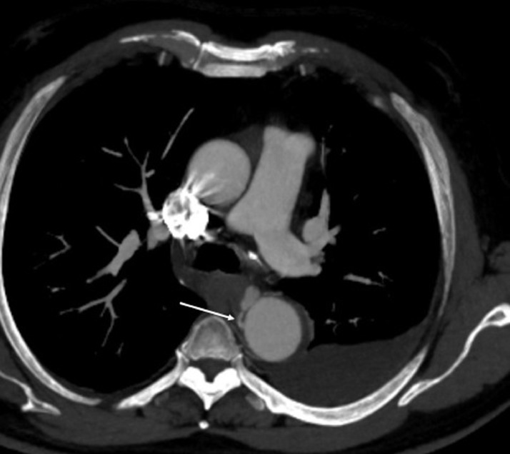
Mediastinal bronchial artery aneurysm is very rare and only few cases have been reported in the literature. The clinical presentations are varied, ranging from an incidental radiological finding to a cataclysmic rupture leading to hemorrhagic shock. Thus, a quick treatment is indicated upon diagnosis. Therapeutic options are various including surgical resection, stent grafting with percutaneous embolization of feeding vessel or transtarterial embolization. Herein we describe a case of an incidental mediastinal bronchial artery aneurysm in a 63-year-old man, managed by transtarterial embolization.
Keywords: Aneurysm; Mediastinal bronchial artery; Transtarterial embolization.
© 2021 The Authors. Published by Elsevier Inc. on behalf of University of Washington.
Figures


References
-
- Tanaka K, Ihaya A, Horiuci T. Giant mediastinal bronchial artery aneurysm mimicking benign esophageal tumor: a case report and review of 26 cases from literature. J Vasc Surg. 2003;38:1125–1129. PubMed: 14603226. - PubMed
-
- Watanabe S, Matayoshi Y, Takeshita H, Nishizawa I, Koh R. Two cases of bronchial artery aneurysm. Hirosaki Med J. 1981;33:512–513.
-
- Abet D, Pietri J. Ruptured bronchial artery aneurysm simulating dissection of the aorta in a patient with bronchopulmonary sequestration. J Chir. 1981;118:743–746. - PubMed
-
- Sancho C, Dominguez J, Escalante E, Hernandez E, Cairois M, Martinez X. Embolization of an anomalous bronchial artery aneurysm in a patient with agenesis of the left pulmonary artery. J Vasc Interv Radiol. 1999;10:1122–1126. - PubMed
-
- Remy-Jardin M, Remy J, Ramon P, Fellous G. Mediastinal bronchial artery aneurysm: dynamic computed tomography appearance. Cardiovasc Interv Radiol. 1991;14:118–120. - PubMed
Publication types
LinkOut - more resources
Full Text Sources
