Case Reports
. 2021 Aug 1;16(10):2882-2885.
doi: 10.1016/j.radcr.2021.07.005. eCollection 2021 Oct.

Miliary brain tuberculosis in an infant

Affiliations
Case Reports

Miliary brain tuberculosis in an infant

Le Anh Duc et al. Radiol Case Rep. .

Abstract

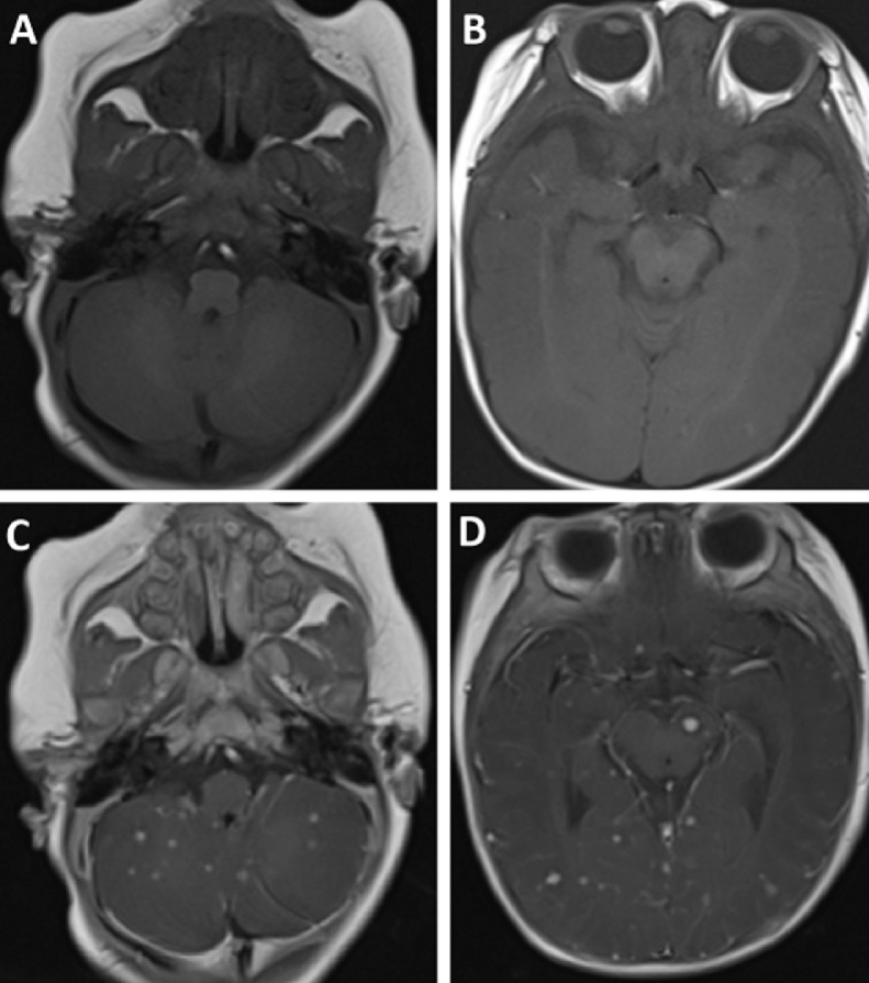
Tuberculosis remains prevalent in developing countries. Central nervous system tuberculosis often occurs secondary to pulmonary tuberculosis, transmitted through the bloodstream, and has a high mortality rate. Meningitis is the most common presentation of central nervous system tuberculosis, followed by tuberculoma, tuberculous brain abscess, and miliary tuberculosis. In this report, we present a case of miliary tuberculosis in a 3 month-old boy. The patient had a fever and was breathless for 1 month. The patient appeared cyanotic, experienced a seizure, and became comatose. Chest computed tomography scan suggested a pulmonary miliary tuberculosis abscess in the right lung and mediastinal lymph node tuberculosis. Brain magnetic resonance imaging showed the lesions were homogeneously enhancing tiny 2-3 mm nodules characteristic of miliary TB. Polymerase chain reaction of the cerebrospinal fluid and sputum samples confirmed tuberculosis. The patient died 1 month after diagnosis.

Keywords: Brain tuberculosis; Central nervous system; Children; Miliary tuberculosis.


Figures

Fig 1 -
Fig. 1
Chest X-ray revealed consolidation in the right lung and diffuse, bilateral nodules, sized 1–3 mm in diameter, in both lungs.
Fig 2 -
Fig. 2
Chest CT scan with contrast (A, C and D) and in lung window (B). Chest computed tomography scan showed consolidation of the right upper lobe with central necrosis (A, arrow). Diffuse bilateral nodules were observed with random distribution, smaller than 3 mm in size (B). Multiple necrotic mediastinal lymphadenopathies were observed (C and D, arrows).
Fig 3 -
Fig. 3
Axial T1 MRI pre-contrast (A and B) and post-contrast (C and D). Diffuse, homogeneous enhancing nodules were observed in both supratentorial and infratentorial regions. These nodules were less than 2 mm in size.

References

    1. Mert A, Arslan F, Kuyucu T, Koç EN, Yilmaz M, Turan D. Miliary tuberculosis: Epidemiological and clinical analysis of large-case series from moderate to low tuberculosis endemic country. Medicine. 2017;96(5):e5875. doi: 10.1097/MD.0000000000005875. - DOI - PMC - PubMed
    1. Vasconcelos G, Santos L, Couto C, Cruz M, Castro A. Miliary brain tuberculomas and meningitis: tuberculosis beyond the lungs. Eur J Case Rep Int Med. 2020 doi: 10.12890/2020_001931. (LATEST ONLINE) - DOI - PMC - PubMed
    1. Cherian A, Thomas SV. Central nervous system tuberculosis. Afr Health Sci. 2011;11(1):116–127. - PMC - PubMed
    1. Goncalves R, Lopes P, Mendes P, Trêpa J, Rua J, Sá R. Miliary tuberculosis with cerebral, liver, prostate and retroesophageal involvement. Idcases. 2017;9:25–27. doi: 10.1016/j.idcr.2017.03.011. - DOI - PMC - PubMed
    1. Khan FY. Review of literature on disseminated tuberculosis with emphasis on the focused diagnostic workup. J Family Community Med. 2019;26(2):83–91. doi: 10.4103/jfcm.JFCM_106_18. - DOI - PMC - PubMed

Publication types

LinkOut - more resources