

"Undiagnosed aortic coarctation with 2 simultaneous acute aortic syndromes: Intramural hematoma and mycotic aneurysm"
- PMID: 34401029
- PMCID: PMC8350181
- DOI: 10.1016/j.radcr.2021.07.014
"Undiagnosed aortic coarctation with 2 simultaneous acute aortic syndromes: Intramural hematoma and mycotic aneurysm"
Abstract
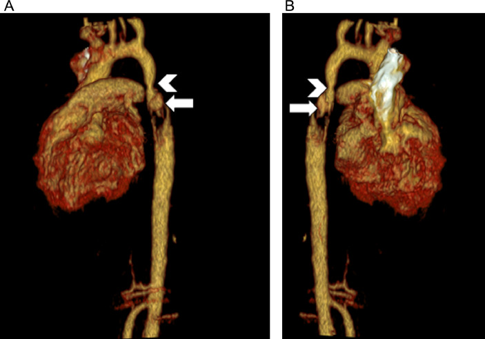
Acute aortic syndrome can be a fatal pathology if not diagnosed and managed early. Although acute aortic syndrome is more often a diagnosis of adulthood, it may occasionally afflict the pediatric patients. We herein present a case of a 5-year-old female that was discovered to have multiple acute and congenital aortic abnormalities after presenting to the emergency department with infectious symptoms and lower extremity pain. Acute aortic syndrome may not be a top differential consideration in children with acute chest pain; however, it is important to consider because delayed diagnosis and management can have fatal implications.
Keywords: Aortic coarctation; Computed tomography angiography; Intramural hematoma; Mycotic aneurysm.
© 2021 The Authors. Published by Elsevier Inc. on behalf of University of Washington.
Figures

References
-
- Revels JW, Wang SS, Richards A. Bilateral renal artery parvus tardus: a sign of aortic coarctation in pediatric patients. Abdom Radiol (NY) 2021;46:841–843. - PubMed
-
- Ishizaka N, Sohmiya K, Miyamura M, Umeda T, Tsuji M, Katsumata T. Infected aortic aneurysm and inflammatory aortic aneurysm–in search of an optimal differential diagnosis. J Cardiol. 2012;59:123–131. - PubMed
Publication types
LinkOut - more resources
Full Text Sources
