. 2021 Jul 7;7(7):e07503.
doi: 10.1016/j.heliyon.2021.e07503. eCollection 2021 Jul.

Coeloglossum viride var. bracteatum extract attenuates staurosporine induced neurotoxicity by restoring the FGF2-PI3K/Akt signaling axis and Dnmt3

Affiliations

Coeloglossum viride var. bracteatum extract attenuates staurosporine induced neurotoxicity by restoring the FGF2-PI3K/Akt signaling axis and Dnmt3

Zhe-Ping Cai et al. Heliyon. .

Abstract

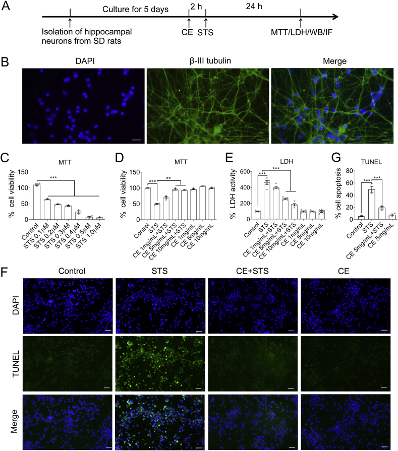
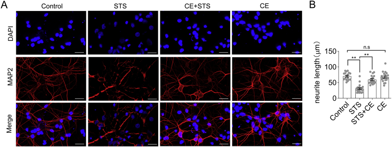
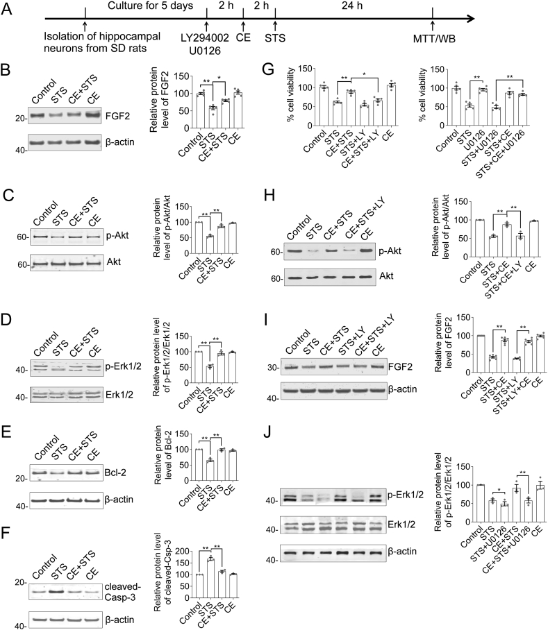
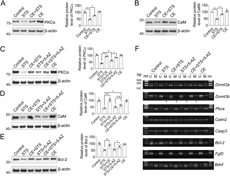
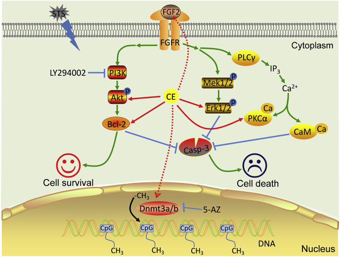
We previously demonstrated the antioxidant activity of Coeloglossum viride var. bracteatum extract (CE) in rat cortical neurons and in mice with chemically induced cognitive impairment. In this work, we established a staurosporine (STS)-induced toxicity model to decipher the neuroprotective mechanisms of CE. We found that CE protected cell viability and neurite integrity in STS-induced toxicity by restoring the levels of FGF2 and its associated PI3K/Akt signaling axis. LY294002, a pan-inhibitor of PI3K, antagonized the activity of CE, although its-mediated restoration of FGF2 was unaffected. In addition, CE restored levels of Bcl-2/Caspase-3, PKCα/CaM pathway, and Dnmt3a and Dnmt3b, two methyltransferases that contribute to de novo DNA methylation. The Dnmts inhibitor 5-azacytidine impaired CE-mediated restoration of Dnmt3 or CaM, as well as the transition of DNA methylation status on the Dnmt3 promoter. These results reveal potential mechanisms that could facilitate the study and application of CE as a neuroprotective agent.

Keywords: CE; Dnmt3a; Dnmt3b; FGF2; PI3K/Akt; Staurosporine.


Conflict of interest statement

The authors declare no conflict of interest.

Figures

Figure 1
Figure 1
CE protects hippocampal neurons from STS-induced toxicity. (A) Experimental timeline. Hippocampal neurons were isolated from neonatal rats and incubated with Coeloglossum viride var. bracteatum extract (CE) or staurosporine (STS). (B) Purity and integrity of neurons were examined by immunofluorescence of neuron-specific β-III tubulin. Scale bar = 25 μm. (C) Neurons were treated with set concentrations of STS. The optimal concentration is 0.2 μM, corresponding to a 40%–50% decreases in cell viability. Cell viability assay (D) and LDH cytotoxicity assay (E) determined the optimal CE concentration (5–10 mg/mL) to protect neurons from STS-induced toxicity. (F–G) TUNEL assay confirmed the effectiveness of CE in protecting neurons from STS-induced cell death (apoptosis). In C-E and G, results are shown as mean ± s.e.m. Statistical significance between groups was tested by one-way ANOVA, post hoc Tukey's tests in D (n = 3), E (n = 5) and G (n = 3). ∗∗, p < 0.05; ∗∗∗, p < 0.001. Non-adjusted images of Figure 1B were presented in Supplementary Material Figure S5.
Figure 2
Figure 2
CE protects neuronal cell body integrity, neurite outgrowth and extension from STS-induced toxicity. (A) Neurons were incubated with STS (0.2 μM) and CE (5 mg/mL) and immuno-labeled with MAP2 antibody. STS compromised cell body integrity and caused neurite shortening, indicating impaired neuronal activity. In contrast, CE rescued neurons from STS-induced toxicity, while CE alone did not introduce observable toxicity. Scale bar = 25μm. Nuclei were stained with DAPI. (B) The length of neurite was calculated by Image J (n > 17). Data are shown as mean ± s.e.m. Statistical significance of difference between groups was examined by one-way ANOVA followed by Tukey's multiple comparison tests. ∗∗, p < 0.01; n.s, not significant. Non-adjusted images were presented in Supplementary Material Figure S6.
Figure 3
Figure 3
CE restores the FGF2-Akt, Erk1/2 signaling axis and Bcl-2/caspase-3 apoptotic pathway to protect neurons from STS-induced toxicity. (A) Experimental timeline. Hippocampal neurons were incubated with LY294002 (PI3K inhibitor) or U0126 (Erk1/2 inhibitor), STS or CE as indicated, followed by Western blot and MTT cell viability assay. (B–F) Representative blots of FGF2, Akt, Erk1/2, Bcl-2, and cleaved-Casp-3. STS induced a decrease in FGF2 levels, inhibited the FGF2-Akt/Erk1/2 pathway and turned on the apoptotic pathway. In contrast, CE restored FGF2 expression, reactivated the Akt/Erk1/2 pathway, and eliminated activation of the apoptotic pathway. Relative protein levels of FGF2, Bcl-2 and cleaved-Casp-3 were normalized to β-actin, whereas p-Akt and p-Erk1/2 were normalized to their total protein (Akt or Erk1/2), respectively. (G) MTT cell viability assay of neurons incubated with STS, CE, LY294002, or U0126 as indicated. LY294002 but not U0126 antagonized the neuroprotective effect of CE on STS-induced toxicity. (H–J) Representative blots of Akt, FGF2, and Erk1/2. Inhibition of the PI3K/Akt pathway (via LY294002) abrogated CE-mediated rescue of p-Akt, but did not affect CE-mediated recovery of FGF2. In this figure, results are expressed as mean ± s.e.m. One-way ANOVA and Tukey's multiple comparison tests were performed to examine statistical significant differences between groups (n = 5). ∗, p < 0.05; ∗∗, p < 0.01. Non-adjusted blots were presented in Supplementary Material Figure S7.
Figure 4
Figure 4
Dnmt3 is involved in CE-mediated neuroprotective effects. (A) Experimental timeline. Hippocampal neurons were incubated with 5-AZ (Dnmts inhibitor), STS or CE, followed by microscopic imaging, Western blot and MTT assay. (B) Images of drug-exposed neurons. Scale bar, 25 μm. (C) MTT assay showing that inhibition of Dnmts did not inhibit CE-mediated protection against STS-induced toxicity. n = 5. (D) Representative blots of Dnmt3b and Dnmt1. Dnmt3b but not Dnmt1 was restored by CE to recover from STS-induced toxicity (CE + STS column). (E) Representative blots of Dnmt3a and Dnmt3b. Inhibition of Dnmts (by 5-AZ) prevented CE-mediated recovery of Dnmt3a and Dnmt3b. In D (n = 5) and E (n = 3), relative protein levels were calculated from the optical density of blots and normalized to β-actin. (C–E) Data are shown as mean ± s.e.m. One-way ANOVA, post hoc Tukey's tests were performed. ∗, p < 0.05; ∗∗, p < 0.01. n.s, not significant. Non-adjusted images and blots were presented in Supplementary Material Figures S8 and S9.
Figure 5
Figure 5
CE ameliorates the PKCα/CaM pathway to protect neurons from STS-induced toxicity. (A–B) Representative blots of PKCα and CaM. CE rescues STS-induced down-expression of PKCα and CaM. (C–E) Inhibition of Dnmts by 5-AZ impairs CE-mediated recovery of CaM but not PKCα and Bcl-2 expression. In A-E, results are shown as mean ± s.e.m. Statistical significance of differences between groups was calculated by one-way ANOVA followed by Tukey's tests, n = 5. ∗, p < 0.05; ∗∗, p < 0.01; n.s, not significant. (F) Methylation-specific PCR suggests that promoter methylation of Dnmt3b is involved in CE-mediated neuroprotection. mr, DNA marker; U, unmethylated; M, methylated. Non-adjusted blots and gels were presented in Supplementary Material Figures S10 and S11.
Figure 6
Figure 6
Schematic representation of CE-mediated neuroprotection against STS-induced toxicity. STS inhibits protein kinases and induces apoptosis and severe cytotoxicity to neurons. CE restores expression of FGF2, maintains the activity of PI3K/Akt, Erk1/2 and PKCα/CaM pathway, and promotes cell survival. The PI3K/Akt signaling pathway plays a role in CE-mediated function, as inhibition of PI3K/Akt by LY294002 is known to abrogate CE activity. Meanwhile, the cleaved-Casp-3 associated apoptotic pathway activated by STS was significantly attenuated by CE. In addition, DNA do novo methyltransferase activity may be involved in CE-mediated regulation of gene expression and can be inhibited by the Dnmts inhibitor 5-AZ, suggesting that epigenetic regulation is involved in CE-mediated neuroprotection processes.

References

    1. Cannella N., Oliveira A.M.M., Hemstedt T., Lissek T., Buechler E., Bading H., Spanagel R. Dnmt3a2 in the nucleus accumbens shell Is required for reinstatement of cocaine seeking. J. Neurosci. 2018;38:7516–7528. - PMC - PubMed
    1. Cheng Y., Li Z., Kardami E., Loh Y.P. Neuroprotective effects of LMW and HMW FGF2 against amyloid beta toxicity in primary cultured hippocampal neurons. Neurosci. Lett. 2016;632:109–113. - PMC - PubMed
    1. Dehmelt L., Halpain S. The MAP2/Tau family of microtubule-associated proteins. Genome Biol. 2005;6:204. - PMC - PubMed
    1. Ford-Perriss M., Abud H., Murphy M. Fibroblast growth factors in the developing central nervous system. Clin. Exp. Pharmacol. Physiol. 2001;28:493–503. - PubMed
    1. Guo Z., Pan R.Y., Qin X.Y. Potential protection of Coeloglossum viride var. Bracteatum extract against oxidative stress in rat cortical neurons. J. Anal. Methods Chem. 2013;2013:326570. - PMC - PubMed