Review
. 2021 Oct 5;22(10):e52445.
doi: 10.15252/embr.202152445. Epub 2021 Aug 17.

Linking transport and translation of mRNAs with endosomes and mitochondria

Affiliations
Review

Linking transport and translation of mRNAs with endosomes and mitochondria

Kira Müntjes et al. EMBO Rep. .

Abstract

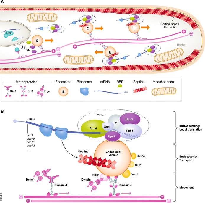
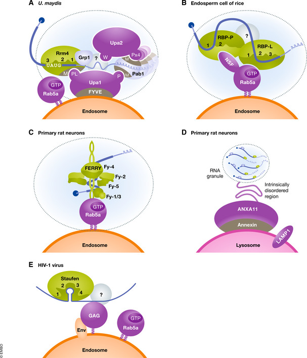
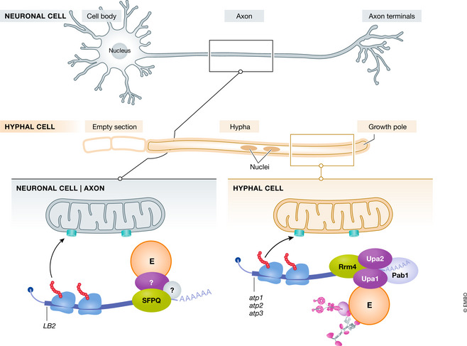
In eukaryotic cells, proteins are targeted to their final subcellular locations with precise timing. A key underlying mechanism is the active transport of cognate mRNAs, which in many systems can be linked intimately to membrane trafficking. A prominent example is the long-distance endosomal transport of mRNAs and their local translation. Here, we describe current highlights of fundamental mechanisms of the underlying transport process as well as of biological functions ranging from endosperm development in plants to fungal pathogenicity and neuronal processes. Translation of endosome-associated mRNAs often occurs at the cytoplasmic surface of endosomes, a process that is needed for membrane-assisted formation of heteromeric protein complexes and for accurate subcellular targeting of proteins. Importantly, endosome-coupled translation of mRNAs encoding mitochondrial proteins, for example, seems to be particularly important for efficient organelle import and for regulating subcellular mitochondrial activity. In essence, these findings reveal a new mechanism of loading newly synthesised proteins onto endocytic membranes enabling intimate crosstalk between organelles. The novel link between endosomes and mitochondria adds an inspiring new level of complexity to trafficking and organelle biology.

Keywords: RNA transport; endosomes; local translation; microtubules; mitochondria; organelle.


Conflict of interest statement

The authors declare that they have no conflict of interest.

Figures

Figure 1
Figure 1. Model depicting endosomal mRNA transport in Ustilago maydis
(A) Overview of an infectious hypha. Endosomes (E) shuttle along antiparallel microtubule bundles (pink). Plus‐end directed transport is mediated by Kin3, whereas dynein transports endosomes to the minus ends. Cargo mRNPs (dashed ovals) are assembled most likely close to the nucleus involving components of the NineTeen complex (NTC, light blue) deposited during splicing. Local translation (ribosomes in dark blue) of cargo mRNAs like septins results in septin complex formation (red octamer). These complexes are transported towards the growth pole to generate higher‐order septin filaments with gradients emanating from the growing tip (red line). Endosomal mRNP transport appears to be also important for import of proteins into mitochondria (see Fig 3). (B) Close‐up of endosomes shown above. Components important for movement, endocytosis, transport and localised translation are shown (further details are given in the text).
Figure 2
Figure 2. Schematic comparison of endosomal RNA transport in fungi, plants and animals
(A–E) On the cytoplasmic surface of transport endosomes or lysosomes, mRNPs are attached by different factors (purple) to endosomes. Key RNA‐binding proteins (green) interact with cargo RNA (blue). mRNAs are symbolised by CAP (blue circle) and a poly(A) tail (B and D are adapted from Tian et al, and Liao et al, , respectively. Panel (C) is adapted from Schuhmacher et al, and Quentin et al, (both preprints). Panels (D, E) are proposed based on current literature (further details are given in the text).
Figure 3
Figure 3. Schematic comparison of endosome‐coupled translation and mitochondrial protein import
At the top, a highly polarised neuron and a fungal hypha are depicted. In both cases, a defined axis of polarity is established with the growth pole indicated on the right. Local translation of mRNAs encoding mitochondrial proteins at the surface of late endosomes (E) in neurons (left) is compared to membrane‐coupled translation at the surface of early endosomes (E) in hyphae (right). The TOM complex is depicted as a pore in the mitochondrial membrane. Symbols are used as in the other figures, and details are given in the text.

References

    1. Abouward R, Schiavo G (2020) Walking the line: mechanisms underlying directional mRNA transport and localisation in neurons and beyond. Cell Mol Life Sci 78: 2665–2681 - PMC - PubMed
    1. Akopian D, Shen K, Zhang X, Shan SO (2013) Signal recognition particle: an essential protein‐targeting machine. Annu Rev Biochem 82: 693–721 - PMC - PubMed
    1. Arlt H, Steglich G, Perryman R, Guiard B, Neupert W, Langer T (1998) The formation of respiratory chain complexes in mitochondria is under the proteolytic control of the m‐AAA protease. EMBO J 17: 4837–4847 - PMC - PubMed
    1. Bassell GJ, Powers CM, Taneja KL, Singer RH (1994) Single mRNAs visualized by ultrastructural in situ hybridization are principally localized at actin filament intersections in fibroblasts. J Cell Biol 126: 863–876 - PMC - PubMed
    1. Basyuk E, Galli T, Mougel M, Blanchard JM, Sitbon M, Bertrand E (2003) Retroviral genomic RNAs are transported to the plasma membrane by endosomal vesicles. Dev Cell 5: 161–174 - PubMed

Publication types