

Late Gadolinium Enhancement of Left Ventricular Papillary Muscles in Patients with Mitral Regurgitation
- PMID: 34402246
- PMCID: PMC8484157
- DOI: 10.3348/kjr.2020.1485
Late Gadolinium Enhancement of Left Ventricular Papillary Muscles in Patients with Mitral Regurgitation
Abstract
Objective: Arrhythmogenic mitral valve prolapse (MVP) is an important cause of sudden cardiac death characterized by fibrosis of the papillary muscles or left ventricle (LV) wall, and an association between late gadolinium enhancement (LGE) of the LV papillary muscles and ventricular arrhythmia in MVP has been reported. However, LGE of the papillary muscles may be observed in other causes of mitral regurgitation, and it is not limited to patients with MVP. This study was to evaluate the association of LGE of the LV papillary muscles or ventricular wall on cardiac magnetic resonance imaging (CMR) and ventricular arrhythmia in patients with mitral regurgitation.
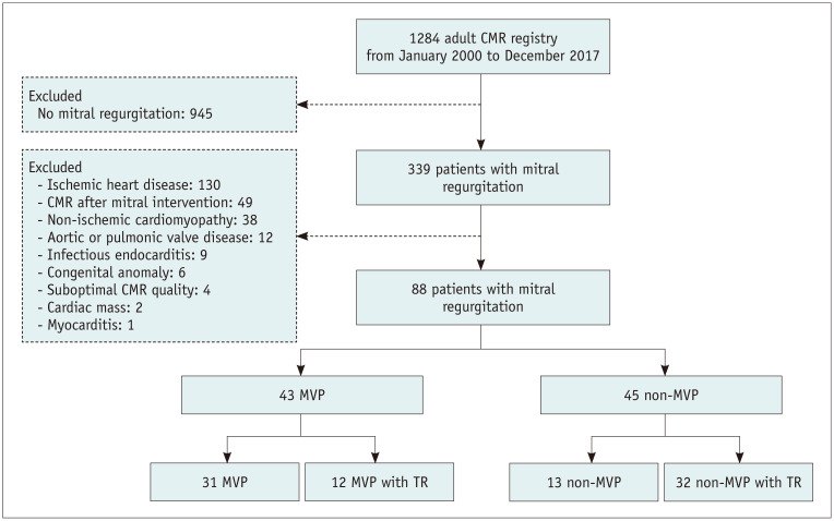
Materials and methods: This study included 88 patients (mean age ± standard deviation, 58.3 ± 12.0 years; male, 42%) with mitral regurgitation who underwent CMR. They were allocated to the MVP (n = 43) and non-MVP (n = 45) groups, and their LGE images on CMR, clinical characteristics, echocardiographic findings, and presence of arrhythmia were compared.
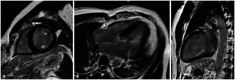
Results: LV myocardial wall enhancement was more frequent in the MVP group than in the non-MVP group (28% vs. 11%, p = 0.046). Papillary muscle enhancement was observed in 7 (7.9%) patients. Of the 43 patients with MVP, 15 (34.8%) showed LGE in the papillary muscles or LV myocardium, including 12 (27.9%) with LV myocardial wall enhancement and 4 (9.3%) with papillary muscle enhancement. One patient with bilateral diffuse papillary muscle enhancement experienced sudden cardiac arrest due to ventricular fibrillation. Univariable logistic regression analysis showed that high systolic blood pressure (BP; odds ratio [OR], 1.05; 95% confidence interval [CI], 1.01-1.09; p = 0.027) and ventricular arrhythmia (OR, 6.84; 95% CI, 1.29-36.19; p = 0.024) were significantly associated with LGE of the papillary muscles.
Conclusion: LGE of the papillary muscles was present not only in patients with MVP, but also in patients with other etiologies of mitral regurgitation, and it was associated with high systolic BP and ventricular arrhythmia. Papillary muscle enhancement on CMR should not be overlooked.
Keywords: Arrhythmia; Late gadolinium enhancement; Mitral regurgitation; Mitral valve prolapse.
Copyright © 2021 The Korean Society of Radiology.
Conflict of interest statement
The authors have no potential conflicts of interest to disclose.
Figures
Similar articles
-
Dark-blood late gadolinium enhancement CMR improves detection of papillary muscle fibrosis in patients with mitral valve prolapse.Eur J Radiol. 2022 Feb;147:110118. doi: 10.1016/j.ejrad.2021.110118. Epub 2021 Dec 25. Eur J Radiol. 2022. PMID: 34972057
-
Replacement Myocardial Fibrosis in Patients With Mitral Valve Prolapse: Relation to Mitral Regurgitation, Ventricular Remodeling, and Arrhythmia.Circulation. 2021 May 4;143(18):1763-1774. doi: 10.1161/CIRCULATIONAHA.120.050214. Epub 2021 Mar 12. Circulation. 2021. PMID: 33706538
-
Left ventricular fibrosis and CMR tissue characterization of papillary muscles in mitral valve prolapse patients.Int J Cardiovasc Imaging. 2024 Feb;40(2):213-224. doi: 10.1007/s10554-023-02985-w. Epub 2023 Oct 28. Int J Cardiovasc Imaging. 2024. PMID: 37891450 Free PMC article.
-
Role of cardiac magnetic resonance in stratifying arrhythmogenic risk in mitral valve prolapse patients: a systematic review and meta-analysis.Eur Radiol. 2024 Nov;34(11):7321-7333. doi: 10.1007/s00330-024-10815-3. Epub 2024 Jun 6. Eur Radiol. 2024. PMID: 38844620 Free PMC article.
-
Transthoracic echocardiography for arrhythmic mitral valve prolapse: Phenotypic characterization as first step.Echocardiography. 2022 Sep;39(9):1158-1170. doi: 10.1111/echo.15439. Epub 2022 Aug 27. Echocardiography. 2022. PMID: 36029124 Review.
Cited by
-
Arrhythmic mitral valve prolapse: valve geometry and traction force quantification by echocardiography.Europace. 2024 Aug 30;26(9):euae224. doi: 10.1093/europace/euae224. Europace. 2024. PMID: 39188205 Free PMC article. No abstract available.
-
How to Clearly and Accurately Report Odds Ratio and Hazard Ratio in Diagnostic Research Studies?Korean J Radiol. 2022 Aug;23(8):777-784. doi: 10.3348/kjr.2022.0249. Epub 2022 May 31. Korean J Radiol. 2022. PMID: 35695319 Free PMC article. No abstract available.
-
Left Ventricular Fibrosis by Cardiac Magnetic Resonance Tissue Characterization in Chronic Mitral Regurgitation Patients.J Clin Med. 2024 Jul 1;13(13):3877. doi: 10.3390/jcm13133877. J Clin Med. 2024. PMID: 38999443 Free PMC article.
-
Left ventricular fibrosis in arrhythmic mitral valve prolapse: quantification and comparison of semi-automated techniques assessed by cardiac magnetic resonance.Int J Cardiovasc Imaging. 2024 Feb;40(2):275-285. doi: 10.1007/s10554-023-03006-6. Epub 2023 Dec 23. Int J Cardiovasc Imaging. 2024. PMID: 38141098 Free PMC article.
-
The Association Between Late Gadolinium Enhancement by Cardiac Magnetic Resonance and Ventricular Arrhythmia in Patients With Mitral Valve Prolapse: A Systematic Review and Meta-Analysis.Clin Cardiol. 2024 Jul;47(7):e24316. doi: 10.1002/clc.24316. Clin Cardiol. 2024. PMID: 38958255 Free PMC article.
References
-
- La Vecchia L, Ometto R, Centofante P, Varotto L, Bonanno C, Bozzola L, et al. Arrhythmic profile, ventricular function, and histomorphometric findings in patients with idiopathic ventricular tachycardia and mitral valve prolapse: clinical and prognostic evaluation. Clin Cardiol. 1998;21:731–735. - PMC - PubMed
-
- Chen HY. Relationship of heart rate turbulence, heart rate variability and the number of ventricular premature beats in patients with mitral valve prolapse and non-significant regurgitation. Int J Cardiol. 2009;135:269–271. - PubMed
-
- Turker Y, Ozaydin M, Acar G, Ozgul M, Hoscan Y, Varol E, et al. Predictors of ventricular arrhythmias in patients with mitral valve prolapse. Int J Cardiovasc Imaging. 2010;26:139–145. - PubMed
MeSH terms
Substances
LinkOut - more resources
Full Text Sources
Miscellaneous
