Review
. 2021 Jul;24(4):316-325.
doi: 10.1111/vop.12923. Epub 2021 Aug 17.

Biomechanics of the optic nerve head and sclera in canine glaucoma: A brief review

Affiliations
Review

Biomechanics of the optic nerve head and sclera in canine glaucoma: A brief review

Shin Ae Park et al. Vet Ophthalmol. 2021 Jul.

Abstract

Glaucoma is a leading cause of irreversible blindness, a progressive optic neuropathy with retinal ganglion cell (RGC) death beginning in the optic nerve head (ONH). A primary risk factor for developing glaucoma is elevated intraocular pressure (IOP). Reducing IOP is the only treatment proven to be effective at delaying disease progression. Nevertheless, even when patients have their IOP reduced, the majority of them continue to lose vision. There are, in both humans and dogs, significant interindividual variabilities in susceptibilities to IOP-induced optic nerve damage. Vision loss progresses much more slowly in Beagles with open-angle glaucoma (OAG) caused by ADAMTS10 mutation. This can be attributed to the mutation-related altered ocular biomechanical properties. The principal site of optic nerve (ON) damage in glaucoma is the ONH. It is suggested that the biomechanical properties of the ONH and the surrounding peripapillary sclera (PPS) contribute to glaucoma development and progression. As far as the beneficial biomechanical properties of the ONH and PPS for a decreased susceptibility and slow progression of glaucoma, data are inconsistent and conflicting. Recent biomechanical studies on beagles with ADAMTS10 mutation demonstrated that the mutant dogs have mechanically weak posterior sclera. This weakness was associated with a reduced collagen density and a lower proportion of insoluble collagen. These changes, observed before glaucoma development, were considered intrinsic characteristics caused by the mutation rather than a secondary effect of IOP elevation. Further studies of ADAMTS10-OAG may elucidate the effects of altered biomechanical properties of ONH and PPS in determining the glaucoma progression.

Keywords: ADAMTS10; dog; neuroprotection; open-angle glaucoma; stiff; viscoelastic.


Conflict of interest statement

CONFLICT OF INTEREST

None.

Figures

FIGURE 1
FIGURE 1
Comparable anatomy of lamina cribrosa (LC) in humans (A) and dogs (B). (C) IOP-induced mechanical compression (arrow) causes axonal swelling and mitochondrial accumulation in a canine ocular hypertension model, suggesting LC as a principal site of glaucomatous optic nerve damage, Panel C: Martins et al., with permission
FIGURE 2
FIGURE 2
Optic disk cupping and loss of optic nerve axons in a dog with end-stage glaucoma (top row) vs. normal control (bottom row). (A and B) Fundus images; (C and D) OCT of the optic nerve head; (E and F) cross sections of the whole nerve for axon counting; (G and H) high magnification (40x) of the optic nerve cross section; (I) histopathology of the ON showing thinning of the prelaminar optic nerve (arrows) tissue in the glaucomatous eye compared to (J) normal; (K) immunohistochemistry of the canine glaucomatous retina shows loss of RGC (NeuN) (L) compared to the normal eye
FIGURE 3
FIGURE 3
(A) Electroretinograms (ERG) show well-preserved retinal function in a Beagle with ADAMTS10-OAG and documented diurnal IOP of 30–65 mmHg for >6 weeks vs (B) essentially non-recordable retinal function in a non-mutant dog with uveitic glaucoma developed post-phacoemulsification and recorded IOP of 62 mmHg for less than 1 day. The globe was enucleated 2 days later and histopathology of the globe showed typical findings of glaucoma including diffuse inner and segmental full-thickness retinal atrophy and the suggested etiology was pre-iridal fibrovascular membrane causing peripheral anterior synechia. No overt inflammation was noted in the posterior segment. (C) Relatively well-preserved RGCs (red arrows) and retinal layers in a dog with ADAMTS10-OAG after elevated IOP >30 mmHg for 14 months (including documented 40–50 mmHg during the 5-month period) vs (D) a dog with angle-closure glaucoma (ACG) showing extensive full-thickness retinal degeneration after measured IOPs of up to 42 mmHg for less than 3 days. (E) Longitudinal mean diurnal IOPs of the dog with ADAMTS10-OAG (black diamond, same dog as C) and individual IOP measurements taken during office visits from a dog with ACG (red square, the same dog as D)
FIGURE 4
FIGURE 4
Intraocular pressure (IOP) induced stress in the eye (A) IOP elevation results in increased translaminar pressure gradient (IOP—retrolaminar tissue pressure: RLTP) that leads to ONH cupping. A circumferential (hoop) stress is applied to the cornea and sclera that is concentrated to the peripapillary sclera (PPS) and contributes to scleral expansion. (B) Directions of circumferential stress on the peripapillary sclera surrounding the LC. Figures modified from Quigley et al., 2013 and Burgoyne et al., 2011
FIGURE 5
FIGURE 5
A schematic of the excision location of the posterior scleral strip. The strip was cut adjacent to the temporal side of the ONH. S, superior; I, inferior; N, nasal; and T, temporal. From Palko et al., 2013
FIGURE 6
FIGURE 6
Exponential fitting of the stress-strain curves from the tensile ramp tests. The two older samples were noticeably stiffer than those from the young dogs. From Palko et al., 2013
FIGURE 7
FIGURE 7
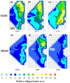
Collagen density maps. Contour maps of relative fibrillar collagen density across the (A–C) carrier and (D–F) affected ADAMTS10 mutant posterior canine scleras. Broken line: border of the mid-posterior (MPS) and PPS regions. Collagen density is visibly reduced in all areas of the affected specimens as compared to carrier controls. Data sampling interval: 0.4 mm. From Boote et al., 2016 with permission

References

    1. Komaromy AM, Petersen-Jones SM. Genetics of canine primary glaucomas. Vet Clin N Am Small Anim Pract. 2015;45(6):1159–1182. v. - PubMed
    1. Plummer CE, Regnier A, Gelatte KN. The Canine Glaucomas. In: Gelatt KN, Gilger BC, Kern TJ, eds. Veterinary ophthalmology. 5th ed. Wiley-Blackwell Publishing; 2013:1050–1145.
    1. Quigley HA, Broman AT. The number of people with glaucoma worldwide in 2010 and 2020. Br J Ophthalmol. 2006;90(3):262–267. - PMC - PubMed
    1. Snyder KC, Oikawa K, Williams J, et al. Imaging distal aqueous outflow pathways in a spontaneous model of congenital glaucoma. Transl Vis Sci Technol. 2019;8(5):22. - PMC - PubMed
    1. Curto EM, Gemensky-Metzler AJ, Chandler HL, Wilkie DA. Equine glaucoma: a histopathologic retrospective study (1999–2012). Vet Ophthalmol. 2014;17(5):334–342. - PubMed

Substances