. 2021 Aug 17;19(1):247.
doi: 10.1186/s12951-021-00992-4.

An in situ tissue engineering scaffold with growth factors combining angiogenesis and osteoimmunomodulatory functions for advanced periodontal bone regeneration

Affiliations

An in situ tissue engineering scaffold with growth factors combining angiogenesis and osteoimmunomodulatory functions for advanced periodontal bone regeneration

Tian Ding et al. J Nanobiotechnology. .

Abstract

Background: The regeneration of periodontal bone defect remains a vital clinical challenge. To date, numerous biomaterials have been applied in this field. However, the immune response and vascularity in defect areas may be key factors that are overlooked when assessing the bone regeneration outcomes of biomaterials. Among various regenerative therapies, the up-to-date strategy of in situ tissue engineering stands out, which combined scaffold with specific growth factors that could mimic endogenous regenerative processes.

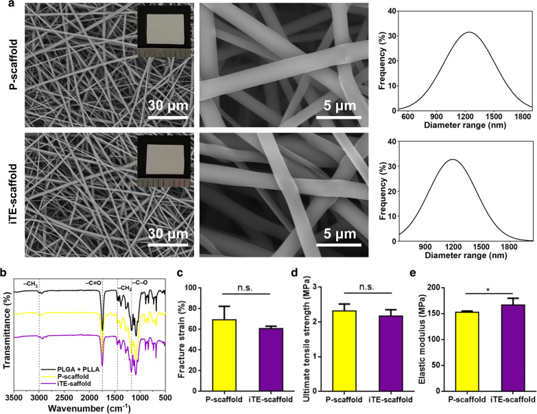
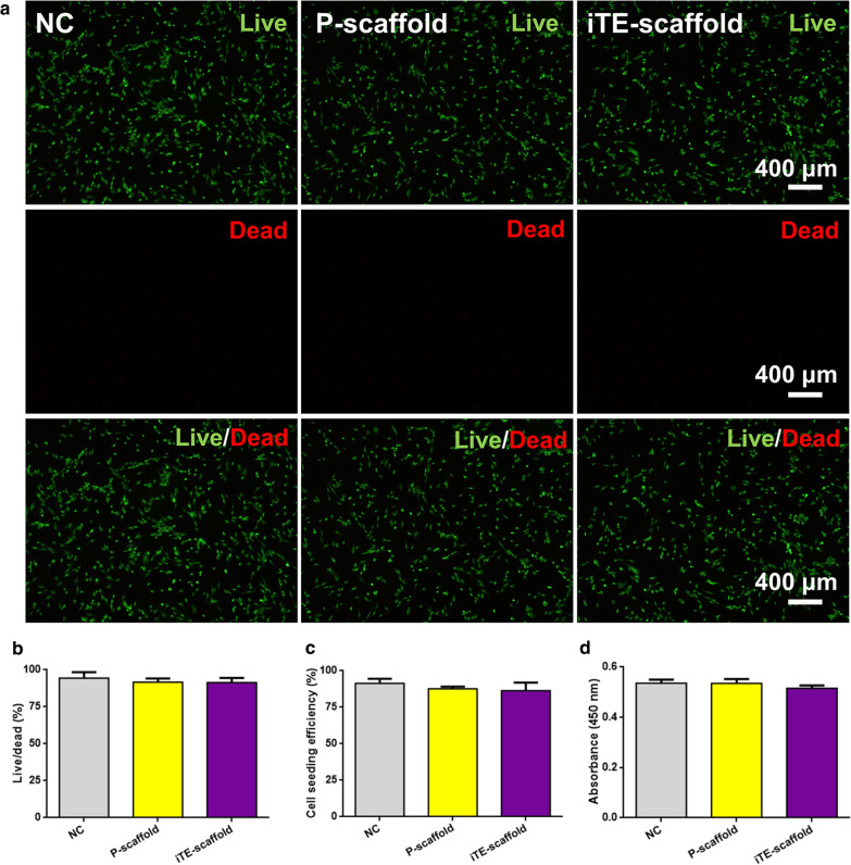
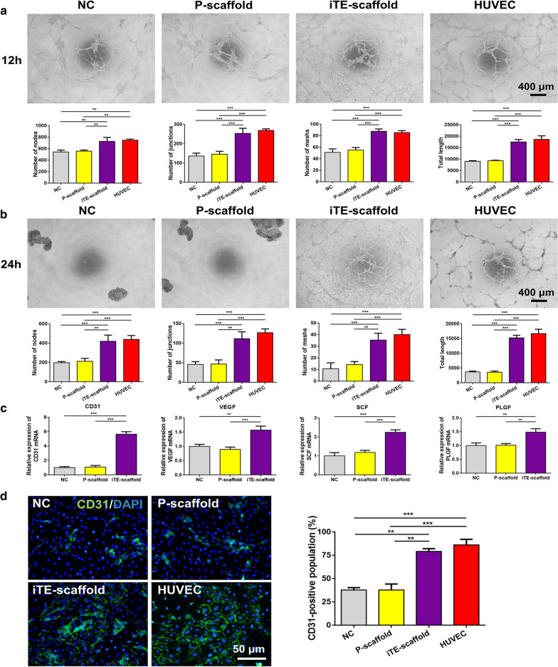
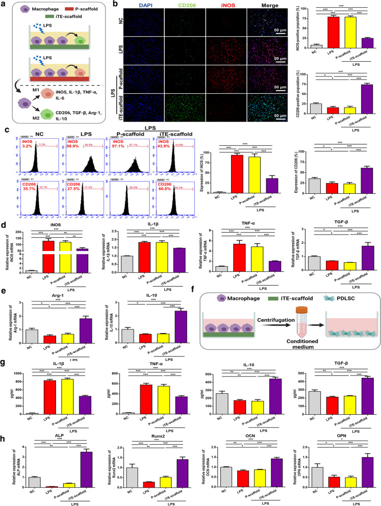
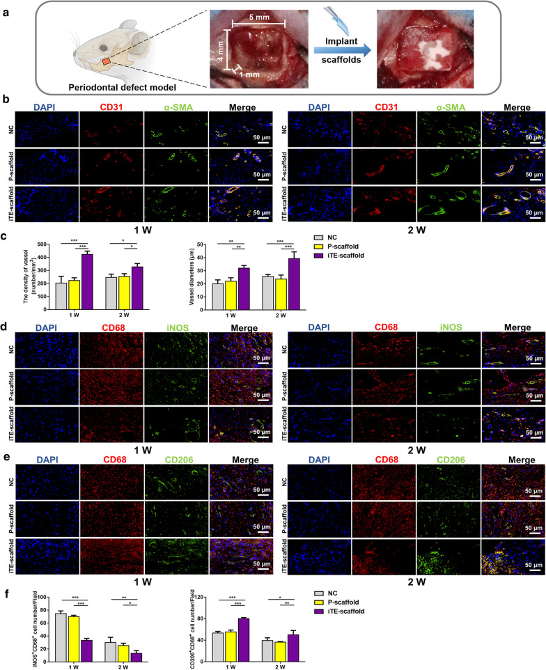
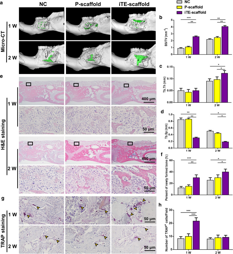
Results: Herein, we fabricated a core/shell fibrous scaffold releasing basic fibroblast growth factor (bFGF) and bone morphogenetic protein-2 (BMP-2) in a sequential manner and investigated its immunomodulatory and angiogenic properties during periodontal bone defect restoration. The in situ tissue engineering scaffold (iTE-scaffold) effectively promoted the angiogenesis of periodontal ligament stem cells (PDLSCs) and induced macrophage polarization into pro-healing M2 phenotype to modulate inflammation. The immunomodulatory effect of macrophages could further promote osteogenic differentiation of PDLSCs in vitro. After being implanted into the periodontal bone defect model, the iTE-scaffold presented an anti-inflammatory response, provided adequate blood supply, and eventually facilitated satisfactory periodontal bone regeneration.

Conclusions: Our results suggested that the iTE-scaffold exerted admirable effects on periodontal bone repair by modulating osteoimmune environment and angiogenic activity. This multifunctional scaffold holds considerable promise for periodontal regenerative medicine and offers guidance on designing functional biomaterials.

Keywords: Angiogenesis; Biomimetic repair; In situ tissue engineering; Osteoimmunomodulation; Periodontal bone regeneration.


Conflict of interest statement

The authors declare no competing financial interest.

Figures

Fig. 1
Fig. 1
Physicochemical structure characterization assessment of the scaffolds. a Representative SEM images, photographs (insert), and diameter distribution analysis of P-scaffold and iTE-scaffold. b FTIR spectra of the scaffolds. c Facture strain of the fibrous scaffolds. d Ultimate tensile strength of the fibrous scaffolds. e Elastic modulus of the fibrous scaffolds. *P < 0.05, n.s. no statistical significance
Fig. 2
Fig. 2
Cytocompatibility study of the scaffolds. a, b Representative images and quantitative analysis of live/dead staining of PDLSCs incubated on the different substrates. c Cell seeding effciency of PDLSCs cultured on the different substrates. d Cell activity on the different substrates
Fig. 3
Fig. 3
Evaluation of angiogenic capability of PDLSCs on the different fibrous scaffolds. a, b Representative images and quantitative analysis of tube-like structure formation at 12 h and 24 h after seeding on the Matrigel. Tube formation parameters: the number of nodes, junctions, meshes and total tube length. c Gene expression levels of VEGF, CD31, SCF, and PLGF at week 1. d Representative immunostaining images and quantitative analysis of CD31 in PDLSCs cultured for 7 days. **P < 0.01, ***P < 0.001
Fig. 4
Fig. 4
Evaluation of the osteoimmunomodulatory function in vitro. a Schematic illustration of biomaterial-mediated macrophage polarization. b Fluorescent staining images of the macrophage phenotypes and quantitative analysis of the CD206/iNOS-positive population. c Flow cytometric analysis of the macrophage phenotypes. d The gene expression level of macrophages, including iNOS, TNF-α, IL-1β, TGF-β, Arg-1 and IL-10. e Schematic illustration of macrophage-mediated osteogenic differentiation of PDLSCs. f The immune factors secreted by macrophages in different groups were detected by ELISA. g qRT-PCR analysis for the gene expression levels of ALP, Runx2, OCN and OPN of PDLSCs cultured in the conditioned medium for 7 days. *P < 0.05, **P < 0.01, ***P < 0.001
Fig. 5
Fig. 5
Fluorescence analysis of the tissue sections at week 1 and 2 post-operation. a Schematic illustration of the operation. b Double immunofluorescence staining of mandible sections. CD31 (red): blood vessel; α-SMA (green): smooth muscle actin. c Quantified neo-vascular density and neo-vascular diameters of different groups. d, e Double-labeled immunofluorescence staining images of macrophage phenotype in the defect areas. CD68 (red): a universal macrophage marker; iNOS (green): an M1-phenotype macrophage marker; CD206 (green): an M2-phenotype macrophage marker. f Statistical summary of the population of iNOS+CD68+ and CD206+CD68+. *P < 0.05, **P < 0.01, ***P < 0.001
Fig. 6
Fig. 6
Evaluation of the bone remodeling at week 1 and 2. a Micro-CT reconstructed 3D images of the rat mandibular bone defects. Green color displayed the newly formed bone in the defect sites. bd Quantification of BS/TV (b), Tb. Th (c) and Tb. Sp (d). e Representative H&E staining images of the mandible sections. The visual fields framed by the black line were magnified in the figures below. f The percentage of new bone formation at two time points. g Representative TRAP staining images of mandibular bone defects. Yellow arrows indicated osteoclasts in the trabecular bone surface. h The number of TRAP+ cells in defect regions. *P < 0.05, **P < 0.01, ***P < 0.001

References

    1. Slots J. Periodontitis: facts, fallacies and the future. Periodontol. 2000;2017(75):7–23. - PubMed
    1. Kinane DF, Stathopoulou PG, Papapanou PN. Periodontal diseases. Nat Rev Dis Primers. 2017;3:17038. doi: 10.1038/nrdp.2017.38. - DOI - PubMed
    1. Cardoso EM, Reis C, Manzanares-Cespedes MC. Chronic periodontitis, inflammatory cytokines, and interrelationship with other chronic diseases. Postgrad Med. 2018;130:98–104. doi: 10.1080/00325481.2018.1396876. - DOI - PubMed
    1. Bosshardt DD, Sculean A. Does periodontal tissue regeneration really work? Periodontol. 2000;2009(51):208–219. - PubMed
    1. Deschaseaux F, Sensebe L, Heymann D. Mechanisms of bone repair and regeneration. Trends Mol Med. 2009;15:417–429. doi: 10.1016/j.molmed.2009.07.002. - DOI - PubMed

Substances

LinkOut - more resources