. 2021 Oct;11(10):2436-2445.
doi: 10.1158/2159-8290.CD-21-0702. Epub 2021 Aug 17.

Parallel Genomic Alterations of Antigen and Payload Targets Mediate Polyclonal Acquired Clinical Resistance to Sacituzumab Govitecan in Triple-Negative Breast Cancer

Affiliations

Parallel Genomic Alterations of Antigen and Payload Targets Mediate Polyclonal Acquired Clinical Resistance to Sacituzumab Govitecan in Triple-Negative Breast Cancer

James T Coates et al. Cancer Discov. 2021 Oct.

Abstract

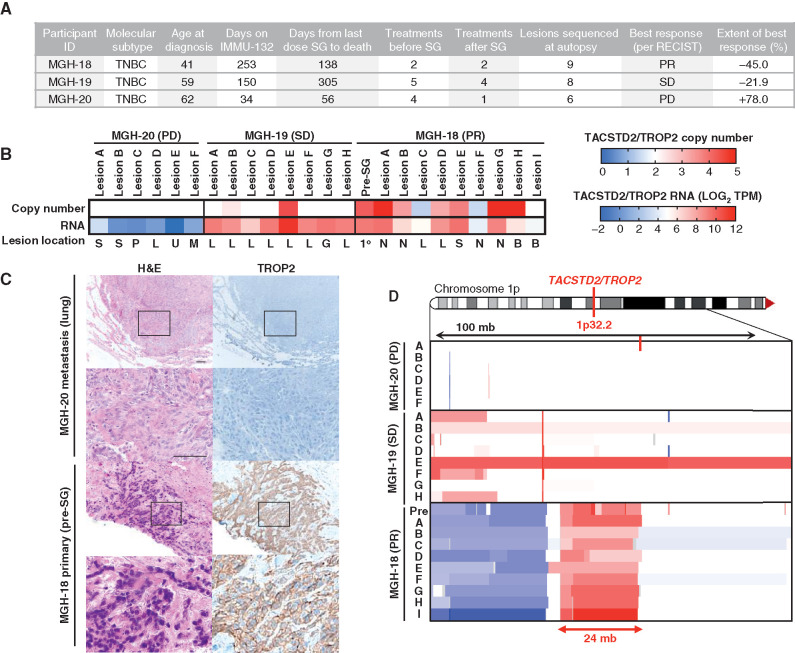
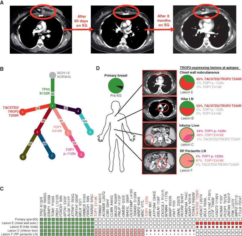
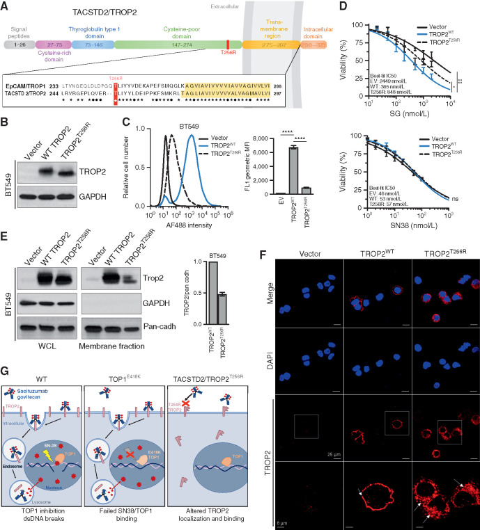
Sacituzumab govitecan (SG), the first antibody-drug conjugate (ADC) approved for triple-negative breast cancer, incorporates the anti-TROP2 antibody hRS7 conjugated to a topoisomerase-1 (TOP1) inhibitor payload. We sought to identify mechanisms of SG resistance through RNA and whole-exome sequencing of pretreatment and postprogression specimens. One patient exhibiting de novo progression lacked TROP2 expression, in contrast to robust TROP2 expression and focal genomic amplification of TACSTD2/TROP2 observed in a patient with a deep, prolonged response to SG. Analysis of acquired genomic resistance in this case revealed one phylogenetic branch harboring a canonical TOP1 E418K resistance mutation and subsequent frameshift TOP1 mutation, whereas a distinct branch exhibited a novel TACSTD2/TROP2 T256R missense mutation. Reconstitution experiments demonstrated that TROP2T256R confers SG resistance via defective plasma membrane localization and reduced cell-surface binding by hRS7. These findings highlight parallel genomic alterations in both antibody and payload targets associated with resistance to SG. SIGNIFICANCE: These findings underscore TROP2 as a response determinant and reveal acquired SG resistance mechanisms involving the direct antibody and drug payload targets in distinct metastatic subclones of an individual patient. This study highlights the specificity of SG and illustrates how such mechanisms will inform therapeutic strategies to overcome ADC resistance.This article is highlighted in the In This Issue feature, p. 2355.


Conflict of interest statement

S. Sun reports grants from NCI, Terri Brodeur Breast Cancer Foundation, and Massachusetts General Hospital during the conduct of the study. I. Leshchiner reports personal fees from PACT Pharma, Inc and Enov1, LLC outside the submitted work. A. Kambadakone reports grants from Philips Healthcare, GE Healthcare, PanCAN, personal fees from Bayer, and other support from Siemens Healthcare outside the submitted work. S.J. Isakoff reports personal fees from Immunomedics, Mylan, Myriad, Puma, Novartis, Seattle Genetics, grants and personal fees from OncoPep and AbbVie, grants from Merck, AstraZeneca, and Genentech outside the submitted work. D. Juric reports grants and personal fees from Novartis, Genentech, Syros, grants from Pfizer, Takeda, personal fees from Relay, PIC Therapeutics, Mapkure, Vibliome, grants and personal fees from Eisai, grants from InventisBio, Arvinas, Ribon Therapeutics, and Infinity Pharmaceuticals outside the submitted work. G. Getz reports grants from IBM during the conduct of the study; and receives research funds from Pharmacyclics. G. Getz is a founder, consultant, and holds privately held equity in Scorpion Therapeutics. A. Bardia reports grants from Genentech, Novartis, Pfizer, Merck, Sanofi, Radius Health, Immunomedics, and Diiachi Pharma/AstraZeneca, and personal fees from Pfizer, Novartis, Genentech, Merck, Radius Health, Immunomedics, Taiho, Sanofi, Diiachi Pharma/AstraZeneca, Puma; Biothernostics Inc., Phillips, Eli Lilly, Foundation Medicine, during the conduct of the study; personal fees from Pfizer, Novartis, Genentech, Merck, Radius Health, Immunomedics, Taiho, Sanofi, Diiachi Pharma/AstraZeneca, Puma; Biothernostics Inc., Phillips, Eli Lilly, Foundation Medicine, and grants from Genentech, Novartis, Pfizer, Merck, Sanofi, Radius Health, Immunomedics, and Diiachi Pharma/AstraZeneca outside the submitted work. L.W. Ellisen reports grants from DOD/CDMRP and other support from Tracey Davis Memorial Fund during the conduct of the study; and sacituzumab (drug only) provided by the sponsor for in vitro experiments as acknowledged in the manuscript. No disclosures were reported by the other authors.

Figures

Figure 1.
Figure 1.
TROP2 expression and gene copy number and response to SG. A, Clinical characteristics, treatment history, and SG response data for the three autopsy series patients. PR, partial response; SD, stable disease; PD, progressive disease. B, TACSTD2/TROP2 copy number (top) and RNA expression (bottom) for all analyzed tumor specimens from each case. Note, white indicates diploid copy number. Key for lesion location: S, subcutaneous; P, pericardium; L, liver; U, lung; M, mediastinum; G, gallbladder; N, lymph node; B, brain; 1o, primary breast tumor. C, Hematoxylin and eosin (H&E) and TROP2 IHC show tumor TROP2 protein expression is absent in MGH-20 but shows intense membrane staining in MGH-18. Scale bars, 50 μm. D, Focal amplification of TACSTD2/TROP2 in pretreatment primary tumor and multiple metastatic lesions from MGH-18. Copy-number scale per B is shown for the indicated region of chromosome 1p of each lesion, indicating up to N = 5 TACSTD2/TROP2 copies. See also Supplementary Fig. S1 and Supplementary Table S1.
Figure 2.
Figure 2.
Parallel and mutually exclusive mutations in TOP1 and TACSTD2/TROP2 in an individual patient with acquired resistance to SG. A, CT radiographs showing deep response and subsequent progression of chest wall lesion (red circle) in MGH-18 under treatment with SG. B, Phylogenetic tree representing the clonal architecture present in primary tumor and metastatic (autopsy) lesions of MGH-18 shown in Fig. 1, using PhylogicNDT (10). Circles indicate numbered clones, and numbers in squares indicate their associated somatic alterations. The primary tumor (green clone) harbors a truncal TP53 mutation, and two major branches harbor TACSTD2/TROP2 and TOP1 mutations. C, Representative clonal and subclonal somatic alterations detected in the indicated tissue specimens. The size of each square represents the estimated tumor proportion of each alteration, with an empty box indicating no detection. D, Clonal composition of primary and metastatic lesions of MGH-18. Layered pie charts represent the likely clonal composition of the indicated specimens, with the color of each subclone matching the color of the respective clone/branch in the phylogenetic tree. The percent of the TACSTD2/TROP2 and TOP1-mutant clones are indicated for each lesion. Note, the TOP1 p.-122fs (frameshift mutation) denotes a subbranch that also harbors the TOP1E418K mutation. CT images show the respective lesions (circled). Note, pie charts and clonality charts for lesions lacking TOP1 and TACSTD2/TROP2-mutant branches are shown in Supplementary Fig. S2. LN, lymph node; RP, retroperitoneal.
Figure 3.
Figure 3.
TACSTD2/TROP2T256R confers resistance to SG via altered localization and decreased binding to hRS7. A, Domain structure and homology between TROP1/EPCAM and TROP2/TACSTD2. EPCAM and TROP2 share 49% sequence identity and 67% sequence similarity. Homology is highest in the membrane-proximal region and transmembrane region (yellow highlight). ★, identical amino acids; •, conservative differences. T256R mutation site indicated in red. B, Western blot of lentivirally expressed control vector or TROP2 cDNAs in TROP2-negative BT549 cells, showing equivalent expression levels of wild-type (WT) and mutant proteins. C, Live-cell flow cytometry of BT549 cells shown in B stained with hRS7, the anti-TROP2 antibody backbone of SG, reveals a marked decrease in signal intensity for cells harboring TROP2T256R compared with WT. Bar graphs to the right represent geometric mean fluorescent intensity (MFI). Data are representative of three independent experiments, and error bars show SD. ****, P < 0.0001. D, TROP2T256R-mutant confers resistance to SG but not SN38. BT549 cells expressing empty vector (EV), WT TROP2, or T256R-mutant TROP2 were treated with SG or SN-38 at the indicated doses for 4 hours and then cultured in fresh medium for 96 hours before assessing cell viability by using CellTiter-Glo. Values plotted represent the mean of quadruplicate wells and an average of three independent experiments. Error bars indicate SD. P value calculated by two-way ANOVA (cell line, concentration). *, P < 0.05; ns, nonsignificant. E, TROP2T256R has significantly decreased plasma membrane association, shown by comparing whole-cell lysate (WCL) and plasma membrane fraction of BT549 cells expressing the vector, WT TROP2, or T256R-mutant TROP2. GAPDH and pan-cadherin serve as controls for cytosol and membrane fractions, respectively. Bar graphs at right represent the mean of at least two independent experiments. Error bars indicate range. F, Confocal immunofluorescence using rabbit monoclonal clone EPR20043 reveals plasma membrane staining of WT TROP2 and intracellular staining of TROP2T256R. Vector, WT, or T256R TROP2-expressing BT549 cells were stained for TROP2 and then examined at 40× using a Nikon A1R confocal microscope. Bottom, magnified images corresponding to white boxes. Scale bars, 25 μm and 8 μm (magnified). G, Model for parallel polyclonal acquired resistance to SG. Left, SG induces tumor cell killing through TROP2-dependent delivery of SN-38, which binds at the TOP1/DNA interface and induces double-strand DNA (dsDNA) breaks. Middle, TOP1E418K has altered DNA binding and sequence specificity, resulting in failure of SN-38 binding and SN-38–induced DNA damage. Right, the novel TROP2T256R mutant shows attenuated cell membrane localization and decreased binding to hRS7, impeding SG binding and SN-38 delivery. See also Supplementary Figs. S3 and S4.

References

    1. Bianchini G, Balko JM, Mayer IA, Sanders ME, Gianni L. Triple-negative breast cancer: challenges and opportunities of a heterogeneous disease. Nat Rev Clin Oncol 2016;13:674–90. - PMC - PubMed
    1. Carey L, Winer E, Viale G, Cameron D, Gianni L. Triple-negative breast cancer: disease entity or title of convenience? Nat Rev Clin Oncol 2010;7:683–92. - PubMed
    1. Anders CK, Zagar TM, Carey LA. The management of early-stage and metastatic triple-negative breast cancer: a review. Hematol Oncol Clin North Am 2013;27:737–49. - PMC - PubMed
    1. Gradishar WJ, Anderson BO, Abraham J, Aft R, Agnese D, Allison KH, et al. Breast cancer, version 3.2020, NCCN clinical practice guidelines in oncology. J Natl Compr Canc Netw 2020;18:452–78. - PubMed
    1. Nicolo E, Zagami P, Curigliano G. Antibody-drug conjugates in breast cancer: the chemotherapy of the future? Curr Opin Oncol 2020;32:494–502. - PubMed

Publication types

MeSH terms