Review
. 2022 Feb;77(2):203-209.
doi: 10.1136/thoraxjnl-2021-217561. Epub 2021 Aug 17.

Respiratory epithelial cell responses to SARS-CoV-2 in COVID-19

Affiliations
Review

Respiratory epithelial cell responses to SARS-CoV-2 in COVID-19

James P Bridges et al. Thorax. 2022 Feb.

Abstract

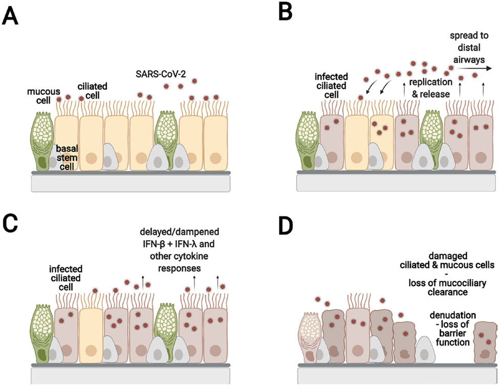
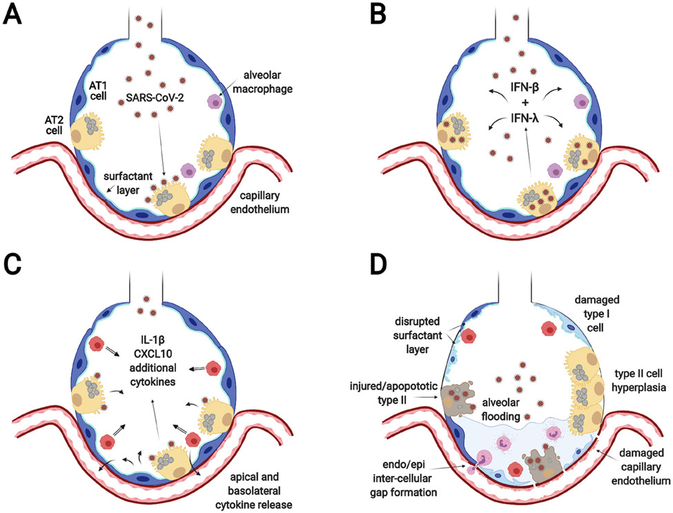
COVID-19 has different clinical stages, and effective therapy depends on the location and extent of the infection. The purpose of this review is to provide a background for understanding the progression of the disease throughout the pulmonary epithelium and discuss therapeutic options. The prime sites for infection that will be contrasted in this review are the conducting airways and the gas exchange portions of the lung. These two sites are characterised by distinct cellular composition and innate immune responses, which suggests the use of distinct therapeutic agents. In the nose, ciliated cells are the primary target cells for SARS-CoV-2 viral infection, replication and release. Infected cells shed their cilia, which disables mucociliary clearance. Evidence further points to a suppressed or incompletely activated innate immune response to SARS-CoV-2 infection in the upper airways. Asymptomatic individuals can still have a productive viral infection and infect others. In the gas exchange portion of the lung, the alveolar type II epithelial cell is the main target cell type. Cell death and marked innate immune response during infection likely contribute to alveolar damage and resultant acute respiratory distress syndrome. Alveolar infection can precipitate a hyperinflammatory state, which is the target of many therapies in severe COVID-19. Disease resolution in the lung is variable and may include scaring and long-term sequalae because the alveolar type II cells are also progenitor cells for the alveolar epithelium.

Keywords: ARDS; COVID-19; airway epithelium.


Conflict of interest statement

Competing interests: None declared.

Figures

Figure 1
Figure 1
SARS-CoV-2 infection of the upper airway epithelium (A) SARS-CoV-2 infects the ACE2 expressing ciliated cells. (B) SARS-CoV-2 replicates, is released apically, and infects neighbouring cells. (C) The ciliated cells initiate the innate immune response by secreting type I and type III interferons and other cytokines. It should be noted that the events in (B) and (C) are shown to illustrate the infection process. However, they occur nearly simultaneously in an infected tissue. The time from infection to initial release of virus is estimated to be about 6 hours. SARS-CoV-2 has the ability to dampen or delay the interferon and cytokine response compared with similar infections with influenza. (D) The infected ciliated cells shed their cilia leading to impaired mucociliary clearance. In some areas, the epithelial barrier is severely damaged. The basal cells are spared from infection and can proliferate and restore the damaged the epithelium.
Figure 2
Figure 2
SARS-CoV-2 infection of the alveolar epithelium (A) SARS-CoV-2 enters the alveoli, infects ACE2 expressing alveolar type II cells and replicates. (B) Infected AT2 cells release virus that infects bystander type II cells and type I (INFβ) and type III (INFλ) interferons are induced to initiate the innate immune response. (C) Infected and bystander cells respond to the interferons by secreting a variety of inflammatory cytokines and chemokines to recruit and activate immune cells. The illustration shows secretion into the alveolar lumen for simplicity, but there is also secretion to the basolateral side to attract inflammatory cells and affect endothelial cells. (D) The resultant effect is diffuse alveolar damage with loss of functional surfactant, alveolar flooding, influx of inflammatory cells, and damage to type I cells and endothelial cells.

References

    1. Ravindra NG, Alfajaro MM, Gasque V, et al. Single-Cell longitudinal analysis of SARS-CoV-2 infection in human airway epithelium identifies target cells, alterations in gene expression, and cell state changes. PLoS Biol 2021;19:e3001143. - PMC - PubMed
    1. Hu B, Guo H, Zhou P, et al. Characteristics of SARS-CoV-2 and COVID-19. Nat Rev Microbiol 2021;19:141–54. - PMC - PubMed
    1. Wiersinga WJ, Rhodes A, Cheng AC, et al. Pathophysiology, transmission, diagnosis, and treatment of coronavirus disease 2019 (COVID-19): a review. JAMA 2020;324:782–93. - PubMed
    1. V’kovski P, Kratzel A, Steiner S, et al. Coronavirus biology and replication: implications for SARS-CoV-2. Nat Rev Microbiol 2021;19:155–70. - PMC - PubMed
    1. Gengler I, Wang JC, Speth MM, et al. Sinonasal pathophysiology of SARS-CoV-2 and COVID-19: a systematic review of the current evidence. Laryngoscope Investig Otolaryngol 2020;5:354–9. - PMC - PubMed

Publication types