

A Deep Learning Approach for Histopathological Diagnosis of Onychomycosis: Not Inferior to Analogue Diagnosis by Histopathologists
- PMID: 34405243
- PMCID: PMC9413660
- DOI: 10.2340/00015555-3893
A Deep Learning Approach for Histopathological Diagnosis of Onychomycosis: Not Inferior to Analogue Diagnosis by Histopathologists
Abstract
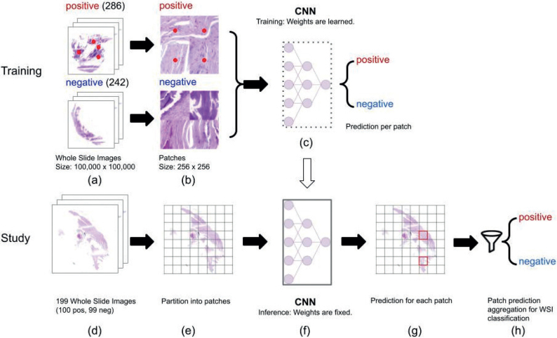
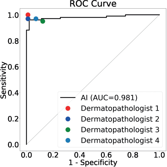
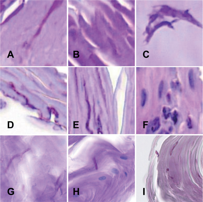
Onychomycosis is common. Diagnosis can be confirmed by various methods; a commonly used method is the histological examination of nail clippings. A deep learning system was developed and its diagnostic accuracy compared with that of human experts. A dataset with annotations for fungal elements was used to train an artificial intelligence (AI) model. In a second dataset (n=199) the diagnostic accuracy of the AI was compared with that of dermatopathologists. The results show a non-inferiority of the deep learning system to that of analogue diagnosis (non-inferiority margin 5%) with respect to specificity and the area under the receiver operating characteristic curve (AUC). The AI achieved an AUC of 0.981. One limitation of this system is the need for a large number of training images. The AI had difficulty recognizing spores and confused serum or aggregated bacteria with fungal elements. Use of this deep learning system in dermatopathology routine might help to diagnose onychomycosis more efficiently.
Keywords: deep learning; dermatopathology; onychomycosis; artificial intelligence.
Conflict of interest statement
Figures
References
-
- Westerberg DP, Voyack MJ. Onychomycosis: current trends in diagnosis and treatment. Am Fam Physician GP 2013; 88: 762–770. - PubMed
-
- Reinel D. Non-dermatophyte fungi in onychomycosis-epidemiology and consequences for clinical practice. Mycoses 2021; 64: 694–700. - PubMed
-
- Lipner SR, Scher RK. Onychomycosis: clinical overview and diagnosis. J Am Acad Dermatol 2019; 80: 835–851. - PubMed
-
- Vanhooteghem O, Szepetiuk G, Paurobally D, Heureux F. Chronic interdigital dermatophytic infection: a common lesion associated with potentially severe consequences. Diabetes Res Clin Pract 2011; 91: 23–25. - PubMed
-
- Lipner SR, Scher RK. Onychomycosis: treatment and prevention of recurrence. J Am Acad Dermatol 2019; 80: 853–867. - PubMed
MeSH terms
LinkOut - more resources
Full Text Sources
