. 2021 Oct 14;42(39):4064-4072.
doi: 10.1093/eurheartj/ehab506.

Immune complexes, innate immunity, and NETosis in ChAdOx1 vaccine-induced thrombocytopenia

Affiliations

Immune complexes, innate immunity, and NETosis in ChAdOx1 vaccine-induced thrombocytopenia

Sverre Holm et al. Eur Heart J. .

Abstract

Aims: We recently reported five cases of vaccine-induced immune thrombotic thrombocytopenia (VITT) 7-10 days after receiving the first dose of the ChAdOx1 nCoV-19 adenoviral vector vaccine against corona virus disease 2019 (COVID-19). We aimed to investigate the pathogenic immunological responses operating in these patients.

Methods and results: We assessed circulating inflammatory markers by immune assays and immune cell phenotyping by flow cytometry analyses and performed immunoprecipitation with anti-platelet factor (PF)4 antibody in plasma samples followed by mass spectrometry from all five patients. A thrombus was retrieved from the sinus sagittal superior of one patient and analysed by immunohistochemistry and flow cytometry. Precipitated immune complexes revealed multiple innate immune pathway triggers for platelet and leucocyte activation. Plasma contained increased levels of innate immune response cytokines and markers of systemic inflammation, extensive degranulation of neutrophils, and tissue and endothelial damage. Blood analyses showed activation of neutrophils and increased levels of circulating H3Cit, dsDNA, and myeloperoxidase-DNA complex. The thrombus had extensive infiltration of neutrophils, formation of neutrophil extracellular traps (NETs), and IgG deposits.

Conclusions: The results show that anti-PF4/polyanion IgG-mediated thrombus formation in VITT patients is accompanied by a massive innate immune activation and particularly the fulminant activation of neutrophils including NETosis. These results provide novel data on the immune response in this rare adenoviral vector-induced VITT.

Keywords: Immune activation; Neutrophils; Thrombus; Vaccine-induced immune thrombotic thrombocytopenia.


Figures

None
Graphical abstract
Figure 1
Figure 1
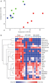
Analysis of immune complexes using immunoprecipitation and mass spectrometry. (A) Principal component analysis of the protein identification and quantification. Patient samples are marked with red, healthy vaccinated controls are marked with blue, and non-vaccinated controls are marked with green. (  B) Heatmap of the proteins that were more abundant (P < 0.05) in patient samples compared to vaccinated controls.
Figure 2
Figure 2
Characteristics of immune mediators in plasma from vaccine-induced immune thrombotic thrombocytopenia patients. Biomarkers in plasma from patients (P, n = 5), unvaccinated controls (HC, n = 11), and healthy vaccinated controls (HV, n = 8). (A) Violin plots of biomarkers, dashed lines indicate median and interquartile range. P-values indicate comparisons between HC and P. (B) Correlogram showing Pearson’s correlation between all plasma molecules detected in non-vaccinated and healthy/non-healthy vaccinated donors. A heat scale where red colour shows positive linear correlation coefficients, and blue colour shows negative linear correlation. The size of the dot corresponds to the r correlation value and the significant correlations are indicated with stars (*P < 0.05, **P < 0.01, P-values were adjusted for multiple testing using the Bonferroni method).
Figure 3
Figure 3
Immune phenotype of vaccine-induced immune thrombotic thrombocytopenia patients. Analysis of 5 cases and 12 vaccinated healthy control blood samples at three time points. (A) Peripheral blood of healthy donors and cryo-preserved PBMC from vaccine-induced immune thrombotic thrombocytopenia patients were surface stained to characterize low-density neutrophils. After the removal of debris and dead cells based on FSC-A and SSC-A, singlets were gated for haematopoietic cells. Granulocytes were defined as CD45+SSCHiCD66b+CCR3CD123 (see also Supplementary material online, Figure S2 for gating). (B) Longitudinal follow-up of neutrophils in surviving vaccine-induced immune thrombotic thrombocytopenia patients. The expression of maturation markers on neutrophils was compared between healthy donors (HD, top panel) and fatal vaccine-induced immune thrombotic thrombocytopenia (Patients 2 and 5, top) or in longitudinal samples for non-fatal cases (Cases 3 and 4 at Day 7, Day 14 or 20 for Time-points 2 and 3, respectively). The intensity of each marker was normalized in the overlaid histograms. See also Supplementary material online, Figure S4 for hierarchical clustering of all time points.
Figure 4
Figure 4
Thrombus from vaccine-induced immune thrombotic thrombocytopenia patients is rich in neutrophils and stain positive for IgG and neutrophil extracellular traps. (A) Haematoxylin and eosin staining of vaccine-induced immune thrombotic thrombocytopenia and control thrombus at 100× and 400× magnifications. (B) Immunofluorescent staining of IgG (red) in vaccine-induced immune thrombotic thrombocytopenia—control venous thrombus and control cerebral arterial thrombus. (C) Double immunofluorescent staining of S100A8 (red) and S100A9 (green) in vaccine-induced immune thrombotic thrombocytopenia thrombus and control venous thrombus; the lower panels are magnifications of selected parts of the original image as indicated, showing the presence of the S100A8/A9 complex outside of cytosol in vaccine-induced immune thrombotic thrombocytopenia thrombus. (D) Double immunofluorescent staining of elastase (green) and citrullinated histone H3 (red) in vaccine-induced immune thrombotic thrombocytopenia thrombus; the image to the right is a magnification of the original image as indicated. DAPI serves as a nuclear DNA counterstain (blue). Scale bars indicate 100 µm. (E) Single-cell suspension was made from the blood clot and analysed by flow cytometry, presented as UMAP plots showing the clustering of all haematopoietic immune cells by tissue origin. Cells composing the clot occupy the upper right region of the two-dimensional map. Clustering is based on the expression of all phenotypic markers assessed. Granulocytes can be identified as SSC-AHI CD45low cells. Automatic clustering was generated by Phenograph. (F) Cluster quantification in samples derived from the fatal Case 5. A heat map represents the frequency of each cluster. See also UMAP phonograph, Supplementary material online, Figure S4.

Comment in

References

    1. Norwegian Institute of Public Health. COVID 19 Ukerapport—uke 10. 2021. https://www.fhi.no/contentassets/8a971e7b0a3c4a06bdbf381ab52e6157/vedleg... August 2021).
    1. Norwegian Institute of Public Health. Vaccination with AstraZeneca Vaccine against COVID-19 Put on Hold. 2021. https://www.fhi.no/en/news/2021/vaksinasjon-med-astrazeneca-vaksinen-mot....
    1. Schultz NH, Sørvoll IH, Michelsen AE, Munthe LA, Lund-Johansen F, Ahlen MT, Wiedmann M, Aamodt A-H, Skattør TH, Tjønnfjord GE, Holme PA. Thrombosis and thrombocytopenia after ChAdOx1 nCoV-19 vaccination. N Engl J Med 2021;384:2124–2130. - PMC - PubMed
    1. Greinacher A, Thiele T, Warkentin TE, Weisser K, Kyrle PA, Eichinger S. Thrombotic thrombocytopenia after ChAdOx1 nCov-19 vaccination. N Engl J Med 2021;384:2092–2101. - PMC - PubMed
    1. Scully M, Singh D, Lown R, Poles A, Solomon T, Levi M, Goldblatt D, Kotoucek P, Thomas W, Lester W. Pathologic antibodies to platelet factor 4 after ChAdOx1 nCoV-19 vaccination. N Engl J Med 2021;384:2202–2211. - PMC - PubMed

Publication types