

Association of 2D and 3D transvaginal ultrasound findings with adenomyosis in symptomatic women of reproductive age: a prospective study
- PMID: 34406269
- PMCID: PMC8341039
- DOI: 10.6061/clinics/2021/e2981
Association of 2D and 3D transvaginal ultrasound findings with adenomyosis in symptomatic women of reproductive age: a prospective study
Abstract
Objective: To evaluate the association of two-dimensional (2D) and three-dimensional (3D) transvaginal ultrasound (TVUS) findings with adenomyosis symptoms.
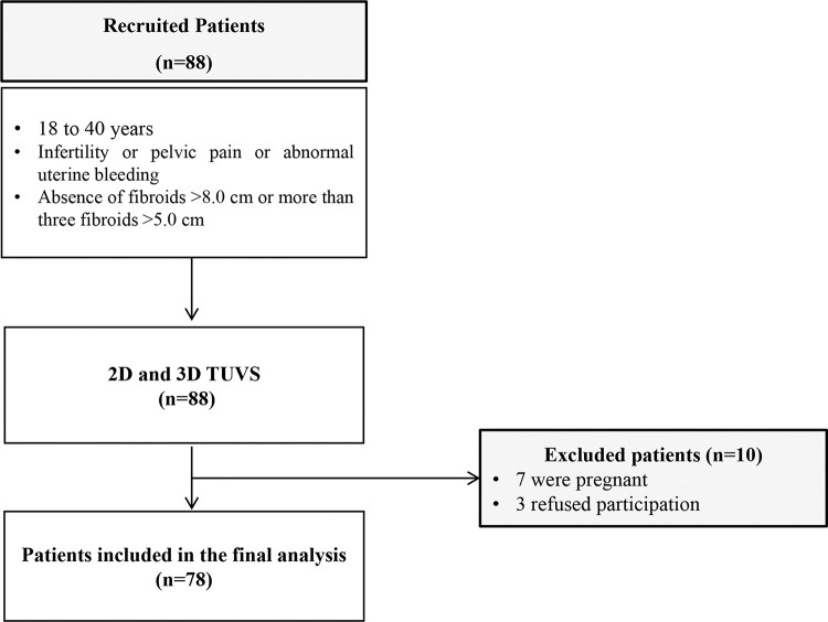
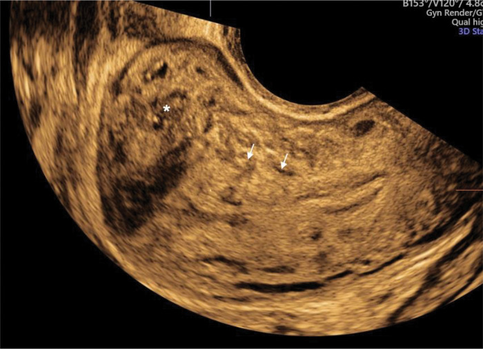
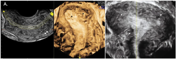
Methods: This prospective study conducted between January and December 2018 enrolled 78 women aged 18 to 40 years with abnormal uterine bleeding (AUB), infertility, and/or pelvic pain. All patients underwent 2D and 3D TVUS. Signs of adenomyosis on TVUS were identified according to the consensus of the Morphological Uterus Sonographic Assessment group.
Results: The prevalence of adenomyosis on TVUS was 55.12%. Patients with adenomyosis were older (p=0.002) and had more dysmenorrhea, AUB, and endometriosis than those without adenomyosis. When comparing the presence of symptoms with each adenomyosis feature, on 2D TVUS, severe dyspareunia was significantly associated with the presence of a poorly defined junctional zone (JZ) (p=0.023) and on 3D TVUS, patients with AUB had a more irregular (p=0.003), poorly defined (p=0.028), and interrupted JZ (p=0.011). After logistic regression analysis, signs of adenomyosis on TVUS remained significantly associated only with age over 30 years (OR: 1.2; 95% CI: 1.0-1.2) and AUB (OR: 7.65; 95% CI: 2-29). Patients with diffuse adenomyosis were older and presented with more infertility and AUB than patients with focal or no adenomyosis.
Conclusion: The findings of adenomyosis by 2D and 3D TVUS showed association with age and AUB. 3D TVUS alterations in the JZ were associated with AUB and dyspareunia. Diffuse adenomyosis was associated with older age, a greater prevalence of infertility, and AUB.
Conflict of interest statement
No potential conflict of interest was reported.
Figures
Similar articles
-
Prevalence of adenomyosis features in women scheduled for assisted reproductive treatment, using the Morphological Uterus Sonographic Assessment group definitions.Acta Obstet Gynecol Scand. 2024 Jun;103(6):1142-1152. doi: 10.1111/aogs.14812. Epub 2024 Feb 27. Acta Obstet Gynecol Scand. 2024. PMID: 38410091 Free PMC article.
-
Transvaginal sonographic features of diffuse adenomyosis in 18-30-year-old nulligravid women without endometriosis: association with symptoms.Ultrasound Obstet Gynecol. 2015 Dec;46(6):730-6. doi: 10.1002/uog.14834. Ultrasound Obstet Gynecol. 2015. PMID: 25728241
-
Inter-rater agreement in the diagnosis of adenomyosis by 2- and 3-dimensional transvaginal ultrasonography.J Ultrasound Med. 2019 Mar;38(3):657-666. doi: 10.1002/jum.14735. Epub 2018 Sep 4. J Ultrasound Med. 2019. PMID: 30182497
-
Diagnosing adenomyosis: an integrated clinical and imaging approach.Hum Reprod Update. 2020 Apr 15;26(3):392-411. doi: 10.1093/humupd/dmz049. Hum Reprod Update. 2020. PMID: 32097456 Review.
-
Transvaginal Ultrasound for the Diagnosis of Adenomyosis: Systematic Review and Meta-Analysis.J Minim Invasive Gynecol. 2018 Feb;25(2):257-264. doi: 10.1016/j.jmig.2017.08.653. Epub 2017 Aug 30. J Minim Invasive Gynecol. 2018. PMID: 28864044
Cited by
-
Prevalence of adenomyosis features in women scheduled for assisted reproductive treatment, using the Morphological Uterus Sonographic Assessment group definitions.Acta Obstet Gynecol Scand. 2024 Jun;103(6):1142-1152. doi: 10.1111/aogs.14812. Epub 2024 Feb 27. Acta Obstet Gynecol Scand. 2024. PMID: 38410091 Free PMC article.
-
New MUSA classification of adenomyosis: correlation of symptoms and clinical severity with direct and indirect features.Prz Menopauzalny. 2024 Dec;23(4):185-191. doi: 10.5114/pm.2024.145950. Epub 2024 Dec 22. Prz Menopauzalny. 2024. PMID: 39811387 Free PMC article.
-
Role of ultrasonography in the evaluation of disease severity and treatment efficacy in adenomyosis.Arch Gynecol Obstet. 2024 Feb;309(2):363-371. doi: 10.1007/s00404-023-07034-4. Epub 2023 Apr 28. Arch Gynecol Obstet. 2024. PMID: 37115275 Review.
-
Association of Uterine Tissue Innervation and Peripheral Nerve Density with Adenomyosis Related Pain. A Systematic Review.Reprod Sci. 2024 Aug;31(8):2137-2149. doi: 10.1007/s43032-024-01587-8. Epub 2024 May 8. Reprod Sci. 2024. PMID: 38720155
-
Adenomyosis and Infertility: A Literature Review.Medicina (Kaunas). 2023 Aug 26;59(9):1551. doi: 10.3390/medicina59091551. Medicina (Kaunas). 2023. PMID: 37763670 Free PMC article. Review.
References
MeSH terms
LinkOut - more resources
Full Text Sources
Medical
